


Abstract
Objective: To determine the clinical significance of skip metastases (pN2/S) in patients with resected pIIIA/N2 NSCLC. The incidence of pN2/S after mediastinal lymph node dissection (MLD) and sampling (MLS) were compared. Method: From 1997 to 2000, 580 lung resections for NSCLC performed at our department. The 151 patients (26.5%) at stage IIIA/N2 (pN2+) were grouped according to their skip metastases status. Group A included the ordinary pN2 (pN2/O) cases (71%) and group B the pN2/S (29%). Age, gender, type of resection, right or left lesion, histology, tumor lobe predilection, MLD or MLS pathologic results, the level and the number of node stations involved and survival were analyzed. Results: In 44 patients (29%) pN2/S disease was present. Statistical analysis revealed significant difference between pN2/O and pN2/S for the following: (1) pN2/S was more common for right-sided lesions (P=0.007); (2) Squamous carcinoma was the main type of pN2/S (P=0.007) and (3) pN2/S was more frequently detected after MLD than after MLS (P=0.001). Although pN2/S involved more often upper mediastinal lymph nodes (Nos. 2, 3) and one station level (pN2/S: 41 vs. pN2/O: 19.6%, P=0.228) was not found statistically significant. pN2/O was more common after right upper lobectomy and pN2/S after right lower lobectomy. The 3-year survival was more favorable for the pN2/S group (A: 24 vs. B: 36%, P=0.07). Conclusion: (1) MLD was found to be more reliable for pN2/S detection than MLS. (2) The presence of pN2/S proved to be a less aggressive form of lymphatic spread that should be taken into account in the future. (3) Strong correlation between right lower lobe tumors and pN2/S was demonstrated. (4) Different routes of cancer lymphatic spread between pN2/S and pN2/O are suggested.
1 Introduction
Surgical resection remains the cornerstone of management for non-small cell lung cancer. Among other factors the prognosis of these patients depends on metastasis to the lymph nodes, especially the ipsilateral (N2) or contralateral (N3) mediastinal lymph nodes. NSCLC with positive for metastases N2 lymph nodes (approximately 20–40% of all patients with NSCLC) shows extremely low survival rates, although highly selected patients with N2 disease achieve better 5-year results in this group [1–3]. Preoperative staging detecting positive N2 lymph nodes renders surgical resection not useful and these patients should be submitted to neoadjuvant therapy and reconsidered for surgical treatment [4,5]. Most clinicians dealing with thoracic oncology agree that patients who suffer from NSCLC with ipsilateral mediastinal lymph node (N2) involvement is a heterogeneous group [6–9]. This heterogeneity involves factors such as preoperative detection, susceptibility to neoadjuvant treatment, clinically unsuspected N2 disease, level/site and/or number of involved mediastinal lymph nodes, etc. [10–12].
The typical pattern of lung's lymphatic drainage suggests a linear model of dissemination malignancy initiating from the tumor, spreading to intrapleural lymph nodes and then to hilar ones (N1). The next station is the ipsilateral mediastinal lymph nodes in a downstream manner, i.e. from the closer nodes to the hilum to the most distant [13]. In extremis, the contralateral mediastinal and the extrathoracic lymph nodes are involved (N3). This is not always the case. Sometimes mediastinal lymph nodes metastases are detected without intraparenchymal and hilar lymph nodes involvement, which is called skip metastasis.
Although several studies have been published during the last few years, the exact incidence, the clinical significance, the pathologic and oncologic interpretation of skip metastases remains to be clarified. The present study was conducted on a fairly large population in order to determine these parameters for skip metastases in patients with resected pIIIA/N2 NSCLC. Several clinicopathologic factors were analyzed and compared in the groups of patients with ordinary N2 disease and with skip metastases.
2 Material and methods
The authors retrospectively analyzed 580 patients who were subjected to lung resection for bronchogenic carcinoma from January 1997 to December 2000. Our group performed all operations. Inoperable cases were excluded (c/pIIIB and c/pIV stage). There were 517 men (89%) and 63 women (11%), whose age ranged from 44 to 78 years (mean 64 years). The types of resection comprised 185 pneumonectomies (31.9%) and 395 lobectomies (68.1%). The patients were preoperatively staged by different means of chest imaging (radiograph, computed tomography, magnetic resonance imaging) and invasive procedures (mediastinoscopy, anterior mediastinotomy, etc.).
Three hundred ninety-five (68%) patients were subjected to systematic mediastinal lymph node dissection (MLD) and 185 patients (32%) to mediastinal lymph node sampling (MLS). MLD was defined as the en-bloc removal of all ipsilateral lymph nodes along with the surrounding fat tissue, while MLS as the systematic sampling from all ipsilateral mediastinal lymph node stations. Since no survival benefit has ever been proved for either MLD or MLS, there was no consensus in our group for a common policy. Indications for the use of either method rely upon each surgeon's opinion.
All the operations performed were radical (pRo). Thirty-five patients (6%) were subjected to postoperative adjuvant chemotherapy. Unfortunately, no common protocol was followed.
Stage pIIIA (all with positive N2 disease) consisted of 151 cases (26%). These were cases of unsuspected N2 disease. Any cN2 disease has been entered to the induction chemotherapy protocol. These cases were excluded from the study. According to their skip metastases status the 151 patients (26%) at stage IIIA/N2 (pN2+) were grouped. Group A included the ordinary pN2 (pN2/O) and group B the positive mediastinal lymph node skip metastases (pN2/S) cases. Age, gender, type of resection, right or left lesion, histology, tumor lobe predilection, MLD or MLS pathologic results, the level and the number of node stations involved and survival were analyzed. Furthermore, the N2 lymph nodes were classified according to their location in association with primary tumor location. The sites of the mediastinal lymph nodes were named according to lymph node map for NSCLC proposed by Naruke et al. [18]. Specifically Nos. 1–6 were designated as upper mediastinal lymph nodes (UMLN) whereas 7–9 as lower mediastinal lymph nodes (LMLN).
As a retrospective study survival analysis referred to the 3-year survival rate, according to primary tumor location, positive mediastinal lymph node location, number of positive mediastinal lymph node stations, and to the combination of metastatic N2 lymph nodes level and primary tumor location, since the study has not matured for 5-year survival estimation.
Statistical analysis was performed using Student's t-test (otherwise the Wilcoxon rank-sum test) and χ2 (Fisher's Exact test when needed) test where appropriate.
3 Results
Patients at stage pIIIA (pN2+) were the target group to be studied. pN2+group consisted of 151 patients (26%) submitted to 49 pneumonectomies (32.5%) and 102 lobectomies. The right side was the most common one of the primary lesion (61%). Among 151 cases with positive mediastinal lymph nodes (pN2+), 44 were skip metastases (29%) (pN2/S) and 107 of ordinary positive N2 status (PN2/0).
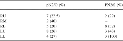
The skip lesions group consisted of 36 men (81.8%) and 8 women (18.2%), whose age ranged from 54 to 72 years (mean 62 years). The patients with pN2/S were subjected to 19 pneumonectomies (42.8%), 14 right (74%) and 5 left ones (26%) and to 25 lobectomies (57.2%), which involved 5 right upper (20%), 14 right lower (35.6%), 4 left upper (16%) and 2 left lower ones (8%). The localization of the primary lesion was right-sided in 31 cases (70.5%) and left-sided in 13 cases (Table 1) . Histology detected 31 squamous carcinomas (70.4%) and 13 adenocarcinomas (29.6%). The grade of malignancy was high in 21 cases (47.7%), moderate in 16 (36.4%) and low in 7 cases (15.9%). MLD was performed in 34 cases (77.2%) while MLS was performed in 10 cases (22.8%).
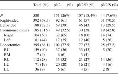
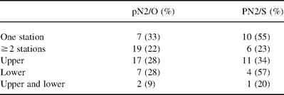
Demographic data of positive N2 lymph nodes
The T status was: T1, none (0%); T2, 25 cases (57%) including 17 cases of them with visceral pleural involvement; T3, 19 cases (43%) including seven cases with parietal pleural involvement and one case with diaphragmatic involvement.
In 25 cases N2 disease was detected during the preoperative clinical staging (cIIIa). These patients were submitted to neoadjuvant chemotherapy and were all good responders with consequent downstaging to N0/1 evaluated with CT scanning or redo mediastinoscopy upon indication, since PET scan was not available at that period of time. In nine cases N2 positive lymph nodes were detected during pathology examination (36%). Two of them (22.2%) proved to be skip lesions (Table 2) .
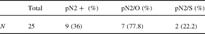
Post-neoadjuvant pN2/S results
The pN2/S lymph nodes level included 32 cases (72%) of upper level involvement; 7 (16%) cases of lower level; 5 cases (12%) of both upper and lower levels (only right-sided lesions). In details: level 2 and/or 3 and/or 4=19 cases (43.2%); level 5 and/or 6=13 cases (29.5%); level 7=5 cases (11.4%); level 8=none; level 9=2 cases (4.6%); 2 and/or 3 and/or 4+7=4 cases (9%); 2 and/or 3 and/or 4+9=1 case (2.3%). Level 7 was less frequent in pN2/S than in pN2/O group (20.4 vs. 39%, respectively, P<0.001) and levels 5 and 6 more common in pN2/S group (29.5 vs. 18%, respectively). One station involvement appeared in 18 cases of pN2/S (41%) and to 21 cases (19.6%) in ordinary pN2 group (Table 3) .
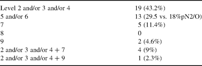
pN2/S lymph node level
The pN2/S group findings can be summarized as follows: Right lung resections predominate in a percentage of 70.5%. The most frequent histologic type was squamous carcinoma (70.4%) and the most common localization of primary tumor that developed mediastinal lymph nodes skip metastases was right lower lobe (57%), followed by right upper lobe (20%). Surprisingly, 77.2% of pN2/S were detected in specimens after MLD. High grade of tumor differentiation was demonstrated in 47.7%. The level distribution among pN2/S lymph nodes revealed a high percentage (41%) of one-station metastases, which might be the explanation of the more favorable survival rates, compared to the one of ordinary N2 disease (36 vs. 24% at 3 years). UMLN (levels 2–4) were the most frequent site of skip metastases (72%).
The N2/S group was compared to ordinary N2 disease group (Table 4) . pN2/S was more common for right-sided lesions (P=0.007) whereas squamous carcinoma was the main type of pN2/S (P=0.007). pN2/S was more frequently detected after MLD than after MLS (P=0.001). Although pN2/S involved one station level more often (pN2/S: 41 vs. pN2/O: 19.6%, P=0.228) this was not found to be statistically significant. On the contrary, pN2/S involved more often UMLN (P=0.003). pN2/O was more common after right upper lobectomy, whereas pN2/S after right lower lobectomy (P<0.001). Tumor differentiation in group B was of higher grade though not statistically significant (28.1 vs. 47.7%, P=0.763). The 3-year survival was more favorable for the pN2/S group (A: 37 vs. B: 24%, P=0.07).

Comparative characteristics of N2 positive lymph nodes
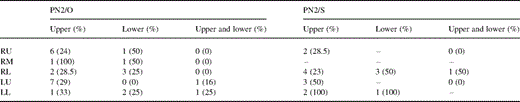
The mediastinal lymph node involvement according to primary tumor location was studied (Table 5) . In the pN2/O group positive lymph nodes belonged to the upper mediastinal group when the primary tumor was located at right or left upper lobes, whereas tumors of right middle, right lower and left lower lobe metastasize more often to the LMLN or less commonly to both UMLN and LMLN. On the contrary, skip metastases were most often detected to the UMLN irrelevantly to the primary tumor location. For right side and both lower lobes' tumors the above comparison found to be statistically significant (P<0.001). One should note that among LMLN, subcarinal location was the most common site for the pN2/O group in contrast to pN2/S group.
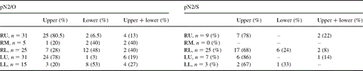
Lymph node involvement according to primary tumor location
A 3-year follow-up was conducted. Survival analysis revealed that in pN2/S group better prognosis should be attributed to the more common finding of one station nodal metastasis (Table 6) since no survival benefit was detected for multiple stations metastases between pN2/O and pN2/S groups. Left side and lower lobe primary tumor location were proved to be favorable prognostic factors (Table 7) . Besides, the level of mediastinal lymph node metastases seemed to influence survival. The analysis of pN2/S to LMLN carried a better 3-year survival rate (Table 6).
Three-year survival according to the number of stations and to the location of positive mediastinal lymph nodes
Three-year survival according to primary tumor location
The study of 3-year survival according to primary tumor location combined with the level of mediastinal lymph node metastases favored pN2/S patients' survival when the primary tumor was located either to the lower lobes or to the left side along with skip metastases confined to the LMLN. In detail, a tumor of either lower lobes with positive pN2/S LMLN or a left-sided tumor demonstrated the best prognosis (Table 8) .
Three-year survival according to primary tumor location in combination with positive mediastinal lymph node location
Neither the type of resection nor the primary tumor histology had any statistically significant impact on survival.
Postoperative chemotherapy did not offer any survival benefit. Thirteen patients subjected to adjuvant therapy demonstrated survival rate of 38.2%, while for the rest 33.3%. This difference was not found statistically significant.
4 Discussion
The presence or absence of lymph node metastasis is the single most important factor for estimating the possibility of disease recurrence and prognosis in surgical treatment of non-small cell lung cancer. The incidence of the recorded skip metastases in the N2 group (29%) is in accordance with that previously reported [14–21]. However, the histology distribution results and the primary tumor location are completely different in other series. In the literature is well documented that skip metastases are usually found in patients with adenocarcinomas of the right upper lobe [14–17]. Although the authors agree that right side is the more frequent one for skip metastases, squamous carcinoma was the most common histologic type of skip metastases in our study. Furthermore, right lower lobe proved to be the most common location of primary tumors that gave birth to skip metastases. To our knowledge, there is only one report in accordance with our results [22]. This might imply that the main factor responsible for skip metastases is the biologic and genetic profile of a tumor rather than the histological type. Tumor differentiation comparison (Table 4) between pN2/O and pN2/S indirectly supports the role of genetic substrate. Also unique anatomical and genetic characteristics of each patient might contribute to skip metastases development.
N2 group is an extremely heterogeneous one and IIIA(N2) stage is characterized by several subgroups with variable survival rates. For example Nos. 5 and 6 N2 nodes have better prognosis, cN2 worse than respective unsuspected pN2, single vs. multiple N2 stations, the extracapsular spread, the presence of subcarinal node metastasis, etc. Among other subcategories pN2/S should be considered as a completely different subpopulation of positive mediastinal lymph nodes. N2/S demonstrates more often UMLN involvement along with more common one-station involvement and less frequent combined UMLN and LMLN metastasis. This finding might be an explanation for the better survival rates of pIIIa (N2/S) patients. Among the positive LMLN skip metastases subcarinal nodes involvement (known for their dismal prognosis) was much less frequent than in the pN2/O group (20.4 vs. 39% respectively, P<0.001), whereas aortopulmonary window lymph nodes (No. 5) were more common in pN2/S group (29.5 vs. 18%, respectively). This also implies the need for TNM modification coupled with molecular biologic staging so that one can include all these discrete oncologic entities and predict outcome for the individual patient. The type of resection proved to have no influence on outcome.
Mediastinal lymph node dissection detected more systematically skip metastases than mediastinal lymph node sampling. This might be interpreted as superiority of MLD for more accurate intraoperative staging. Apart from micrometastasis detection skip metastases commonly involving only one positive mediastinal lymph node are another example of MLD importance. It is beyond any argument that macroscopic intraoperative evaluation of lymph node's metastasis is an unreliable method. Enlarged nodes may be benign in up to 40% of cases and ‘normal-looking’ ones may be malignant in almost 25% of cases. Under these circumstances sampling might lead to loss of a skip lesion and consequently to erroneous pathologic interpretation (understaging). This has deleterious effects on decision-making based on estimated prognosis, life expectancy and on evaluating the possibility for adjuvant therapies. It might be suggested that complete hilar and mediastinal lymphadenectomy should be done because of the frequent incidence of skip metastasis, which is actually a case of hidden, unsuspected N2 disease, unless radioguided sentinel node detection is undertaken which is now accurate in 85–96% [22,23]. Almost 20% of patients with cI or cII stage were clinically downstaged because of undetected mediastinal lymph node malignant involvement (N2) [24]. This postoperative upstaging should be attributed to skip metastases in up to 33% [15].
Riquet et al. have reported direct lymph passages from each lobe to mediastinum [25]. More commonly these communications were observed in the upper lobes. However, in our study, skip metastases were strongly related to primary lesion of right lower lobe although not statistically significant. Since at least one more report [22] supports this correlation, it is concluded that great variability exists concerning the patterns of lymphatic drainage from bronchopulmonary segments to mediastinal lymph nodes. One should not take for granted that cancer lymphatic spread follows a linear model from intraparenchymal nodes to hilar, mediastinal and extrathoracic ones. The lymphatic network draining the lung is extensive and variability is probably the rule. This provides multiple pathways for dissemination creating a complicated model to be used for clinical assessment. It is accepted that one should not underestimate the genetic profile of the primary tumor, which might keep a central role to the mode of tumor's lymphatic spread [16].
Favorable survival of patients with skip metastases should be attributed mainly to the high percentage of one station mediastinal lymph node involvement as well as of high tumor differentiation and to the lower detection of subcarinal nodes metastases. However, the lack of survival benefit for pN2/S group when compared to pN2/O for multiple stations of the involved N2 nodes suggests that skip metastases can be considered as a sign of the systemic nature of NSCLC. One station skip metastasis should be evaluated as a solitary intrathoracic metastasis with good prognosis if it is removed along with the primary tumor.
Further prospective studies should be conducted using immunohistochemical node examination in order to detect micrometastases and to define the exact incidence of skip metastases. One should also proceed to more ‘sophisticated’ staging methods in order to obtain more efficient treatment algorithms for such an obscure and complicated clinical entity as lung cancer.
In conclusion, the presence of pN2/S proved to be a good prognostic factor that should be taken into account in the future. Strong correlation between right lower lobe tumors and pN2/S was demonstrated. MLD was found to be more reliable for pN2/S detection than MLS. Different routes of cancer lymphatic spread between pN2/S and pN2/O are suggested.
Appendix A Conference discussion
Dr Yildirim (Turkey): Would it be wise to stop searching for skip metastases upon finding no metastases in the sentinel lymph nodes? Would you please comment on this?
Dr Misthos: I beg your pardon, I did not understand exactly what you mean.
Dr Yildirim: Would it be wise to stop searching for skip metastases when you find no metastases in the sentinel lymph nodes?
Dr Misthos: Well, since their incidence is 29% among N2 disease, they obviously do exist. That means that even though our finding for intrapleural lymph nodes is negative, that is not something that reassures us. On the other hand, I might add to that something that is very, very controversial. I mean benign-looking lymph nodes are about 25% positive, and enlarged nodes, suspected, are 40% benign. So for more accurate intraoperative staging and so as to come to a conclusion for multimodality treatment, I think we should proceed to mediastinal lymph node dissection even though our findings of N1 lymph nodes are negative.
Dr W.S. Walker (Royal Infirmary of Edinburgh, Scotland): I have just one question. You've done a lot of univariate testing here. Have you thought of correcting for that in your analysis?
Dr Misthos: I'm sorry, I didn't get your question.
Dr Walker: It's just a small statistical point. You have done a lot of single comparisons.
Dr Misthos: Yes. You mean whether it is unbiased or not? Your question is to statistical analysis, I think. You are talking about a multivariate analysis between the two groups?
Dr Walker: I just wondered whether you might have corrected for multiple testing. It was just a question, not a big issue.
Dr Misthos: Okay.
