


Case summary
We present the case of a 68-year-old woman with a giant aneurysm of the sinus of Valsalva of the non-coronary aortic cusp leading to heart failure, diagnosed with transthoracic echocardiography. The patient was successfully operated with resection of the aneurysm and reconstruction of the sinus with xenopericard and recovered completely.
Case description
An active 68-year-old woman was referred to our hospital with acute heart failure with New York Heart Association (NYHA) functional classification Class III. Her medical history revealed arterial hypertension and surgical closure of an atrial septal defect (ASD) and correction of partial anomalous pulmonary venous connection at the age of 23.
Echocardiography showed a dilated left ventricle (end-diastolic diameter 56 mm) with good systolic function, a dilated right ventricle (end-diastolic diameter 51 mm) with good systolic function, moderate mitral regurgitation, and severe tricuspid regurgitation with an estimated pulmonary artery pressure of 62 mmHg. Furthermore, a giant aneurysm of the sinus of Valsalva of the non-coronary aortic cusp was seen with rupture to the enlarged left atrium (Figure 1). Colour Doppler flow showed a continuous shunt (Figure 2, Supplementary material online, Video S1) between the aneurysm and left atrium with a gradient of 70 mmHg (Supplementary material online, Figure S3). The rupture was earlier not appreciated on neither cardiac computed tomography (Supplementary material online, Figure S4) nor magnetic resonance imaging. The patient was successfully operated with resection of the aneurysm, reconstruction of the sinus with xenopericard, and mitral and tricuspid valve repair. She recovered completely.
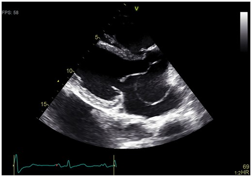
Transthoracic echocardiography showing a giant aneurysm of the sinus of Valsalva of the non-coronary aortic cusp.
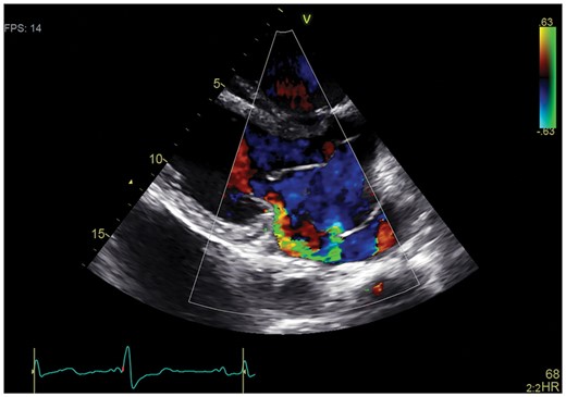
Transthoracic echocardiography showing colour Doppler flow with a shunt between the aneurysm and left atrium.
A sinus of Valsalva aneurysm (SVA) is a cardiac malformation that stems from incomplete fusion of the aortic media and the aortic valve annulus. Congenital defects that can be associated with SVA are ventricular septal defects (31%) and bicuspid aortic valves (9%).1 ASD has been reported four times in literature as coexistent congenital defect.2 Rupture can lead to a large shunt causing congestive heart failure and cardiac tamponade. Uncorrected, average life expectancy is poor with a median survival of 1–2 years.1 Treatment of choice is surgical correction but percutaneous closure can be an alternative in selected cases with high surgical risk.3 The potentially dramatic consequences of rupture of a Valsalva aneurysm justify immediate echocardiography if suspected.
Consent: The author/s confirm that written consent for submission and publication of this case report including image(s) and associated text has been obtained from the patient in line with COPE guidance.
Conflict of interest: none declared.

Comments