


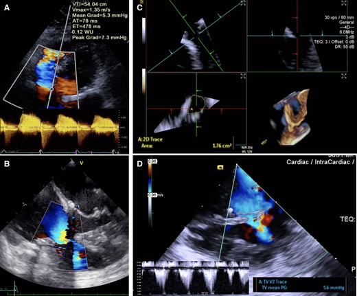
A 37-year-old female with atrioventricular septal defect had undergone patch repair and left-sided atrioventricular (AV) valvuloplasty at 2 years of age. She developed significant regurgitation of both AV valves 2 years ago, which were surgically repaired including annular ring insertions. However, 18 months later, she complained of progressive dyspnoea and ankle swelling. Transthoracic echocardiogram showed an increase in right-sided AV valve gradient to a mean of 5 mmHg with mild regurgitation. Flow acceleration over the right-sided AV subvalvular region was suspected (Panel A).
A transesophageal echocardiogram was performed, but assessment was limited by artefact from the previous annuloplasty ring (Panel B, see Supplementary data online, Video S1). Therefore, three-dimensional (3D) intracardiac echocardiogram (ICE) was performed under local anaesthesia through the right femoral vein. From the right atrium with slight anterior tilt of the ICE probe, the right-sided AV valve was visualized and 3D images showed restricted motion of the septal and posterior leaflets with a valve orifice of 1.76 cm2 as measured by planimetry (Panel C, see Supplementary data online, Video S2). The ICE catheter was further advanced into the inlet portion of the right ventricle, which revealed multiple fibrous strands at the right-sided AV subvalvular apparatus causing severe stenosis (Panel D, see Supplementary data online, Video S3). The patient was referred for surgery during which the findings of ICE were confirmed.
Right-sided subvalvular AV valve obstruction is rare. ICE is useful in imaging the right-sided valvular structures, especially with the presence of intracardiac prosthesis, which often obstruct the images from transesophageal echocardiogram.
Supplementary data are available at European Heart Journal - Cardiovascular Imaging online.
Data availability
No new data were generated or analysed in support of this research.
Author notes
Conflict of interest: None declared.
