


Case history
A 32-year-old woman was referred with a diagnosis of acute kidney injury (AKI). She had initially presented to a regional district hospital with a short history of feeling generally unwell with flu-like symptoms, headache as well as conjunctivitis and photophobia. She had no previous medical history whatsoever and was on no medication. She then developed thrombocytopenia (platelet count 37 × 109/L [normal 140–440 × 109/L]), AKI (serum creatinine 449 μmol/L [normal 45–84 μmol/L]) and mildly deranged liver function tests (ALT 81 U/L [<41 U/L]). Lactate dehydrogenase was elevated at 900 U/L (240–480 U/L). Hepatitis and HIV serology were negative. A haematologist did not see any fragmented red blood cells in repeated films of peripheral blood, which were also negative for malaria and ehrlichiosis. Intravenous ceftriaxone was begun to cover meningoencephalitis although a subsequent sample of cerebrospinal fluid was normal. A computed tomography of the brain was also normal. On arrival here, her vital signs were stable and she was no longer photophobic. There was no neck stiffness. The remainder of the clinical examination was essentially unremarkable. The patient had not travelled abroad and all her family were well. She worked as a zookeeper in a wildlife park. During the last weeks, she had worked with a variety of captive birds, kangaroos, and primates, such as lemurs (Figure 1). The patient did not recall exposure to sick animals but had cleaned kennels recently. Urine dipstick was positive for blood and leucocytes but negative for protein and nitrite. Virology and autoimmune screen was negative. Ultrasound showed normal sized kidneys with no evidence of obstruction. Plans were made for an urgent renal biopsy but serum creatinine improved to 221 μmol/L on Day 4.
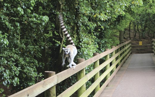
Ring-tailed lemur (Lemur catta), June 2012. This species is endemic on the island of Madagascar. It is perhaps the best known of all lemur species due to the fact that it reproduces readily in captivity and is therefore the most popular lemur in zoos worldwide.
Question
What is the most likely diagnosis?
Answer
The most likely diagnosis is leptospirosis, based on the patient's occupational history, the presentation with headache and conjunctivitis with concurrent thrombocytopenia, AKI and elevated liver function tests. A positive serology was received for Leptospira (IgM 1:1280 and micro-agglutination test [MAT] 1:320). A subsequent sample was positive for Leptospira IgM at 1:2560, strongly suggesting acute infection. Attempts at speciation failed.
Discussion
Leptospirosis [1] is a zoonotic disease caused by a microorganism of the Leptospira genus, an obligate aerobic spirochete of worldwide distribution, with two species: L. interrogans (pathogenic) and L. biflexa (non-pathogenic and saprophytic). The rat is the major reservoir of Leptospira in urban areas although a variety of other animals [1] including lemurs [2] also harbour the disease. Transmission to humans occurs through contact with blood, tissues or urine of infected animals or when injured skin is exposed to contaminated water. The mean incubation period is 15 days.
Clinical syndromes vary from self-limiting illness to fatal disease. The disease generally begins with the abrupt onset of fever and rigors as well as myalgia and headache [1]. Conjunctivitis is common but often overlooked [1]. Myalgia, splenomegaly, lymphadenopathy, pharyngitis and skin rash may also occur. Laboratory tests often show thrombocytopenia and elevated liver function tests although jaundice is only seen in a minority of cases within the severe end of the disease spectrum (Weil's disease) [1]. Other extra-renal complications include uveitis, pulmonary haemorrhage, acute respiratory distress syndrome, myocarditis and rhabdomyolysis [1].
Renal manifestations of leptospirosis are due to acute interstitial nephritis and acute tubular necrosis [3]. The spectrum of renal involvement ranges from asymptomatic abnormalities in the urine sediment to AKI requiring dialysis [3]. In milder cases, renal function usually recovers spontaneously in a few days or 1 week. In more severe cases, normalization of the renal function often occurs in the second week of the disease concomitantly with the increase in the number of platelets and decrease in bilirubin levels.
A high degree of suspicion is required to make the diagnosis and detailed history taking is of particular importance. Most patients will have exposure to animal urine through either occupation (farmers, abattoir or sewage plant workers) or recreational activities (fresh water swimming, canoeing etc.) [1]. A definitive diagnosis of leptospirosis is established by isolation of the Leptospira, but it is a difficult technique that is rarely performed outside of a research laboratory. Detection of IgM antibodies by use of enzyme immunoassay has high sensitivity and specificity (∼90% for both), although sensitivity is lower (39–72%) in the acute phase. Antibodies can also be detected by the MAT. Ideally, both these tests are undertaken on acute and convalescent serum samples collected with 10- to 14-day interval. A 4-fold rise in titre over this period is regarded as significant and, by this criterion, the rate of false-negative results was 13% [4]. If acute serum is not available, a single high titre sample is regarded as significant. Polymerase chain reaction on serum is helpful in the acute phase (first 6 days of illness) before antibodies become detectable.
The differential diagnosis includes hantavirus haemorrhagic fever, viral or bacterial meningitis, malaria and ehrlichiosis, viral hepatitis and dengue. Films of peripheral blood showed no evidence of either malaria or ehrlichiosis although both seemed somewhat unlikely since the patient had not travelled and recalled no tick bites. Once we appreciated the patient's occupational exposure to animals (and their urine), we strongly suspected either Leptospira infection or hantavirus, but hantavirus serology was negative. Her exposure to primates led us to consider the possibility of other primate-borne zoonotic diseases [5], including flavivirus, and the case was discussed in great detail with the in-house microbiology staff. Urgent advice was therefore sought from the UK National Reference Authority for rare pathogens, the Health Protection Agency Centre for Emergency Preparedness and Response at Porton Down. However, we were all relieved when tests for dengue, West Nile virus and rickettsiae were all negative.
In conclusion, our young patient with acute kidney injury led us into a veritable zoo of ever more esoteric diagnostic considerations. The fact that the National Centre for Bioterrorism and Rare Pathogens became involved made our excursion quite a bit more exiting, and we now enjoy visits to the zoo with more robust knowledge of primate species and their pathogens.
Conflict of interest statement
None declared.
Acknowledgements
We are indebted to Dr Gail Thomson, Consultant in Infectious Diseases, at the Health Protection Agency Centre for Emergency Preparedness and Response at Porton Down, UK, for advice and to the Leptospira Reference Unit, Department of Microbiology, County Hospital, Hereford, UK, for help with serology.


Comments