


(.)
At the end of a 3-week trip throughout Peru, a 24-year-old Belgian woman developed a sudden swelling of the right lower eyelid. The patient’s history was unremarkable except for a short episode of gastritis the week before. She was unaware of a local insect bite. A preseptal cellulitis was suspected and treatment with amoxicillin-clavulanic acid was administered for 2 weeks. This improved the overall swelling; however, a underlying firm nodule became discernible.
On examination 1 month after return, a painless nodule in the medial part of the right lower eyelid remained distinguishable (Figure 1). There were no signs of inflammation. Laboratory results revealed no systemic inflammation nor eosinophilia. A magnetic resonance imaging scan showed a thin walled cystic lesion (4 × 10 mm) with a signal intensity corresponding to fluid (Figure 2). After contrast injection, enhancement of the peripheral wall was noticed.
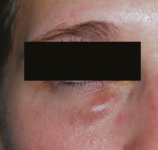
Swelling of the right lower eyelid.
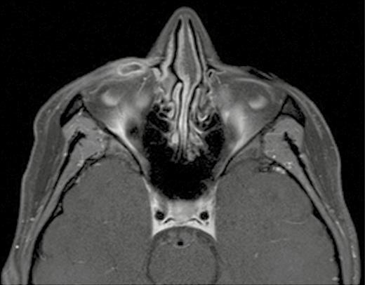
Contrast-enhanced T1-weighted magnetic resonance imaging scan of the orbit shows a thin walled cystic lesion.
What is your diagnosis?

