


A 27-year-old woman with no significant medical history presented with 12 months of progressive left knee pain and was found to have a large lesion of the distal femur on magnetic resonance imaging. This was identified as a desmoplastic fibroma on biopsy. Due to significant pain and inability to bear weight, she subsequently underwent wide resection and distal femur replacement. An indwelling catheter was placed at the time of surgery and was retained for 4 days. The patient's surgery and immediate postoperative course were uncomplicated.
Approximately 4 weeks after surgery, she re-presented with a short history of increasing pain associated with fevers and breakdown of the surgical wound.
On examination she was afebrile, but a small area of wound dehiscence with surrounding erythema and a significantly reduced range of motion of the affected knee were noted. Laboratory findings were significant for a c-reactive protein level of 209 mg/L (normal range, <5 mg/L). The white blood cell and neutrophil counts were within normal limits at 9.1 × 10⁹/L and 7.2 × 10⁹/L, respectively. No organism was isolated from blood cultures after 5 days of incubation.
She was admitted to hospital and underwent surgical washout of the joint on the same day; approximately 400 mL of purulent fluid was drained and sent for microscopy and culture. Five periprosthetic tissue specimens were also sent for culture and histopathology. No antibiotics had been administered before surgery. No organism was seen on Gram stain of the tissues and she was treated empirically with ceftriaxone and vancomycin while awaiting culture results. All 5 periprosthetic tissues showed a histiocytic reaction with neutrophil infiltration on histopathological examination.
After 72 hours of incubation (5% carbon dioxide at 37°C) small pinpoint colonies were seen on the chocolate agar plates from 4 of the 5 tissues and both fluids (Figure 1). A Gram stain was performed on the colonies, but no organisms were seen. The organism could not be reliably identified by matrix-assisted laser desorption/ionization–time of flight mass spectrometry. Subculture onto specialized agar allowed direct microscopic examination of the colonies with the distinctive colony morphology (Figure 2), revealing the organism's identification.
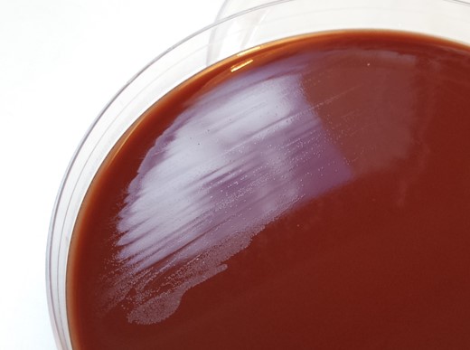
Small translucent colonies on chocolate agar after 72 hours in 5% carbon dioxide at 37°C.
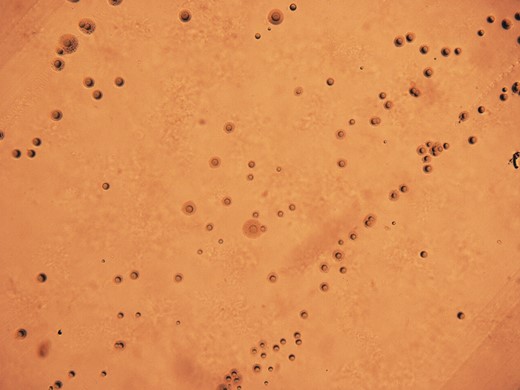
Colony morphology of isolate from periprosthetic tissue, after 24 hours of anaerobic incubation on specialized agar.
What is your diagnosis?

