


Diagnosis: Acute hemorrhagic edema of infancy.
Biopsy of purpuric lesions showed leukocytoclastic vasculitis, and direct immunofluorescence studies showed vascular wall fibrinogen deposition without immunoglobulin A (IgA) deposition, findings consistent with acute hemorrhagic edema of infancy (AHEI) (Figure 1).
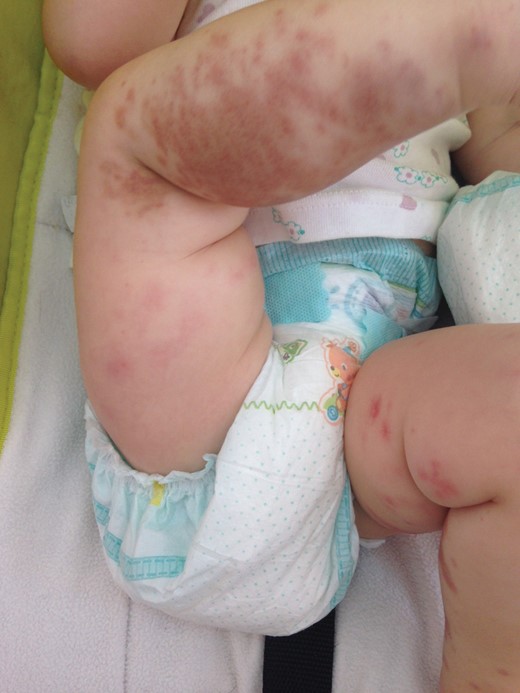
Acute hemorrhagic edema of infancy. Further spreading of purpuric rash with edema over limbs.
The first case of AHEI was reported in 1913, and later Seidlmayer and Finkelstein characterized the disease [1]. The precise incidence of AHEI remains unknown because of its rarity and confusion with other diseases. Based on a recent review, the typical patient is a boy (67%), 6–24 months of age, without racial predominance [2]. However, 1 case has been reported at birth [3]. There seems to be a seasonal variation as most cases of AHEI tend to present during winter [4].
The usual clinical appearance of a patient with AHEI includes nontoxic presentation, low-grade fever, abrupt onset of large purpuric skin lesions, and edema in face and extremities [4]. The rapid appearance of the hemorrhagic rash, together with the excellent general condition of the patient, makes it necessary to recognize this disease and differentiate it from other severe diseases such as meningococcal septicemia. Although it is a self-limited disease and the prognosis is excellent, there are reports of relapses [5, 6]. Complete recovery usually occurs within 1–3 weeks [1]. However, some rare complications have been described such as renal and intestinal involvement, epididymo-orchitis, and necrotic ulcers of the purpuric lesions [7].
According to existing literature, AHEI is associated with some viruses and bacteria (adenovirus, cytomegalovirus, herpes simplex virus, varicella zoster virus, tuberculosis, streptococci, staphylococci, pneumococcus), but the etiology of this disease remains unknown [1, 8]. Many cases of AHEI are triggered by infection (respiratory infection, urinary tract infection, bacteremia), vaccination (H1N1, measles-mumps-rubella, BCG, diphtheria-tetanus-pertussis, polio, Haemophilus influenzae), or drug intake (penicillin, cephalosporin, trimethoprim-sulfamethoxazole, acetaminophen), which reveal a possible immune-mediated pathophysiology of AHEI [4, 9]. Our patient had enterovirus isolated from the throat, and hepatitis B vaccine was given 2 weeks before admission, signifying a possible association with AHEI.
AHEI has nonspecific blood laboratory results. There might be thrombocytosis, leukocytosis, eosinophilia or lymphocytosis, and increased erythrocyte sedimentation rate or C-reactive protein. Proteinuria and microscopic hematuria have been reported, but urinalysis is usually normal [10]. Serum complement and immunoglobulin levels are frequently normal. Diagnosis is based on clinical features but if diagnosis is unclear, a skin biopsy of the rash will be helpful. The most common histopathological description is perivascular neutrophilic infiltration with nuclear fragments in the vascular wall and fibrinoid necrosis [4]. IgA deposition may be detected in up to one-third of cases, but the absence of IgA deposition with c1q deposition may be pertinent to distinguish AHEI from other leukocytoclastic vasculitis such as Henoch–Schönlein purpura (HSP) [1].
Topical or systemic corticosteroids, oral antihistamines, anti-inflammatory drugs, and antibiotics have been used for treatment [5, 6]; however, no specific therapy seems to be highly effective.
Many authors have regarded AHEI as a variant of HSP, but there are some differences between these clinical entities [5]. HSP usually occurs in children >3 years of age, whereas AHEI mostly occurs in infancy. The purpuric rash of HSP concerns the lower extremities and buttocks; in contrast, large purpura of AHEI appears on face, ankles, and wrists. Moreover, HSP includes arthralgia, renal, and gastrointestinal involvement whereas visceral involvement in AHEI is typically absent [5]. Differential diagnosis also includes Sweet syndrome, erythema multiforme, purpura fulminans, Kawasaki disease, and meningococcal septicemia [1, 6].
In conclusion, AHEI is a benign, self-limited leukocytoclastic vasculitis of small vessels affecting children <2 years of age that must be distinguished from other severe diseases to avoid unnecessary investigations and treatments.
Note
Potential conflicts of interest. All authors: No reported conflicts.
All authors have submitted the ICMJE Form for Disclosure of Potential Conflicts of Interest. Conflicts that the editors consider relevant to the content of the manuscript have been disclosed.

