


 https://orcid.org/0000-0002-9362-0465

Abstract
Coeliac Disease (CeD) is a chronic gastrointestinal inflammatory disease initiated by dietary gluten in genetically predisposed individuals. While the inflammatory processes which drive tissue destruction in the coeliac duodenum have been extensively characterized, an increased oxidative stress (OS) response has also been suggested to contribute to CeD pathogenesis. However, the precise mechanisms which regulate OS in the coeliac mucosa and whether they impact inflammation remain ill defined. The master anti-oxidant transcriptional regulator Nuclear factor erythroid 2-related factor 2 (Nrf2), and its inhibitor, Kelch like ECH-associated protein 1 (Keap1) have been implicated in chronic gastrointestinal inflammatory diseases, such as ulcerative colitis but have been largely unexplored in the context of CeD. To investigate redox balance in the CeD duodenum, we utilized single cell transcriptomics to assess overall OS and cytoprotective Nrf2 activation across cell subsets in duodenal biopsies from CeD patients. OS induced gene expression was broadly increased across multiple cell subsets in the CeD mucosa. Simultaneously, specific markers of Nrf2 activation were decreased in cell subtypes central to pathogenesis of CeD, including activated CD4+ T cells and intraepithelial T lymphocytes, indicating a distinct redox imbalance in these cells. Furthermore, pharmacological activation of Nrf2 significantly decreased gliadin induced IFNG expression in CeD duodenal biopsies. Taken together, our findings demonstrate that redox imbalance represents a therapeutic opportunity for the modulation of proinflammatory responses that drive the pathogenesis of CeD.
Introduction
Coeliac disease (CeD) is an immune-mediated enteropathy triggered by dietary gluten in genetically susceptible individuals, leading to chronic inflammation and structural damage of the duodenal mucosa [1]. As a consequence of gluten induced immune activation, a complex network of cross-talk between stromal and immune cell subsets occurs which drives mucosal atrophy and villous blunting characteristic of CeD pathogenesis.
In the duodenum, partially digested gluten peptides are deamidated by tissue transglutaminase (tTG), enhancing their binding to HLA-DQ2/DQ8 molecules on antigen-presenting cells, and promoting the activation of gluten-specific CD4+ T cells in the lamina propria [2, 3]. This drives a TH1-skewed immune response characterized by the release of proinflammatory cytokines such as interferon-γ (IFN-γ) and interleukin (IL)-21, promoting downstream B-cell activation and autoantibody production against tTG and deamidated gliadin peptides [3–5].
Concurrently, innate immune mechanisms amplify tissue damage. IL-15 produced by intestinal epithelial cells in response to stress, and proinflammatory cytokines released by gliadin-specific CD4+ T cells cooperate to give rise to highly proliferative intraepithelial T lymphocytes (T-IELs) with natural killer (NK)-like properties [6–8]. These cytotoxic T-IELs mediate epithelial cell apoptosis and contribute directly to villous atrophy, promoting tissue injury in the CeD duodenum [8, 9].
Oxidative Stress (OS) is characterized by an imbalance between the production of reactive oxygen and nitrogen species (ROS and RNS), and endogenous antioxidant defences, and is implicated in CeD pathogenesis. Several in vitro studies have demonstrated that gliadin exposure can induce OS in duodenal cells derived from CeD patients [10, 11]. Moreover, elevated plasma markers of OS in CeD patients have been reported to correlate with duodenal atrophy, indicating that OS functions in the pathogenesis of CeD [12, 13]. Furthermore, inflammatory cytokines and activated immune cells may exacerbate OS, creating a self-perpetuating cycle of tissue damage and impaired mucosal healing [14–16]. However, whether OS is dysregulated in the CeD duodenal mucosa, and if so, in which cell types, remains to be fully investigated.
To restrict OS induced tissue damage, endogenous antioxidant mechanisms act in a cytoprotective fashion. One such antioxidant defence pathway is mediated by Nuclear factor erythroid 2-related factor 2 (Nrf2), and its inhibitor, Kelch like ECH-associated protein 1 (Keap1) [17]. Mechanistically, Keap1 sequesters Nrf2 in the cytoplasm thereby inhibiting nuclear translocation and pathway activity. Increased ROS releases Keap1 from Nrf2, allowing Nrf2 nuclear entry and regulation of cytoprotective gene transcription through binding to Antioxidant Response Elements [17, 18]. While Nrf2 activity has emerged as an important regulator of mucosal barrier integrity and modulator of inflammation in the context of different gastrointestinal diseases such as ulcerative colitis, its function in the context of CeD is largely unexplored [19, 20]. It is noteworthy however that among its immunomodulatory functions, Nrf2 has been reported to act as a selective inhibitor of TH1 type responses, a key driver of CeD pathogenesis [21, 22].
To address some of these outstanding questions, we first investigated whether elevated OS is evident in the inflamed duodenum of CeD patients, utilizing a single cell transcriptomic atlas we have recently generated [23]. Significantly heightened levels of OS were detected across multiple cell subsets of the inflamed duodenum in CeD. In contrast, the expression of a specific defined panel of Nrf2 dependent genes was found to be reduced in several cell types, including activated CD4+ T cells, and T-IEL subsets which contribute to elevated IFN-γ production and are central to mucosal tissue damage in CeD [23]. Based upon these findings, we assessed whether pharmacological activation of Nrf2 might alter pathogenic responses in the CeD mucosa. In this context, Nrf2 activation suppressed gliadin peptide induced IFNG expression in CeD duodenal explants while promoting antioxidant gene expression. Taken together, these data identify a specific redox imbalance in the CeD duodenal mucosa which may play an important orchestrating role in disease pathogenesis. Understanding the interplay between OS and immune responses in CeD may open new avenues for therapeutic interventions aimed at restoring redox homeostasis alongside strict adherence to a gluten-free diet.
Materials and methods
Patient information
Duodenal Biopsies from CeD and control patients were recruited as part of the Science Underlying CoeliaC Evolution: Explanations, Discoveries, and Solutions (SUCCEEDS) Study, at the gastroenterology unit at Children's Health Ireland (Crumlin, Ireland), approved by the local Research Committee (REC-104-22). All biopsy samples were obtained at the point of diagnostic endoscopy, following informed consent/assent by individuals. All participants in the SUCCEEDS Study were consuming a full gluten-containing diet at the time of endoscopy. Cases were identified based on positive coeliac serology with subsequent histological confirmation and Marsh scoring. Recruited control subjects were undergoing endoscopy for investigation of abdominal symptoms. Controls were only included if no other inflammatory disease was identified at endoscopy, and normal duodenal histology was confirmed. Endoscopists and reporting pathologists were blinded to research participation status. Additional participant characteristics of the cohort used in ex vivo investigation can be found in Table 1. Information on patients included in the single cell RNA sequencing (scRNAseq) analysis can be found in our previous report [23].
Patient information for CeD duodenal biopsy explant studies CeD, coeliac disease.
| Characteristics n (%) | Cases | Controls |
|---|---|---|
| n | 7 | 4 |
| Age at Diagnosis (mean ± SD years) | 10.8 ± 3.4 | 9.0 ± 4.6 |
| Sex | ||
| Female | 5 (72) | 2 (50) |
| Male | 2 (28) | 2 (50) |
| Macroscopic CeD changes | ||
| Duodenal Cap | 7 (100) | |
| Most severe histology (any site) | ||
| Marsh 1 | 0 (0) | |
| Marsh 2 | 0 (0) | |
| Marsh 3a | 2 (29) | |
| Marsh 3b | 2 (29) | |
| Marsh 3c | 3 (42) | |
Data acquisition
We utilized our previously processed scRNAseq dataset, consisting of paediatric duodenal biopsies from 21 active CeD patients and 11 control patients, and can be accessed through the Gene Expression Omnibus (GEO) accession number, GSE277276 [23]. We used a recent bulk RNAseq dataset from adult duodenal biopsies obtained from 11 CeD patients and 5 control patients in our analysis and can be accessed through the GEO accession number, GSE146190 [24]. Gene sets to determine OS and Nrf2 responsive gene module scores were obtained from gene lists reported by Xu et al. and Morgenstern et al., respectively [25, 26].
scRNAseq data analysis
The scRNAseq dataset was loaded into a Seurat Object in R for initial processing [27]. We utilized UCell to generate OS and Nrf2 activity module scores, and graphed module scores for each cell type and compartment using ggplot2 [28]. Information on initial data processing of the scRNAseq dataset used in analysis can be found in our previous report [23].
Bulk RNAseq data analysis
Differential analysis was executed using DESeq2 [29]. Genes classified as differentially expressed matched the criteria of adjusted P-value <0.01 and absolute log2Fold Change > 0.5. Adjusted P-values were determined by the false discovery rate. Differential expression of genes was visualized using pheatmap.
Duodenal biopsy stimulation
Duodenal biopsies isolated from paediatric CeD or control patients were either left unstimulated or stimulated with gliadin peptides (CSBio®, Shanghai, China) with or without Ki696 (MedChemExpress®, NJ, USA), in Dulbecco's Modified Eagle's Medium (Gibco™, MA, USA) supplemented with 10% foetal bovine serum (Corning, Inc., NY, USA), and 0.1% each of Penicillin-Streptomycin (Gibco™, MA, USA), Gentamycin B (Sigma-Aldrich, MO, USA), Amphotericin B (Gibco™, MA, USA), and HEPES (Sigma-Aldrich, MO, USA) for 18 hours at 37°C with 5% CO2. Information on gliadin peptides used in stimulation can be found in Table 2.
Gliadin peptides used for duodenal biopsy stimulation.
| Peptide | Source | Length | Amino acid sequence | Concentration |
|---|---|---|---|---|
| Nex-01 | a-gliadin/Nexvax2 | 16 | ZLQPFPQPELPYPQPQ-NH2 | 5 µM |
| Nex-02 | w-gliadin/Nexvax2 | 15 | ZQPFPQPEQPFPWQP-NH2 | 5 µM |
| Nex-03 | B-hordein/exvax2 | 16 | ZPEQPIPEQPQPYPQQ-NH2 | 5 µM |
RNA isolation and reverse transcription
ISOLATE II RNA Mini Kit (Meridian Bioscience®, OH, USA) was used according to manufacturer's instructions for RNA isolation from duodenal tissues. RNA was placed on ice once eluted and RNA concentration was measured using a DeNovix DS-11 nanodrop spectrometer (DeNovix®, DE, USA). All RNA samples were stored at −80°C for future use. RNA was reverse transcribed into cDNA using the High-Capacity cDNA Reverse Transcription Kit (Applied Biosystems™, MA, USA) according to manufacturer's instructions and using a VeritiPro™ 96-well Thermal Cycler (Applied Biosystems™, MA, USA).
Quantitative PCR
Quantitative polymerase chain reaction (qPCR) was performed with TaqMan™ Fast Advanced Master Mix (Applied Biosystems™, MA, USA) on a QuantStudio™ 3 (Applied Biosystems™, MA, USA). Cycle thresholds were used to calculate the relative expression of target genes using . Primers used in this report are outlined in Supplementary Table S3.
Statistical analysis
Ex vivo data analysis was performed using GraphPad Prism® version 10 (GraphPad Software, Inc., California, USA) and is presented as mean ± SEM. According to normality, and whether data was paired, student t-test, and Mann-Whitney U test was applied for statistical analysis, as stated in figure legends.
Results
Oxidative stress is elevated in the CeD duodenal mucosa
To determine whether OS was elevated in the CeD duodenum, we investigated whether an OS gene expression signature was altered in specific cell subtypes using our previously described scRNAseq dataset [23]. OS module scores were generated for cell subsets using UCell based on a previously reported OS signature panel of 438 genes differentially expressed in the intestinal mucosa of Crohn's disease patients [26].
OS module scores were increased across broad cellular compartments in the CeD duodenum compared with control, with the exception of the epithelial cell compartment (Fig. 1a). At the cell subset level, OS gene expression signatures were elevated in multiple T-cell subsets, including T-IEL subsets, CD4+ T-cell subsets, T follicular helper cells and regulatory T cells (Tregs) (Fig. 1b) In addition, OS activation was also evident among myeloid cell subsets (DC1, monocyte-MNPs and macrophages) (Fig. 1c), B cells and plasma cells (Fig. 1d), enteroendocrine cells, and endothelial cells in the CeD duodenum compared with control (Fig. 1e and f). Collectively, these findings demonstrate that OS is broadly elevated across a multiple of cell types at sites of mucosal injury in the CeD duodenum.
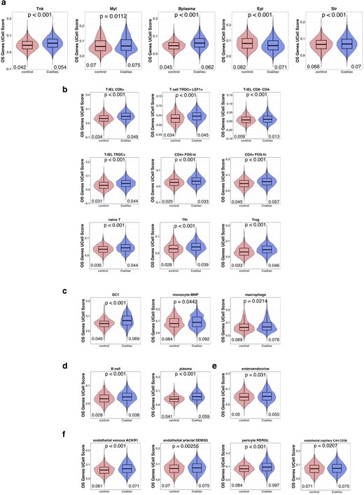
Oxidative stress module scores are increased across multiple cell types in CeD compared with control. Oxidative Stress (OS) related genes were stored as a module and OS module scores were generated using UCell. OS module scores between CeD and control were compared by cellular compartment (a), and OS module scores for each cell type were generated for cells within the T and NK (b), myeloid (c), B plasma (d), epithelial (e), and stromal compartments (f). Cell types shown have significantly increased OS-module scores in CeD compared with control. OS, Oxidative Stress. Statistical analysis performed using Mann-Whitney U test.
Nrf2 induced gene expression is decreased in the CeD duodenum
A redox imbalance between damaging prooxidant and antioxidant responses is an established characteristic of chronic mucosal inflammatory conditions [12, 13, 30, 31]. Given the broad increase in OS across multiple cell types in the CeD duodenum, we next investigated whether the activity of the antioxidant master transcriptional regulator, Nrf2 is similarly altered in the CeD mucosa. Nrf2 is constitutively expressed but post-transcriptionally regulated by Keap1 [17]. Thus, to assess Nrf2 activity, we employed a validated panel of established downstream Nrf2-dependent genes, determined by Morganstern et al. [25].
We first analysed a publicly available bulk RNAseq dataset (GSE146190), consisting of duodenal biopsies from adult CeD patients and control patients. We analysed relative expression levels of genes previously implicated in mucosal inflammation in CeD as well as expression levels of, NFE2L2 (Nrf2), KEAP1, and the robust panel of Nrf2 activation marker genes, described above [25]. As expected, CeD duodenal biopsies exhibited increased IL15, IFNG and KLRK1 (NKG2D) expression compared with control biopsies (Fig. 2a). Interestingly, and in contrast with the elevated OS signature, expression levels of NFE2L2, KEAP1, and established Nrf2 activation markers (NQO1, GCLC, GCLM, HMOX1, and TXNRD1) were significantly downregulated in the CeD mucosa compared with controls (Fig. 2a).
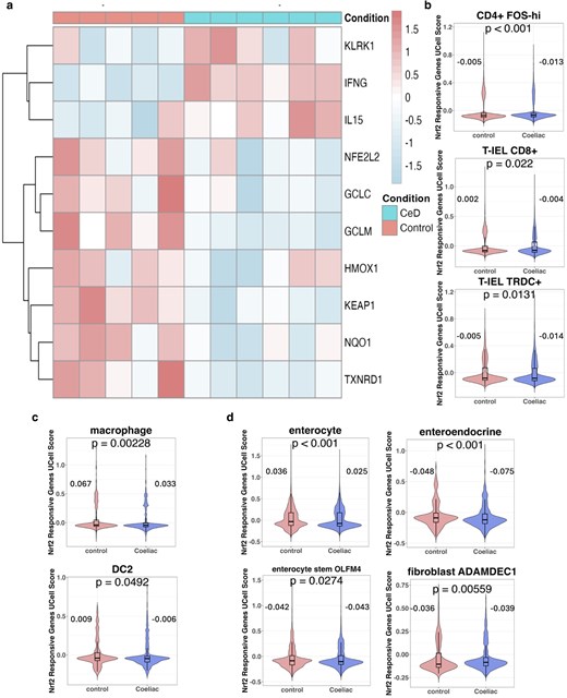
Nrf2 is downregulated at the global transcriptional level, and in cell types central to CeD pathogenesis. Expression of CeD related genes and Nrf2 activity related genes was determined for GSE146190, consisting of bulk RNAseq data from duodenal biopsies from HC (n = 5) and CeD (n = 11) (a). Nrf2 activity responsive genes were stored as a module and UCell module scores for Nrf2 responsive genes were generated for cells in our scRNAseq dataset. Nrf2 responsive gene module scores of CeD and controls were compared for cells within the T and NK (b), myeloid (c), and epithelial (d) compartment. Cell types with significantly reduced Nrf2 module scores in CeD compared with control are shown. Mean UCell values for control and CeD are annotated. Statistical analysis was performed by Mann-Whitney U test.
Given this imbalance between prooxidant and antioxidant responses, we next investigated whether reduced Nrf2 activity was evident in specific cell subsets in the CeD duodenum. To address this, UCell module scores using the Nrf2 activity gene panel were generated across all identified cell types in our scRNAseq dataset [25]. Intriguingly, while most cell types from CeD patients displayed no decrease in Nrf2 activity (Supplementary Figure S1), activated CD4+ T cells (CD4 FOShi), T-IEL CD8+ and T-IEL TRDC+ cells from the duodenum of CeD patients had reduced Nrf2 activity module scores when compared with control patients (Fig. 2b). Similarly, macrophages, DC2 cells, enterocytes, enteroendocrine, tuft, enterocyte stem OLFM4+ and fibroblast ADAMDEC+ cells also had reduced Nrf2 activity signature scores in CeD patients compared with controls (Fig. 2c and d).
These data demonstrate a redox imbalance in the CeD duodenum, characterized by elevated OS, and reduced Nrf2 activation across several cell subsets central to CeD pathogenesis.
Pharmacological activation of Nrf2 inhibits gliadin peptide induced inflammation in the CeD duodenum
Having established that redox imbalance is characteristic of the CeD duodenal mucosa and that Nrf2 activity is reduced in several cell types that play central roles CeD pathogenesis, we next investigated whether enhancing Nrf2 activation in the CeD mucosa may offer a therapeutic opportunity. To address this, we analysed the effects of a Keap1 inhibitor tool compound, Ki696, in CeD duodenal biopsies stimulated with gliadin peptides to elicit a tissue antigen specific response which models the effects of gluten exposure in vivo.
Gliadin peptide stimulation significantly increased IFNG expression, while significantly decreasing the expression of Nrf2 activation marker, GCLC in CeD duodenal biopsies (Fig. 3a and b). Treatment with Ki696 restored Nrf2 activity in CeD biopsies, evidenced through the induction of the Nrf2 target genes, NQO1 and GCLC (Fig. 3b and c). Strikingly, Ki696 significantly attenuated IFNG expression in CeD biopsies stimulated with gliadin peptides, indicating that restoring Nrf2 activity and redox imbalance can modulate a key pathogenic pathway in CeD (Fig. 3a). These findings provide initial translational validation to support the targeting of redox pathways as a potential treatment for CeD patients.
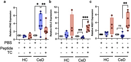
Activation of Nrf2 inhibits gluten mediated expression of IFNG in CeD biopsies ex vivo. Duodenal biopsies from individuals with CeD (n = 7) or healthy control (n = 4) were stimulated with or without gliadin peptides in the presence or absence of Ki696 (TC) for 18 hours, and subsequently analysed for IFNG (a), GCLC (b) and NQO1 (c) mRNA expression by qPCR. Statistical analysis was performed by paired student t-test.
Discussion
CeD is a chronic immune mediated enteropathy of the small intestine triggered by exposure to dietary gluten. The pathogenic mechanisms which drive antigen specific mucosal tissue destruction in the duodenum of CeD patients involve antigen deamidation and subsequent autoactivation of CD4+ T helper cells which orchestrate mucosal inflammation. Although, OS activation has been implicated in these events, the status of the OS response across distinct cell subsets has not been established [10–13]. Moreover, whether OS can impact pathogenic inflammation has not been addressed.
Previous reports have demonstrated elevated OS markers in CeD duodenal biopsies and serum [12, 30–32]. In addition, several studies have shown that exposure to gliadin peptides can induce OS in epithelial cells in vitro [12, 15]. While our scRNAseq analysis has demonstrated that an OS signature is increased across the majority of major cell types in the CeD mucosa (Fig. 1), this signature was found to be decreased in the broader epithelial cell compartment. Although these data may indicate distinct differences in the status of the OS response, among distinct cell subsets, in the inflamed CeD duodenum in vivo, the role of gliadin induced OS in primary duodenal epithelial cells warrants deeper investigation.
As Nrf2 is established as a master regulator of the antioxidant response, and is known to regulate mucosal inflammation in the large intestine [19, 20], we asked whether Nrf2 activity might be similarly altered across cell subsets. A robust panel of Nrf2 gene expression biomarkers were found to be decreased in the CeD duodenum and this decrease was evident across distinct cell subsets, including proinflammatory T cells as well as myeloid and epithelial cell types (Fig. 2). Importantly, decreased Nrf2 activity occurred despite elevated OS activation among these cells indicating a specific redox imbalance. Significantly, this imbalance was evident in activated CD4+ FOShi T cells, as well as γδT-IELs and CD8+ T -IELs subsets in the inflamed mucosa. Activated gliadin-specific CD4+ T cells are well established as central to CeD pathogenesis through the production of proinflammatory cytokines, such as IFN-γ [4, 33]. Further, gliadin-specific CD4+ T cells cooperate with IL-15, produced by stress-induced intestinal epithelial cells to promote NK-like receptor expression, such as NKG2D, in T-IELs to generate proinflammatory T-IELs which mediate intestinal epithelial injury in CeD [6–8, 23, 34]. Notably, we, and others, have previously identified γδT-IELs and CD8+ T-IELs, as well as CD4+ T cells, as major sources of IFNG expression in the CeD duodenum [23]. Together, these findings suggest that impaired Nrf2 activity occurs in IFN-γ producing cells that are central to CeD pathogenesis, and may contribute to proinflammatory signalling and mucosal injury.
To further assess the ex vivo effects of Nrf2 activation on gluten induced inflammation, we stimulated CeD duodenal biopsy explants with gliadin peptides in the presence or absence of the Keap1 inhibitor, Ki696. As expected, the addition of Ki696 led to a robust induction of the Nrf2 responsive genes GCLC and NQO1 in this setting. Interestingly, the gliadin peptides used in this study function specifically to activate HLA-DQ2.5+ gliadin-specific CD4+ T cells and the small, albeit significant, reduction in GCLC expression observed in CeD biopsies stimulated in the absence of Ki696, suggests that antigen-specific CD4+ T-cell activation alone may impair the Nrf2 transcriptional activity in duodenal CeD biopsies. Alternatively, and consistent with previous reports, gliadin peptides may act independently of CD4+ T-cell activation to alter redox balance in different cell types such as epithelial cells [12, 15]. Further investigation, will be necessary to determine the mechanistic implications of these observations.
Strikingly, inhibition of Keap1 reduced gliadin-induced IFNG expression in CeD duodenal biopsies. Having demonstrated that Nrf2 transcriptional activity is reduced in principal IFN-γ producing populations, specifically activated CD4+ T cells, CD8+ T-IELs, and γδT-IELs in the CeD duodenum, these findings suggest that restoration of Nrf2 activity dampened IFNG expression in these cells in response to gliadin stimulation. It is unclear whether these effects occur specifically in the T-cell subsets described, or as a result of the induction of Nrf2 activation among other cell types in the mucosal tissue. Indeed, our analysis has also demonstrated that Nrf2 activity is reduced among other cell types, most notably dendritic cells and myeloid cell subsets. However, it is also noteworthy that Nrf2 activation has previously been reported to directly suppress IFN-γ expression under Th1 differentiation conditions in vitro [21]. A further limitation of our analysis is relatively low patient numbers and consequent reliance on RNA level transcriptomic readouts throughout. A larger scale, adequately controlled, analysis of protein level expression and activity in this context would add significantly to this study.
Collectively, these data demonstrate the breadth of OS activation across distinct cell subsets in the inflamed CeD duodenum and provide evidence for a distinct redox imbalance across key pathogenic cell subsets. Importantly, this data also indicates that a restoration of Nrf2 activation can reduce proinflammatory processes central to CeD pathogenesis.
Acknowledgements
The authors wish to thank all the patients and their parents who have participated in this study.
Author contributions
P.T.W., D.R., S.H., S.C.B., and H.L. designed the study and performed background investigation. S.C.B., H.L., and C.F. performed experiments and analysed data. H.L. and P.T.W. wrote the manuscript.
Supplementary material
Supplementary material is available at Clinical and Experimental Immunology online.
Funding
This work was supported through funding by Taighde Éireann, Research Ireland (https://ror.org/010t7sr36, 21/FFP-P/10135), J&J Innovative Medicine, and the Children’s Health Foundation, RSFG-21-ACC07.
Data availability
Raw scRNAseq data utilized in this analysis can be found through the GEO accession number, GSE277276 https://www.ncbi.nlm.nih.gov/geo/query/acc.cgi?acc=GSE27276. Bulk RNAseq data can be accessed through the GEO accession number GSE146190 (https://www.ncbi.nlm.nih.gov/geo/query/acc.cgi?acc=GSE146190).
Ethical approval
Duodenal biopsies used in this study were obtained as part of the SUCCEEDS study at Children’s Health Ireland, approved by the local Research Committee (REC-104-22).
Permission to reproduce
Not Applicable.
Clinical trial registration
Not Applicable.
References
Author notes
Conflicts of interest: D.R. is an employee of Johnson & Johnson Ltd. All other authors declare that they have no conflict of interest.


