


 https://orcid.org/0000-0002-2475-4638

Abstract
Systemic autoimmune diseases have been traditionally studied with a special focus on the immune system and less attention was paid to the roles of target tissues that are being exposed to the immune assault. For many common autoimmune diseases accumulating data unravel and highlight the potential role of the target tissues as orchestrators of the autoimmune responses. In this selective review, using Sjögren’s disease (SjD) as a paradigm, we discuss the role of salivary gland epithelial cells (SGEC) not as innocent bystander targets of autoimmune responses, but rather as initiators and amplifiers of the inflammatory reactions. In fact, SGEC patients with Sjögren’s disease are characterized by a unique phenotype which is capable of initiating and perpetuating both innate and adaptive immune responses in the glandular microenvironment. Aberrant expression and function of TLRs and IFN pathways, lymphocyte activating proteins as well as rewired cellular metabolism and antigen-presenting features, shape this distinct auto-antigenic phenotype of SGEC. These discoveries and ideas regarding the regulatory potential of the target SGEC in Sjögren’s disease add a new dimension to our concept of regulatory circuits in autoimmunity.
Introduction
For decades, autoimmune diseases were traditionally studied with a special focus on the immune system and less attention was paid to the roles of target tissues that are being exposed to the immune assault. The ferocity of the immune response, evident once clinical manifestations become visible, made it initially difficult to imagine that an apparently uncontrolled attack on self could be regulated or even initiated by the target tissue. For many common autoimmune diseases, accumulating data unravel and highlight the potential role of the target tissues as orchestrators of the autoimmune responses from early onset to late stages of the diseases.
Salivary epithelium in Sjögren’s disease (SjD) [1, 2], fibroblast-like synoviocytes in rheumatoid arthritis [3, 4], dermal fibroblasts and keratinocytes in Systemic Lupus Erythematosus [5–7], and thyroid follicular cells in autoimmune thyroid disease [8] are only a few examples of such target tissues with major role in regulating autoimmune responses. There is accumulating evidence that the target tissues of these diseases are not innocent bystanders of the autoimmune attack but rather participate in a deleterious dialog with the immune system that leads to their own demise.
In this selective review, using SjD as a model autoimmune disease, we discuss the role of salivary gland epithelial cells (SGECs) as orchestrators of the autoimmune responses that take place in the lesion’s microenvironment. We begin by introducing SGEC and their core molecular differences between health and disease. We then discuss recent evidence that demonstrates how their altered phenotype is translated into autoimmune antigenicity; we focus on the interaction of SGEC with established inflammatory and epigenetic pathways that underlie SjD autoimmunity. Finally, we reflect on the limitations of current evidence and look toward future directions to better understand the unique contribution of SGEC to autoimmune reactivity and potential for treatment interventions. The literature covered here is not exhaustive but, rather, highlights key findings that characterize the current state of the field with respect to salivary gland epithelium.
Sjögren’s disease
SjD is one of the most common autoimmune diseases affecting women around menopause and is characterized by dysfunction and destruction of exocrine glands, mainly of the salivary and lacrimal glands. SjD is featured by a broad spectrum of clinical symptoms ranging from disease confined to the exocrine glands (organ-specific exocrinopathy) to various extraglandular manifestations (systemic disease) and B-cell lymphoma development [1].
A major characteristic of SjD is the presence of dense lymphocytic infiltrating lesions around the epithelial tissue in the salivary glands and the polyclonal/oligoclonal B cell hyperactivity, as suggested by the production of various autoantibodies in the blood (such as antinuclear antibodies, antibodies to Ro/SSA, and La/SSB ribonucleoproteins, to histones and to single-stranded DNA, rheumatoid factors, and cryoglobulins) [9, 10]. These lymphocytic infiltrations are the histological hallmark of SjD [11], and the infiltration score is a central part of the differential diagnosis in SjD [12]. The periepithelial infiltrates consist mainly by CD4+ T cells in early disease and B cells in later stages [13]. Other immune cells such as macrophages, plasmatocytoid, and follicular dendritic cells (DCs) are also often seen at lower percentage [14]. Regarding autoantibodies, anti-SSA/Ro and less often anti-SSB/La antibodies are present in the majority of patients with pSS, and they have been linked to glandular immune activation [15].
To date, it is widely accepted that exposure to specific environmental factors (e.g., a viral infection) in susceptible individuals may play a crucial role, triggering the dysregulation of the immune system and SjD occurrence. The innate immune pathways are activated, involving a subsequent pro-inflammatory cytokine production response and the interferon (IFN) pathway activation [16]. In parallel, the adaptive immune system has a central role in SjD development, as the activation of B-cells and the proliferation of Th1 and Th17 cells contribute to its progression [17]. In this context, disruptions of this delicate balance between SGEC and immune cells leads to salivary gland dysfunction, reduced secretory capacity, and alterations in the glandular microenvironment.
The role of viral infections in SjD
Viral infections can influence the immune system and stimulate the cascade of autoimmunity and the formation of autoantigens through various mechanisms, such as molecular mimicry, bystander activation, epitope spreading, and production of superantigens. Several viruses are suggested to have an effect on autoimmunity—among them are Epstein–Barr virus (EBV), hepatitis C (HCV) virus, and human immunodeficiency virus [18]. SGEC, in particular, have been well known to be targeted by various viruses [19].
EBV DNA has been found increased in the salivary gland as well as in lacrimal gland, tear specimens, and parotid biopsy specimens of patients with SjD [20, 21]. EBV can persist in the human organism in a latent form, allowing the virus to hide in the host SGEC and reactivate under favorable conditions. An EBV microRNA, the ebv-miR-BART13, has been shown to be transferred from B cells to SGEC, where it affects the fluid secretion [22]. Other theories about the effect of EBV on the autoimmune process of SjD include molecular mimicry between the viral EBNA-2 protein and the Ro-60 antigen or the viral EBER-1 and EBER-2 proteins and the La antigen.
The best example of a viral causative role of SjD is probably HCV, a virus with a specific predilection for infecting and replicating in SGEC [23]. A striking association between HCV infection and SjD as well as lymphocytic sialadenitis (Sjogren’s-like disease) has been proven, as anti-HCV was shown to be more prevalent in patients with SjD or chronic sialadenitis than in general population and histological evidence of SjD has been found in patients with chronic HCV infection [24, 25]. Moreover, HCV-RNA has been detected in saliva and in salivary glands from patients with sialadenitis, indicating that HCV infects and replicates in the SGEC of these patients [26].
In a population of HIV-positive patients, about 1/3 had symptoms of dry eyes and/or mouth, while few of them had monocellular infiltrates in their minor salivary gland biopsy meeting the criteria of focus score in SjD [27]. Furthermore, it has been shown that antibodies to proteins of HIV are present in patients with SjD [28].
The B4 Coxsackie viruses (CB4) and C1 (CB1) serotypes are being researched as potential risk factors for developing autoimmune diseases. A previous study has provided evidence that minor salivary gland cells contain viral RNA as well as the main antigenic capsid protein VP1 in SjD patients and not in patients with secondary SjD and controls [29]. Moreover, a cross-reaction between antibodies to the major epitope of Ro60 kD autoantigen and a homologous peptide of Coxsackie virus 2B protein was also shown [30].
Core salivary gland epithelium differences between health and SjD
Although the pathogenic pathways underlying SjD have not been yet elucidated, it is well-established that SGEC are central regulators of local autoimmune responses [1]. In recent decades, research has been focused on epithelial cells functions and several lines of immunohistopathologic and in vitro evidence during the last 20 years support the hypothesis that SGEC are not only the target of autoimmunity in SjD patients but may also participate in the initiation and maintenance of the inflammation [31]. Thus far, the importance of the salivary gland epithelium in regulating immune responses in SjD has not been adequately appreciated and the detailed mechanisms merit further investigation.
A milestone in understanding the role of SGEC in autoimmunity was the ability to isolate, expand and long-term culture primary cells derived from salivary gland biopsies during diagnostic work up. In 2002, Dimitriou et al. [32] presented a protocol for the establishment of human non-neoplastic SGEC lines (of ductal type) from a single lobule of labial minor salivary glands for the study of the physiology and pathophysiology of these cells, in a culture free of the influence of other cell types, thus, allowing the evaluation of the constitutive epithelial phenotype and function. In the following years several studies have used this ex vivo approach to show that SGEC under long-term culture conditions retain the ‘activated’ phenotype seen in situ in the patients’ salivary glands’ lesions. To date, it has been shown that SGEC derived from SjD patients are characterized by a unique pro-inflammatory phenotype. Large-scale discovery studies using state-of-the-art technology have provide us with a holistic view of SGEC and their core differences between health and disease.
Moreover, the ‘activated’ SGEC phenotype is characterized by significant morphological and functional changes in SGEC are strongly associated with epithelial cell dysfunction and may serve as key initiators of immune activation in SjD. In comparison to normal SGEC, SjD-SGEC exhibit reduced mitochondrial content, swollen and elongated mitochondria, as well as fewer and aberrant cristae [33]. These alterations (Table 1), regardless of the underlying cause, can induce an immunogenic phenotype and are accompanied by pronounced morphological alterations in situ [34 ]. As explained below, proteomic and transcriptomic analysis has offered insights into the transformation process of SGEC from SjD patients into innate immune cells while uncovering translational modifications associated with metabolic remodeling.
Alterations of SGEC in SjD.
| Category | Alterations in salivary epithelial cells | Functional/pathophysiological implications |
|---|---|---|
| Immune-related alterations | Interactions with T cells – Upregulation of MHC I and II molecules, antigen presentation to CD8+ and CD4+ T cells—Expression of co-stimulatory molecules (CD40, CD80, CD86) supporting T cell activation—Production of IL-6, IL-7, CXCL9, CXCL10, and CXCL11 promoting Th1 and Th17 cell recruitment and retention Interactions with B cells: – Secretion of BAFF and APRIL enhancing B cell survival and autoantibody production—Expression of CD40L–CD40 signaling components facilitating local B–T cell collaboration Interactions with innate immune cells: – Release of type I interferons (IFN-α/β) and upregulation of interferon-stimulated genes (ISGs; IFI27, MX1, ISG15)—Secretion of chemokines (CCL2, CCL5) attracting monocytes and dendritic cells—Activation of TLR3, TLR7, and TLR9 pathways, amplifying innate immune activation. Auto-reactive mechanisms: – Exposure of autoantigens (Ro/SSA, La/SSB) following apoptosis—Autoantigen-loaded exosome secretion—Upregulated Fas/FasL and NF-κB signaling sustaining chronic epithelial activation | SGECs become active participants in local immunity, acting as antigen-presenting and cytokine-producing cells that sustain chronic inflammation, promote ectopic germinal center formation, and drive autoantibody production. |
| Metabolic alterations | – Metabolic rewiring—Mitochondrial dysfunction and damage—Altered lipid metabolism, oxidative stress and DNA damage | Energy imbalance and oxidative stress exacerbate inflammation and secretory dysfunction. |
| Stress-related alterations | – Expression of adrenergic receptors linking sympathetic stress to local responses—Activation of ER stress and unfolded protein response (XBP1, ATF6, CHOP) | Sustained stress signaling promotes autoimmune activation, apoptosis and tissue remodeling. |
From altered phenotype to autoimmune antigenicity—innate immune function
Similar to other epithelial tissues, SGEC have been proved able to mediate local innate immune responses in the salivary gland epithelium in SjD [35, 36]. Multiple innate immune pathways are likely dysregulated, including the nuclear factor-κB pathway, the inflammasome, and IFN signaling. Indeed, beyond its secretory role, SGEC fulfil innate immune features, which are mainly mediated by the expression of pattern recognition receptors, for example, CD91 molecules [35] and TLRs [36] and the secretion of cytokines [37]. Although the exact initial events that cause innate immune activation of SGEC in SjD are not known, some possibilities include the involvement of pathogen-associated molecular patterns or/and danger/damage-associated molecular patterns or the aberrant expression of endogenous factors (e.g., retroelements) [38–40]
The determination of several types of epithelial tissues that express TLR molecules, such as gastrointestinal, bronchial, and urinary epithelia, supports the notion that the epithelium acts as a frontline defense of the innate immune system [41–43]. Moreover, recent studies reveal that TLRs located on the cell surface also play an essential role in the development of autoimmunity, as demonstrated in rheumatoid arthritis [44], systemic lupus erythematosus [45], and inflammatory bowel diseases [46]. While TLR signaling is required for several different autoimmune diseases, its contribution to SjD initiation and progression remains poorly understood.
TLR signaling seems to have a significant role in the activated phenotype and survival of SGEC. Studies have shown that TLR2, TLR4, and TLR6 are highly expressed in situ by salivary glands of SjD patients but not in controls [47]. Cultured non-neoplastic SGEC derived from SjD patients have been shown to express constitutively high levels of several TLRs, such as TLR1, −2, −3, and −4 compared to control-SGEC lines, as attested by the up-regulation of surface ICAM-1 (intercellular adhesion molecule-1), CD40, and MHC-I expression following treatment with the respective TLR-ligands [36], a fact which supports the intrinsic epithelial activation in SjD [31]. In particular, TLR3 expression by SGEC deserves special attention, since it is significantly higher than other TLRs and is mainly located on the membrane surface [36]. Furthermore, TLR3 signaling has been described to induce apoptotic death of epithelial cells through anoikis [48], production of cytokines that promote inflammatory responses, such as type-I IFNs and BAFF [49, 50], whereas it also augments the expression of Ro/SSA and La/SSB autoantigens in SGEC, suggesting that it might be implicated in autoantigen presentation [49]. The significance of TLR3 signaling in SjD pathogenesis is further supported by in vivo findings in experimental mouse models, where it leads to significant salivary gland hypofunction accompanied by up-regulation of type-I IFN responses and recruitment of B cells, DCs and NK-cells in the submandibular glands [51, 52].
Type I IFN signature and SGEC
IFNs have been implicated in the disruption of the salivary gland epithelium and in pathogenesis of SjD, as reflected by the elevated transcript levels of interferon-stimulated genes in SjD patients [53, 54]. Both type I and II IFNs have been observed in SjD patients versus HC (healthy controls) and SC (sicca controls), respectively, with a predominance of type I IFN signature in peripheral blood and a type II IFN signature in MSG tissues [53]. As mentioned above, epithelial cells themselves can produce type I interferon after stimulation via pattern recognition receptors, as demonstrated in studies of mice and of human SGCEs lines. Activation of the cells by TLR3 ligands could be a major source of type I IFNs within the gland [36]. Differentiated human submandibular gland cells (although debatable of being salivary origin or HELA) were stimulated with mucins or oligosaccharide residues, which were recognized by TLR4, initiating a pro-inflammatory response, cytokine, and interferon production (XCL8, TNF-a, IFN-a, IFN-b, IL-6, and IL-1b) [55]. Moreover, poly(I:C) treatment rapidly up-regulated the mRNA levels of type I IFN and inflammatory cytokines in the submandibular glands of Female New Zealand Black⁄White F1 mice, through TLR3 signaling [56]. Furthermore, TLR-3 stimulation in SGEC causes an immediate and indirect IFN-β-dependent up-regulation of Ro52/TRIM21, creating large quantities of the intracellular autoantigens [49]. Thus, as discussed above, TLR-3 is a molecule at the crossroads of innate and adaptive immunity, as its ligation provides not only signals for IFN production but also active synthesis of the intracellular autoantigens that can eventually prime adaptive immune responses.
Microarray data of salivary gland biopsies from SjD patients revealed an upregulation of interferon I signaling pathway and an increased expression of IFN-inducible genes, including BAFF and IFN-induced transmembrane protein 1, OAS1, IFIT3, IFI6, BST2, TAP1 genes [14, 54]. Thus, in the glandular lesion apart from the infiltrating immune cells, IFNs are found upregulated in SGECs, significantly accounting for the interferon signature of the glands. Similarly, IFN signatures have been observed in other rheumatic diseases, including dermatomyositis (DM), polymyositis, scleroderma, and systemic lupus erythematosus (SLE) [57]. In the salivary glands of patients with SjD, IFNβ is also abundantly produced by immune cells and the salivary gland tissue often has a mixed type I and type II interferon signature [58, 59]. Moreover, IFNλ2 is upregulated in salivary glands of SjD patients compared to individuals with non-SjD sicca symptoms. However, none of the type III IFNs was expressed constitutively in resting SGECs, but only after TLR-3 stimulation, suggesting that the in situ epithelial expression can be attributed to local microenvironment [60]. Whether SGEC-derived type III IFNs notably contribute to salivary gland pathology, either independently or in synergy with other IFNs, remains to be elucidated.
Adaptive immune function of the epithelium: from altered phenotype to lymphocyte activation
As in every tissue, in a normal salivary gland, immune surveillance is supported by the presence of tissue resident immune cells, mainly macrophages and CD8+ T cells [61]. By comparison, the histological hallmark of SjD is lymphocyte infiltration of the salivary gland comprised of CD4+ T cells in early diagnosed cases and B cells in later stages during disease progression. A logical question that has not been fully answered during the last decades is the following: what is driving the immune cells to invade the microenvironment of the salivary gland in SjD? Classical immunology suggests that several immune ‘invitations’ need to be sent systemically and ‘invite’ immune cells to infiltrate the target salivary gland tissues. In SjD, it can easily be postulated that SGECs, through their altered phenotype play this orchestrating role, by expressing and secreting pro-inflammatory molecules.
DCs and SGEC
The timeline of salivary gland infiltration indicates that DCs are among the first cells to infiltrate the tissue of SjD patients at the early stages of the disease [62]. This fact highlights their pivotal role in the immunological landscape of SjD, serving as key players in both the initiation and perpetuation of the autoimmune responses. Their interactions with SGEC, T and B cells, alongside their cytokine production, underline their central role in disease progression.
In the salivary gland microenvironment, SGEC produce pro-inflammatory cytokines (e.g. type I FNs, IL-6, TNF-α) and chemokines (CCL21, CXCL13) that recruit and activate DCs, promoting inflammation [63–65]. They also upregulate adhesion molecules (ICAM-1, vascular cell adhesion molecule-1—VCAM-1) [36] to facilitate DC migration into glandular tissue. Uniquely, SGEC can uptake and present autoantigens via MHC molecules to DCs, activating autoreactive T cells and sustaining autoimmunity. Through toll-like receptors (TLRs), SGEC recognize pathogen-associated molecular patterns and damage-associated molecular patterns [36, 55], further enhancing cytokine production and type-2 conventional dendritic cells (cDC2) recruitment.
In the same glandular milieu, studies have shown that SGEC undergo apoptosis due to chronic inflammation. Following, cDC2s can engulf large amounts of cellular debris released by apoptotic SGEC, leading to the activation of autoimmune responses. The presence of a cDC2 gene signature in the inflamed salivary glands was recently reported by transcriptomic analysis of minor salivary glands from SjD patients. This cDC2 signature was strongly associated with CD4+ T cells [66, 67]. Transcriptional alteration of cDC2s and their aberrant antigen and autoantigen uptake and processing, together with increased proliferation of tissue CD4+ cells, indicate altered antigen presentation of SjD cDC2s. On a functional level, these alterations have been strongly linked to anti-SSA positivity and the presence of type I IFNs [68]. In another similar study, it has been reported that in SjD patients, circulating pDCs have distinct transcriptional profile and are primed to enhance pro-inflammatory cytokine production [69].
While all these findings indicate a strong role for DCs in SjD, what triggers this activated phenotype remains unknown. Thus far, robust research data indicate that SGEC possess full capability of priming DCs toward autoimmune responses. Functional in vivo studies could uncover this potential.
SGEC: crosstalk with CD4+ T cells
The interaction between CD4+ T cells and SGECs is a dynamic immunological dialogue involving multiple cell types, cytokines, and signaling pathways. The initial phase of the crosstalk is mediated by the activation of CD4+ T cells in the periphery, leading to their migration into the salivary glands. Activated CD4+ lymphocytes migrate to salivary glands via chemokine gradients and adhesion mechanisms. Within the gland, SGECs—expressing MHC II, co-stimulatory molecules (CD80/CD86) [70], and cytokines— are able to act as antigen-presenting cells, initiating local immune responses. In turn, CD4+ T cells release pro-inflammatory cytokines like IFN-γ, Il-2, and IL-10, amplifying inflammation and recruiting additional immune cells [63].
CD4+ T subsets
Among the different subsets of CD4+ T cells, Th1, Th2, and Th17 cells are particularly prominent in the development of salivary gland lesions. SjD was originally considered a Th1 signature autoimmune disease, but later it was observed that both Th1 and Th2 cells are drivers of the disease depending on the stage. Evidence suggests that in SjD type-1 and type-2 cytokines are in dynamic balance. Type II microenvironment prevails in low-grade infiltration, while type I pattern increases in patients with definite SjD and patients with advanced lymphocytic infiltration [71].
SGECs can attract Th1 cells through a combination of mechanisms involving chemokine secretion and adhesion molecule expression. SGECs produce chemokines that attract Th1 cells by interacting with chemokine receptors expressed on these cells. Key chemokines expressed by SGEC include CXCL9, CXCL10 [72]. Most of the CD3+ infiltrating lymphocytes in periductal foci express CXCR3, the receptor of these chemokines. SGECs also secrete cytokines like IL-1a, IL-6 and TNF-alpha [63], which promote a Th1 environment. All these molecules create a gradient that guides Th1 cells to the SGECs contributing to salivary gland infiltration by lymphocytes. An additional mechanism used by SGEC to attract Th1 cells is the expression of adhesion molecules that facilitate Th1 cell attachment and migration. These include ICAM-1 and VCAM-1 which Interact with Th1 cells, enhancing adhesion and extravasation into tissue [2, 73]. Th1 cells largely produce IFN-γ and TNF-α, cytokines that in turn activate macrophages, NK cells and CD8+ T cells and maintain cell-mediated immunity [74], as well as IL-18, higher levels of which were noticed in the saliva of SjD patients and NOD mice [75].
Th2 cells, in contrast, are generally thought to promote humoral immunity and are characterized by the secretion of cytokines such as IL-4, IL-5, and IL-13 [76]. These cytokines encourage the activation of B cells and contribute to the chronic inflammation seen in SjD. IL-4, in particular, is a very critical cytokine since a knockout of IL-4 in NOD and NOD.B10-H2b mice was found to restore their salivary gland function [77]. SGECs contribute to Th2 cell recruitment by producing chemokines such as CCL17, CCL22 [78], which bind the Th2-associated receptors CCR4 and CCR8. They also secrete cytokines like IL-33, TSLP, and IL-25 that promote Th2 polarization through direct activation or DC priming [79]. In autoimmune conditions, SGECs expressing MHCII may engage CD4+ T cells in a Th2-skewed cytokine environment, supporting Th2 differentiation. In SjD, this Th2 bias may drive tissue remodeling or fibrosis, highlighting SGECs’ role in shaping immune responses. Understanding the balance between Th1 and Th2 recruitment by SGECs can help elucidate disease mechanisms and uncover therapeutic approaches targeting these pathways.
Th17 cells, which secrete IL-17 and other pro-inflammatory cytokines such as TNF-α, IL-22, and IL-26, are also found in increased numbers in the salivary glands of SjD patients [80]. These cells are particularly implicated in the recruitment of neutrophils and the promotion of an inflammatory environment that exacerbates glandular damage. IL-17 produced by Th17 cells induces the production of other pro-inflammatory cytokines and chemokines, further contributing to the recruitment of immune cells and the development of persistent inflammation. SGECs secrete chemokines like CCL20 and CCL25 forming a gradient guiding Th17 cells into inflamed salivary glands [80]. Several studies have showed elevated levels of IL-17 protein and mRNA in minor salivary glands of SjD patients compared with healthy individuals [81, 82], while IL-17 knockout mice models of SjD did not develop the disease and the elimination of IL-17 in B6.NOD-Aec1Aec2 mice restored normal secretory function and reduced sialadenitis primarily in female mice [83].
Follicular helper T cells (Tfh), which play a pivotal role in B cell activation and differentiation in lymphoid structures, can also be primed by SGEC. Co-cultures of naïve CD4+ T cells and SGECs from both, patients with SjD and controls, was reported to induce naïve CD4+ T differentiation into Tfh cells, as evidenced by their acquisition of a specific phenotype, characterized by Bcl-6, ICOS, and CXCR5 expression and IL-21 secretion, but also by their main functional feature: the capacity to enhance B lymphocytes survival [84]. It has also been reported that in SjD patients, a large number of circulating Tfh cells express CXCR3, which helps them migrate to the inflamed salivary glands, where CXCL10 is expressed [80]. In blood and glandular tissues of SjD patients, the frequency of Tfh cells is higher than in healthy individuals suggesting that these cells contribute to the pathogenesis of the disease by promoting the maturation of B cells [85].
Regulatory T cells (Tregs): The interaction between CD4+ lymphocytes and SGECs also involves regulatory T cells (Tregs), a subset of CD4+ T cells responsible for maintaining immune tolerance and suppressing autoimmune responses. In SjD, there is evidence of a decline in Treg function, leading to an imbalance between effector T cells and Tregs. Treg function declines through mechanisms that disrupt their recruitment, survival, or suppressive activity. This decline contributes to the unchecked activation of autoreactive CD4+ T cells, which in turn exacerbates the autoimmune attack on SGECs [86].
In summary, the interplay between SGEC and the full spectrum of CD4+ subsets, including Th1, Th2, Th17, Tfh, and Tregs, is crucial in the pathogenesis of salivary gland lesions in SjD, with each subset contributing to different aspects of the autoimmune response, tissue damage, and disease progression.
‘Invitation’ to B lymphocytes by SGCEs
B cells are a major immune population of the autoimmune processes in SjD. The fact that autoantibodies are present years (up to 18–20) before the disease onset and the occurrence of monoclonal serum components such as hypergammaglobulinemia and cryoglobulins, underlie the importance of B cells in the disease pathogenesis [87]. The crosstalk between B lymphocytes and SGECs in SjD begins with the recruitment and activation of B cells in the periphery, followed by their infiltration into the salivary glands under the influence of chemokines and adhesion molecules.
By producing various chemokines, including CXCL12 (stromal cell-derived factor-1) and CXCL13 (B lymphocyte chemoattractant), SGEC attract B lymphocytes to the inflamed salivary glands [88]. As mentioned earlier in this article, SGEC can act as antigen-presenting cells. They express major histocompatibility complex (MHC) class II molecules and are capable of presenting autoantigens to B lymphocytes.
A previous study has shown that patients with SjD and anti-La/SSB antibodies presented an active synthesis of the La/SSB protein, which was evidenced by the detection of La/SSB mRNA in the acinar cells of their minor salivary gland biopsies, in contrast to patients lacking these antibodies or those with non-specific sialadenitis [89]. Intriguingly, in SjD, the La/SSB protein undergoes cellular redistribution within acinar epithelial cells, shifting from the nucleoli to the nucleoplasm, cytoplasm, and often to the cell membrane [90, 91]. Moreover, immunogenic apoptosis of epithelial cells has been shown to induce relocalization of Ro/SSA and La/SSB autoantigens to cell surface and apoptotic blebs [92]. This aberrant localization of the two major autoantigens on the cell surface and apoptotic bodies may trigger a localized autoantigen-driven immune response, contributing to the initiation and perpetuation of autoantibody production by B lymphocytes. In line with this, the sustained overexpression of La/SSB in epithelial cells of the affected exocrine glands is thought to underlie the continuous and elevated production of anti-La/SSB autoantibodies in patients with SjD.
Once within the glandular microenvironment, B lymphocytes interact with SGECs expressing immune-related molecules such as MHC II and co-stimulatory molecules facilitating antigen presentation, where SGECs present self-antigens to B lymphocytes, initiating an antigen-specific immune response within the salivary glands. This process activates B lymphocytes, which undergo clonal expansion and differentiation, leading to the production of autoantibodies, particularly against Ro (SSA) and La (SSB) antigens.
As B cells produce autoantibodies against self-antigens, immune complexes deposit in the salivary glands, directly leading to SGEC damage and triggering a cycle of further immune cell recruitment, perpetuating the autoimmune response. Many patients with SjD have been found with elevated circulating immune complexes (CICs) of different immunoglobulin classes showing strong correlation with severe or systemic disease and extraglandular manifestations [93]. Moreover, it has been shown that higher CICs were associated with decreased salivary flow [94]. In another study, however, serum ICs did not correlate with the degree of lymphocytic infiltration in labial salivary glands of SjD patients [95]. Some immune complexes (ICs) can form locally (saliva, glands) and lead to glandular damage. A more recent study has shown that ICs with specific antigens can be identified in saliva from SjD patients [96]. Examples of known SjD autoantigens in saliva ICs of SjD patients are spectrin beta chain, non-erythrocytic 2 and La-related protein 1. Moreover, new IC-antigens were detected in SjD saliva, some of which included neutrophil intracellular proteins (e.g. neutrophil defensin 1, myeloperoxidase, neutrophil elastase, and cathepsin G), which suggests that repeated destruction of neutrophils due to abnormal autoimmunity may lead to exposure of autoantigens. It was also shown that there is limited overlap between saliva and serum IC-antigens, implying that many salivary ICs are formed locally and specifically, rather than being derived from serum or systemic circulation. Last, some IC-antigens showed sequence homology with proteins from the oral microbiome, suggesting that exposure to microbial proteins might lead to cross-reactivity and generation of ICs. Moreover, studies of patients with SjD have shown that TRIM21 can be expressed on the cell surface of antigen-presenting cells, enabling transport of aggregated immunoglobulins and immune complexes into the cell, further inducing type I interferon production and triggering B cell hyperactivity, thereby constituting a vicious inflammatory loop perpetuating disease progression [97].
Furthermore, SGEC produce cytokines such as BAFF (B-cell activating factor) and APRIL (a proliferation-inducing ligand), which can promote the survival, proliferation, and differentiation of B lymphocytes [98, 99]. These cytokines contribute to the formation of ectopic lymphoid structures within the salivary glands, where B lymphocytes organize into germinal centers and perpetuate the autoimmune response. In a study that employed SGEC and B cell cocultures, poly(I:C) induced the secretion of soluble factors by SGEC derived from SjD patients, a fact that enhanced B cell survival [54].
In healthy individuals, B lymphocytes are regulated by various mechanisms to maintain immune tolerance and prevent the production of autoantibodies against self-antigens. In SjD, any impairment of these regulatory mechanisms leads to unchecked B cell activation and differentiation.
The proteomic landscape of SGEC points innate immune phenotype
The first proteomic analysis of primary cultured SGEC was recently performed and revealed important insights about the distinct phenotypes between SjD and control at the protein level [33]. Gene ontology analysis revealed that the protein-cluster with highly abundant proteins in SjD-SGEC was enriched in pathways associated with membrane trafficking, exosome-mediated transport, and exocytosis as well as innate immunity related mainly to neutrophil degranulation. In contrast, the low abundance protein cluster in SjD-SGEC was enriched for proteins regulating the translational process of proteins related to metabolic pathways associated to mitochondria. Collectively, it is clear that SGEC proteome has major differences between SjD and control. These findings substantiate the metamorphosis of SGEC into an innate immune cell and reveal that these cells are translationally shifted toward metabolism rewiring. These metabolic alterations are related mainly to mitochondria and are mirrored in situ with heavy morphological changes [33].
The transcriptomic landscape of SGEC
Deregulation of the salivary gland epithelium in SjD is further supported by significant data at the transcriptome level of SGEC. A comparative transcriptome analysis of gene expression profiles in long-term cultured SGEC lines derived from SjD patients and controls [100] revealed that SGEC lines from SjD patients with severe histopathologic lesions are characterized by a distinct pro-inflammatory molecular signature. Several differentially expressed genes were identified between the SjD-SGEC lines and the control SGECs. Among them, the expression of IL-1b, VERSICAN, and PYCARD/ASC was found elevated, while that of NFKBIA (IkBa) was reduced. The pathway analysis of DEGs using ingenuity pathway analysis (IPA), revealed the perturbation of various inflammation-related pathways, including those of interferon signaling, inflammasome, IL-1b, IL-6, IL-8 signaling, NF-κB and pattern recognition receptors, as well as metabolism-related signaling pathways primarily in the SjD-SGEC lines, such as adipogenesis pathway, AMPK and PPAR signaling [101].
In another similar study, total RNAseq profiling was performed using sorted SGEC from SjD patients and control subjects. IFI6, VGLL2, and ZNF879 were the most significantly upregulated genes, and CCL22 was one of the most significantly downregulated genes. The functional enrichment analysis highlighted an over-representation of the IFN signaling pathway, including OAS1, IFIT3, IFI6, BST2, TAP1 genes, B-cell development pathway (BAFF-R, (HLA-DRA), and IL-7 signaling pathway (including IL-7 and STAT1 genes) [54]. These molecules are known to mediate lymphoid cell homing, antigen presentation, neovascularization, and the amplification of epithelial–immune cells interactions. Together, these data clearly indicate that SGECs’ activated phenotype is sustained in vitro, outside the tissue microenvironment and this strongly suggests that the cells are capable of initiating and regulating the autoimmune response in salivary glands.
Abnormal epigenetic modifications in SjD
Over the last few years, epigenetic alterations including changes in DNA methylation, histone modifications, and microRNA expression have been described in SjD and suggest a key role for the pathogenesis of the disease [102, 103]. During the progression of the disease, significant epigenetic modifications have been observed in both immune cells and SGEC, indicating a robust association with autoimmune responses [104]. An epigenetic analysis of labial salivary glands in SjD patients revealed distinct methylation patterns at CpG sites with differential methylation between SjD subgroups, providing evidence for the involvement of epigenetic factors in the heterogeneity of SjD [105]. In a previous study, it was shown that the decreased mRNA levels of the proteins involved in the IRE1alpha/XBP-1 pathway was attributed to the hypermethylation of their promoters and was consistent with chronic ER stress, which may explain the impaired salivary gland function in SjD patients [106]. In another study, DNA methylation reduction was associated with lymphocyte infiltration, and demethylated acini were predominantly found in anti-SSB/La positive SjD patients, as a result of the differential DNA methylation status at the SSB gene promoters between anti-SSB/La positive and negative patients. This led to SSB overexpression at both transcriptional and protein levels in SGCEs of the patients [107]. These data highlight the importance of DNA methylation in the pathophysiology of the SjD and raises questions about the epigenetic deregulation in SjD patients.
Stress and SGEC
The influence of psychological stress on the development or the course of autoimmune disorders has been discussed for a long time and has aroused increasingly widespread interest and concern in the medical community. Indeed, based on epidemiological studies, stress has been suggested to precede AIDs occurrence and to exacerbate symptoms. There is rich literature showing that stress is associated with disease onset, and disease exacerbations in SjD [108–111]. Experiencing more than one negative stressful life event in the year preceding the onset of Sjögren’s syndrome increases 4-fold the risk of disease onset compared to healthy controls. In a recent study, Hsu et al.[112] showed that subjects with a history of PTSD were significantly more likely to develop autoimmune conditions. In fact, they were six times more likely to develop SjD compared to those without PTSD. A major question that remains unanswered is: What are the mechanisms that translate stress into autoimmune responses?
In an attempt to address this question, our group published recently a study investigating the involvement of epinephrine, a major stress hormone, in the pathophysiology of SGEC in SjD [113]. One major finding was that SGEC overexpress beta adrenergic receptors in the salivary gland lesion of SjD patients compared to SGEC from controls. The high expression levels of these receptors by SGEC along with elevated intracellular cAMP suggest an altered adrenergic signaling pathway in SjD. In fact, when SjD SGEC were stimulated with epinephrine, they secreted IL6. Importantly, the effects of adrenergic stress on SGEC are mediated by the cell’s endoplasmic reticulum (ER) stress machinery.
ER is a major part of SGEC phenotype in SjD and data show that ER stress can induce immunogenic apoptosis leading to cell surface and apoptotic blebs relocalization of Ro/SSA and La/SSB autoantigens [92]. The ER chaperone ERdj5, a key molecule for the processing of misfolded proteins in the ER, has been also found elevated in the minor salivary glands of SjD patients, with stain intensity correlated to inflammatory lesion severity and anti-SSA/Ro positivity [114]. Remarkably, ablation of ERdj5 in mice results in an activated ER-stress response, evident by the upregulation of the alternatively spliced form of XBP1 within the salivary glands [115].
The effects of psychological stress on autoimmunity are still poorly defined and SjD appears to be a robust model for this investigation. The published evidence so far indicates that SGEC are pivotal components of this mechanism and represent a strong candidate for further studies in SjD.
Unanswered questions and future directions
The emergence of new molecular biology tools and technologies like single-cell omics, has provided a transformative, high-definition view of the cellular populations that exist in the salivary glands of SjD patients but has also generated many unanswered questions for the research community:
What are the mechanisms of epithelial cell dysfunction? Future research in Sjögren’s needs to focus on comprehensive elucidation of the molecular and cellular mechanisms by which SGECs contribute to autoimmunity. We need a better understanding of the disease in respect to glandular microenvironment in a more detailed and chronological order. It is necessary to identify the very early events in order to design precision and efficient therapies before the full spectrum of the disease develops. This includes investigating how these cells initiate and perpetuate immune responses through the expression of autoantigens, pro-inflammatory cytokines, and adhesion molecules. Understanding the role of SGECs in initiating autoimmunity and activation of autoreactive lymphocytes could reveal novel therapeutic targets to halt disease progression, beyond current immunotherapies.
How do cells in the contemporary single-cell landscape reflect those detected by traditional microscopy methods? Advances in single-cell RNA sequencing and spatial omics have already provided unprecedented insights in various diseases, predominantly in cancer. In SjD, there is an urgent need for mapping the glandular microenvironment and generating a Sjogren’s salivary gland atlas of different stages of the disease progression to help us uncover the full spectrum of events from early beginning to more severe lesions. This spatiotemporal information about SGECs and their interactions with immune cells in the salivary gland microenvironment will help identify specific epithelial cell subsets that drive inflammation and fibrosis, as well as uncover signaling pathways involved in glandular dysfunction. This knowledge could lead to personalized treatment strategies tailored to distinct disease endotypes.
How much significance should we place and what is the importance of focusing on SGEC-targeting therapies and regenerative approaches? Currently, SjD research exists in silos, which hampers our ability to elucidate the interplay and underlying molecular mechanisms by which target SGECs yield biological changes. Most biological therapies are targeted against immune cells and less, if any, are designed to target the host salivary gland tissue. A key future direction is the development of therapies that directly modulate SGEC function to restore glandular homeostasis. This includes exploring biologics, small molecules, or gene therapies that can suppress aberrant immune activation while promoting epithelial cell survival and regeneration. Additionally, stem cell-based approaches to repair or replace damaged salivary gland tissue may offer long-term solutions for restoring saliva production and improving patients’ quality of life.
Conclusion
The crosstalk between SGEC and immune cells in SjD is a complex and dynamic process involving multiple signaling pathways, cytokines, and cell populations (Fig. 1). Understanding the molecular mechanisms governing this interaction is essential for developing targeted therapies to halt or reverse the progression of the disease. By targeting specific molecules or signaling pathways involved in this crosstalk, it may be possible to restore immune tolerance and ameliorate the devastating consequences of Sjögren’s. Further research in this area will undoubtedly shed light on new therapeutic avenues for patients suffering from this challenging autoimmune disorder.
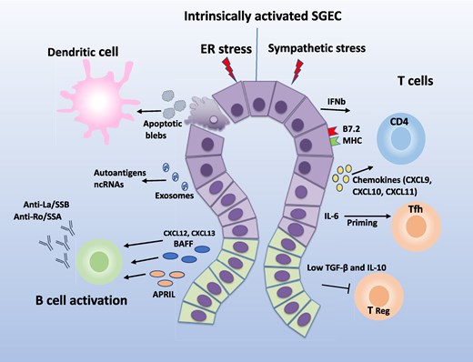
Crosstalk between salivary gland epithelial cells (SGECs) and immune populations in Sjögren’s disease. Intrinsically activated SGEC release IL-6, IFN-β, and chemokines to activate CD4 lymphocytes and prime T follicular helper (Tfh) cells, while low TGF-β/IL-10 suppresses Treg functions. SGECs secrete exosomes and apoptotic debris containing ncRNAs and autoantigens (e.g. Ro/SSA, La/SSB), while activating dendritic cells. Chemokines (CXCL9-11) recruit innate immune cells, while BAFF/APRIL from SGECs drive B cell activation. Autoantibody production by B cells is facilitated by autoantigens derived from apoptotic SGECs and exosomal cargo. Intrinsically activated SGECs are vulnerable to sympathetic stress promoting ER stress and IL-6 release. Together, these interactions both trigger and sustain glandular inflammation and epithelial damage (Abbreviations: ER, endoplasmic reticulum; Tfh, T follicular helper; Treg, regulatory T cell; ncRNA, non-coding RNA).
Acknowledgements
Not applicable.
Author contributions
Maria Filika (Conceptualization, Writing—original draft, Writing—review and editing), and Stergios Katsiougiannis (Conceptualization, Funding acquisition, Writing—review & editing)
Funding
European Alliance of Associations for Rheumatology (EULAR). 2025 Research Voucher Award, ID:Q424RSV203.
Data availability
Not applicable for this review article.
Ethical approval
Not applicable.
References
Author notes
Conflicts of interest: The authors declare no conflicts of interest.


