


 https://orcid.org/0000-0002-9815-3616

Abstract
Reported prevalence estimates of Lewy body pathology (LBP) vary widely, often without considering brain regional distributions or demographic influences. Large, population-representative autopsy cohorts are needed to provide estimates and clarify the distribution and clinical implications of LBP.
Neuropathological, genetic and clinical data were pooled from nine community- or population-based brain autopsy cohorts in the USA (n = 6), Brazil, Austria and Finland (n = 7309 total; 59% women; mean age of death 84.2 years). Cognitive status was available for 6166 participants: dementia (38.5%), mild cognitive impairment (14.8%) and cognitively unimpaired (47.0%). Frequency of LBP was examined by anatomical distribution (neocortical, limbic, brainstem, amygdala, olfactory) and stratified by covariates. Prevalence was calculated in meta-analysis, and mixed-effect logistic regression examined associations with sex, cognitive status, Alzheimer’s disease neuropathological change (ADNC) and limbic-predominant age-related transactive response DNA-binding protein (TDP)-43 encephalopathy neuropathologic change (LATE-NC) co-pathology.
Overall, 27.3% of participants had LBP: neocortical 8.4%, limbic 6.8%, brainstem 4.8%, amygdala 3.7% and olfactory 3.6%. Neocortical LBP was associated with dementia [present in 15%; odds ratio (OR) = 4.06 (95% confidience interval (CI) 3.24–5.10)] and with increased odds of ADNC [OR = 2.30 (95% CI 1.88–2.81)] and LATE-NC [OR = 2.03 (95% CI 1.69–2.44)]. Sex differences were not observed in the frequencies of neocortical or limbic LBP. However, amygdala-predominant LBP was more frequent in women [OR = 1.53 (95% CI 1.06–2.21)] whereas brainstem-predominant LBP was more common in men [OR = 0.64 (95% CI 0.49–0.84)]. Additionally, amygdala-predominant LBP was associated with increased odds of comorbid ADNC [OR = 12.7 (95% CI 5.30–30.6)] while brainstem-predominant LBP was not associated with greater odds of ADNC co-pathology [OR = 0.88 (95% CI 0.67–1.16)].
Pooling data from large, international community-based autopsy cohorts allows for more robust estimates of region-specific LBP prevalence and their associations with cognitive status. The observed sex- and co-pathology-specific differences in brainstem and amygdala-predominant LBP highlight potential biological heterogeneity and suggest that distinct disease pathways may underlie these patterns.
Introduction
Lewy body pathology (LBP) is characterized by the accumulation of intracellular α-synuclein fibrils forming Lewy bodies (LB) and Lewy neurites. These pathologic hallmarks are enriched in a spectrum of clinically defined conditions, including Parkinson’s disease (PD), Parkinson’s disease dementia (PDD) and dementia with Lewy bodies (DLB), the latter two often combined as Lewy body dementia (LBD). Prior studies have reported wide variations in the prevalence of LBD. In a systematic review of community-based cohorts, DLB accounted for 4.2% of dementia cases,1 but some studies have reported prevalence estimates of up to 16%.2 However, these estimates are largely based on clinical diagnoses without neuropathological confirmation, and LBD is believed to be substantially underdiagnosed, with frequent misclassification and diagnostic delay.3,4 Presumed preclinical LBP, often of lower severity, is also found in some individuals without clinical symptoms, and may be referred to as ‘incidental Lewy body disease’.4-7 The reported population prevalence of LBP varies widely, ranging from 6% to 61%, reflecting heterogeneity in study design, neuropathological assessment protocols, and the demographic and cognitive characteristics of the studied populations.8-14
Several different criteria have been proposed for the staging of LBP. The original Braak PD pathologic grading model hypothesized a six-stage caudo-rostral progression of LBP from the brainstem (medulla oblongata) to the neocortex,15 whereas more recent consensus criteria classify LBP by its predominant anatomical distribution (neocortical, limbic, brainstem, amygdala or olfactory only).16,17 Of the LBP subtypes, neocortical and limbic-predominant LBP are more often associated with dementia, with prior neuropathological studies attributing 15%–20% of dementia cases to LBP, while brainstem-predominant LBP is the main substrate for clinical PD and differing results have been reported for the clinical correlates of amygdala-predominant LBP.13,17-21 Moreover, most prior prevalence studies have not reported the regional distribution of LBP, limiting insight into the clinical correlates of these subtypes.22
Many existing prevalence estimates of LBP derive from clinic-based studies, which overrepresent atypical or severe phenotypes and underrepresent mixed pathologies, limiting their generalizability.23 Larger, population-representative and community-based cohorts are needed to provide relatively unbiased estimates of prevalence and to clarify the frequency, anatomical distribution and clinical correlates of LBP.24 Furthermore, the associations between LBP and basic demographic parameters also remain unclear. Although several studies report higher rates of LBP with advancing age,9,14,25 others have not found an association with age.11,20,26 Similarly, sex differences are inconsistent across studies; while clinical DLB and PDD cohorts typically have more men, findings from population-based and neuropathological studies are mixed.27-32
LBP rarely occurs in isolation and neuropathological heterogeneity is common.33 Depending on the criteria being used, up to 50% (or even more) of cases with LBP have concurrent Alzheimer’s disease neuropathological change (ADNC).34 Other co-pathologies including limbic age-related transactive response DNA-binding protein (TDP)-43 encephalopathy neuropathological change (LATE-NC) and cerebrovascular disease (CVD) are also common and increase in frequency with age.19,35-43 The coexistence of LBP and ADNC in particular is associated with accelerated cognitive decline, greater burden of neuropsychiatric symptoms, and lower clinical diagnostic accuracy than either pathology alone.34,44-50 It is not clear how the joint prevalence of co-pathologies may differ with the distribution of LBP, but prior clinical studies demonstrate higher prevalence of co-morbid ADNC in DLB than in PD.37 Other studies have suggested increased ADNC co-pathology in amygdala-predominant LBP,51 possibly reflecting a greater spread of ADNC in ‘brain first’ spread of LBP, but this needs further characterization.52,53
The true prevalence of LBP remains unknown and represents a major unmet need for improving early detection, diagnostic accuracy and therapeutic trial design. Robust prevalence estimates from large, geographically diverse, community- and population-based autopsy cohorts are needed to better define the population burden of LBP and its associated risk factors. Although previous reports from single cohorts and consortia have provided important insights,22 no prior study has combined data from multiple community-based cohorts to examine the prevalence of LBP by anatomical distribution and explore associations with co-pathology, demographic and cognitive characteristics. Addressing this need, we examined the prevalence and regional distribution of LBP in nine well-characterized, community-based autopsy cohorts spanning North America (6), South America (1) and Europe (2). By pooling data across cohorts, we aimed to determine how frequently LBP was observed post-mortem, its associations with age, sex and cognitive status, and how clinical and pathological covariates differed across regional LBP distributions.
Materials and methods
Cohorts and data sources
Summary data were collected for participants with neuropathological assessment of LBP from nine well-characterized community- or population-based brain ageing cohorts: the Adult Changes in Thought (ACT) study54; Baltimore Longitudinal Study of Aging (BLSA)55,56; Biobank for Aging Studies from the University of Sao Paulo (BAS-USP)57-59; Duke University/University of North Carolina Alzheimer’s Disease Research Center (Duke/UNC-ADRC)60; Rush University Religious Orders Study/Memory and Aging Project (ROS-MAP)61,62; University of California, Irvine 90+ Study (UCI 90+)63,64; University of Kentucky Alzheimer’s Disease Research Center (UKy-ADRC)65,66; Vantaa 85+ Study67; and Vienna Trans-Danube Aging (VITA) Study.12,68,69 Data from eight of these cohorts have been published previously as a consortium, but data on LBP were not reported.70
Characteristics of these cohorts are summarized in Supplementary Table 1, with additional specific cohort details provided in Supplementary Table 2. Each study obtained approval from its institutional review board or ethics committee, and participants (or next of kin) provided informed consent. Most analyses were performed on de-identified summary data (i.e. cohort-level data) and no additional ethical approval was required.
Population characteristics
Several included cohorts were population-based, recruiting from defined geographic areas with the aim of sampling broadly from the underlying population. Others were large community-based convenience cohorts, often recruiting through community outreach activities such as media advertisements, health fairs and local presentations. While lacking the unbiased sampling of population-based studies, these cohorts are generally more representative than clinic- or hospital-based series, given their size and the absence of pre-selection into clinicopathological groups. Additional details regarding recruitment are provided in Supplementary Table 2.
Neuropathological assessment
Structured data requests for standardized neuropathological measures were sent to each cohort. LBP was staged in all participants: eight cohorts used the McKeith criteria (classifying neocortical-, limbic-, brainstem- or amygdala-predominant LBP),16 and one cohort (UCI90+) applied only the Braak PD staging (0–VI).15 Three cohorts also reported ‘olfactory-only’ LBP in line with updated consensus recommendations17 (Supplementary Table 1). The ROS-MAP cohort used an earlier modified version of the McKeith classification including only neocortical-, limbic- and brainstem-predominant categories.71,72 All participating sites performed α-synuclein immunohistochemistry, with site-specific antibody technical details described in Supplementary Table 1. There was some variation in tissue processing, neuropathological methods and data completeness across cohorts, described in Supplementary Table 2.
Additional requested measures from each cohort included Braak neurofibrillary tangle (NFT) staging (0–VI scale)73 performed using anti-phospho-Tau antibodies; the Consortium to Establish a Registry for Alzheimer’s Disease (CERAD) neuritic amyloid plaque density scores (graded as ‘None’, ‘Sparse plaques’, ‘Moderate plaques’ or ‘Frequent plaques’)74 and Thal amyloid-β (Aβ) phases (a 0–5 scale based on anatomic distribution of Aβ plaques detected with Aβ immunostaining).75 Overall ADNC severity was classified for each case according to the National Institute on Aging and Alzheimer's Association (NIA-AA) criteria, based on combined Thal Aβ phase, Braak NFT stage and CERAD neuritic plaque score.76 TDP-43 proteinopathy distribution provided the bases of LATE-NC staging, all cohorts sampled the amygdala, hippocampus and middle frontal gyrus.77
Clinical assessment
Summary data for sex, age, Apoliprotein (APOE) ɛ4 allele status, cognitive status at final assessment prior to death [dementia, mild cognitive impairment (MCI), cognitively unimpaired (CU)] were also provided. Cognitive status data was available from seven cohorts, primarily through clinical evaluations in life with diagnoses made via consensus conference, except in the BAS, where status was determined post-mortem from validated next-of-kin interviews.78,79 Diagnostic criteria for dementia varied across studies (detailed in Supplementary Table 1); for example, the VANTAA 85+ cohort used Diagnostic and Statistical Manual of Mental Disorders, Third Edition, Revised criteria to identify dementia and did not classify MCI and in the UCI 90+ study MCI was classified according to the cognitive impairment, no dementia (CIND) criteria which is less stringent than MCI classification.80
Three cohorts (Duke/UNC-ADRC, UKy-ADRC and UCI90+) identified a subgroup of participants who had clinical diagnoses of PDD or DLB made during life. Four cohorts also provided data on core clinical features of DLB, but assessment methods of these symptoms were not standardized; BAS-USP relied on informant ratings from the Tanner scale for parkinsonism and Neuropsychiatric Inventory items for sleep and hallucinations, whereas participants were clinically assessed in other cohorts.
Statistical analyses
Random-effects meta-analyses were performed using restricted maximum likelihood estimation to account for between-cohort heterogeneity for each regional subtype of LBP. Study-specific proportions were pooled using inverse-variance weighting and are presented as pooled proportions with 95% confidence intervals (CI). For the purposes of the analyses the Braak PD staging was approximated to neocortical (stage 5–6), limbic (stage 3–4) and brainstem (1–2) distributions.
Joint distributions of co-pathologies (Braak NFT stage, Thal Aβ phase, CERAD neuritic plaque score and LATE-NC stage) were calculated for each anatomical subtype of LBP (neocortical, limbic-, brainstem- and amygdala-predominant) summed across the cohorts. Olfactory-only was not included in this analysis because it was not reported across a sufficient number of cohorts. ADNC was defined according to recent consensus recommendations, using combined Thal (Aβ), Braak (NFT) and CERAD (neuritic plaques) criteria to generate overall prevalence estimates.81
Demographic characteristics, including mean age at death, sex distribution and APOE ɛ4 frequency, were summarized as cohort-specific estimates weighted by sample size. Univariable meta-regression analyses examined associations between the prevalence of LBP subtypes with mean age at death or APOE ɛ4, and correlations were explored with Spearman’s correlation. The VITA and BLSA cohorts were excluded from APOE analyses because genotype data were not available. To explore other potential sources of heterogeneity, subgroup analyses and meta-regression examined differences by study design (population-based or community-based cohorts) and geographic location (USA versus non-USA cohorts).
Prevalence estimates of LBP by regional distribution were aggregated across cohorts and stratified by cognitive status and sex, with differences across groups evaluated with χ2 tests. Overall prevalence and between-cohort heterogeneity were further examined using random-effects meta-analysis. Associations between LBP distribution and cognition or sex were assessed using mixed-effects logistic regression models for each LBP region, adjusted for mean age at death and sex distribution, with a random intercept for cohort with results presented as odds ratios (OR). Similar models estimated the odds of ADNC and LATE-NC across LBP regions. Statistical significance was assessed at the 5% level with Bonferroni correction applied to these models. Variability in Braak NFT staging across cohorts was examined in meta-analysis to identify sources of heterogeneity for each LBP subtype. Furthermore, to examine whether co-pathology contributed to sex differences in LBP, subgroup descriptive analyses were performed across three cohorts which provided data stratifying co-pathology across different LBP distributions by sex and cognitive status. Multinomial logistic regression, adjusted for the individual sex of participants and cohort, was used to examine associations between sex, cognitive status and both ADNC and LATE-NC with results presented as relative risk ratios (RRR).
Clinical data were summarized across three cohorts (Duke/UNC-ADRC, UKy-ADRC and UCI90) to describe the distribution of LBP subtypes in participants with clinical diagnoses of PDD or DLB. Participants from cohorts were only included in this analysis if clinical diagnoses of PDD or DLB were made during life. Additionally, the distribution of core clinical features among participants with each anatomical subtype of LBP was described. All statistical analyses were performed in Stata/SE version 18.5 (StataCorp).
Results
Across the nine cohorts there was a total of 7309 participants included in the current study. The numbers of participants contributing to the data from each cohort are described in Table 1 along with demographic, clinical, genetic and summary neuropathologic data. The median number of included participants per cohort was 356 (range 264–2208). The mean weighted age at death across cohorts was 84.2 years (range of mean 72.9–97.2) and 59% of included participants were women. The APOE genotype was known in 5655 participants and 27.5% of these participants had an APOE ɛ4 allele. The cognitive status was reported in seven cohorts for a total of 6616 participants; 47.0% were classified as CU, 14.8% had a diagnosis of MCI and 38.5% were diagnosed with dementia at their last clinic visit before death.
Summary demographic and neuropathological characteristics of each cohort
| Cohort characteristics | ACT | BAS-USP | BLSA | Duke-UNC ADRC | ROS-MAP | UCI 90+ | UKy- ADRC | Vantaa 85+ | VITA | Total sample |
|---|---|---|---|---|---|---|---|---|---|---|
| Country | USA | Brazil | USA | USA | USA | USA | USA | Finland | Austria | – |
| Sample size, n | 1008 | 2208 | 356 | 280 | 2089 | 461 | 336 | 264 | 307 | 7309 |
| Average age at death, years (SD) | 89 | 72.9 (13.9) | 87 | 84 (10.8) | 89.5 (6.8) | 97.7 (3.6) | 83.9 (9.6) | 92.6 (3.6) | 83.2 (3.3) | 84.2 |
| Women, % (n) | 58.6% (591) | 49.3% (1091) | 37.9% (135) | 59.6% (167) | 68.8% (1437) | 66.6% (307) | 56% (188) | 82.6% (218) | 58.6% (180) | 59% (4314) |
| APOE status known, n | 1008 | 1354 | – | 271 | 1980 | 461 | 317 | 264 | – | 5655 |
| APOE ɛ4 allele present, % (n) | 27.5% (277) | 29.0% (393) | – | 30.3% (82) | 25.5% (505) | 18.4% (85) | 40.4% (128) | 32.2% (85) | – | 27.5% (1556) |
| Last cognitive status before death, % (n) | ||||||||||
| Cognitively unimpaired | 50.4% (508) | 67.2% (1476) | – | 36.6% (101) | 34% (714) | 28.9% (133) | 26.9% (87) | 34.8% (92) | – | 47% (3111) |
| Mild cognitive impairment | 2.1% (21) | 9.5% (209) | – | 18.8% (52) | 23% (477) | 28.4% (131) | 15.8% (51) | – | – | 14.8% (941) |
| Clinical dementia | 45.7% (461) | 23.3% (511) | – | 44.6% (123) | 43% (897) | 42.7% (197) | 57.3% (185) | 65.2% (172) | – | 38.5% (2546) |
| Lewy body pathology, % (n) | ||||||||||
| Neocortical | 8.7% (88) | 2.1% (46) | 3.4% (12) | 7.1% (20) | 14% (292) | 14.3% (66) | 10% (34) | 14.4% (38) | 3.3% (10) | 8.3% (606) |
| Limbic-predominant | 5.9% (59) | 1.8% (40) | 1.7% (6) | 8.6% (24) | 7.4% (155) | 5.2% (24) | 12.5% (42) | 12.9% (34) | 8.1% (25) | 5.6% (409) |
| Brainstem-predominant | 3.2% (32) | 1.8% (40) | 2.2% (8) | 3.6% (10) | 1.8% (38) | 10.2% (47) | 7.1% (24) | 7.2% (19) | 8.5% (26) | 3.3% (244) |
| Amygdala-predominant | 5.3% (53) | 0.8% (17) | 1.1% (4) | 6.1% (17) | – | – | 7.1% (24) | 3.4% (9) | 3.9% (12) | 2.9% (136) |
| Olfactory only | 0.9% (9) | – | – | – | – | – | 5.4% (18) | – | 5.3% (14) | 2.5% (41) |
| Thal phase > 3 | 48.3% (487) | 16.5% (325) | 34% (121) | 44% (123) | 66% (1379) | 53% (242) | 57% (149) | 70.5% (186) | 12.4% (38) | 43.6% (3050) |
| CERAD Moderate | 26.8% (270) | 13.7% (303) | 36.5% (130) | 32.9% (92) | 37% (776) | 12% (57) | 12% (41) | 53.8% (142) | 22.1% (68) | 24.6% (1795) |
| CERAD Frequent | 28.9% (291) | 10.5% (231) | 19.1% (68) | 8.9% (25) | 30% (636) | 52% (239) | 43% (143) | 12.5% (33) | 14.3% (44) | 22.7% (1655) |
| Braak NFT V–VI | 38.8% (391) | 12.4% (272) | 18.2% (65) | 20.0% (56) | 26% (543) | 38% (173) | 50% (161) | 35.6% (94) | 11.4% (35) | 24.6% (1790) |
| LATE-NC stage >1% | 27.6% (278) | 4.9% (91) | 33.1% (118) | 11.1% (31) | 35% (685) | 30% (140) | 31% (93) | 41.7% (110) | 10.4% (32) | 23.2% (1578) |
ACT = the Adult Changes in Thought (ACT) study; BAS-USP = Biobank for Aging Studies from the University of Sao Paulo; BLSA = Baltimore Longitudinal Study of Aging; Duke-UNC ADRC = Duke University/University of North Carolina Alzheimer’s Disease Research Center; ROS-MAP = Rush University Religious Orders Study/Memory and Aging Project; UCI 90+ = University of California, Irvine 90+ Study; UKy-ADRC = University of Kentucky Alzheimer’s Disease Research Center; VITA = Vienna Trans-Danube Aging Study. APOE = Apolipoprotein E; CERAD = Consortium to Establish a Registry for Alzheimer's Disease; NFT = neurofibrillary tangle; LATE-NC = Limbic-predominant age-related TDP-43 encephalopathy neuropathological change; SD = standard deviation; TDP = transactive response DNA-binding protein.
Prevalence of LBP
Meta-analysis of the overall frequency of LBP across the community-based cohorts separated by regional distribution is shown in Fig. 1. Across the whole sample 27.3% of participants had LBP: neocortical LBP was reported in 8.4% of participants (95% CI 5.2%–11.7%), limbic-predominant LBP was reported in 6.8% (95% CI 4.3%–9.3%) and brainstem-predominant LBP was reported in 4.8% (95% CI 2.8%–6.8%). The prevalence of amygdala-predominant LBP was 3.7% across seven cohorts (95% CI 1.9%–5.6%) and olfactory-only LBP was identified in 3.6% (95% CI 0.5%–6.7%) of participants in the three cohorts reporting this subtype. There was heterogeneity across cohorts in the prevalence of LBP, ranging from 2.1% to 14.4% for neocortical LBP, 1.7%–12.9% for limbic-predominant LBP, 1.8%–10.2% brainstem-predominant LBP, 0.8%–7.1% amygdala-predominant LBP and 0.9%–5.4% for olfactory-only LBP (Fig. 1).
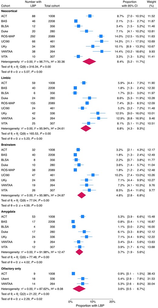
Meta-analysis of proportion of participants with Lewy body pathology for each anatomical distribution. ACT = the Adult Changes in Thought (ACT) study; BAS-USP = Biobank for Aging Studies from the University of Sao Paulo; BLSA = Baltimore Longitudinal Study of Aging; Duke-UNC ADRC = Duke University/University of North Carolina Alzheimer’s Disease Research Center; ROS-MAP = Rush University Religious Orders Study/Memory and Aging Project; UCI 90+ = University of California, Irvine 90+ Study; UKy-ADRC = University of Kentucky Alzheimer’s Disease Research Center; VITA = Vienna Trans-Danube Aging Study. CI = confidence interval; LBP = Lewy body pathology.
Prevalence of LBP by cognitive status
Analyses were conducted using data from the seven cohorts that reported cognitive status. For each LBP subtype, meta-analyses were performed stratified by cognitive status across the community-based cohorts. The overall prevalence and 95% confidence intervals are presented in Table 2, with corresponding meta-analyses shown in Supplementary Figs 1–4.
Participants with each subtype of LBP in stratified by final cognitive status
| LBP subtype | Dementia | Mild cognitive impairment | Cognitively unimpaired | P-value | |||
|---|---|---|---|---|---|---|---|
| Total number across cohorts (N = 2546) | Meta-analysis proportion (95% CI) | Total number across cohorts (N = 941) | Meta-analysis proportion (95% CI) | Total number across cohorts (N = 3111) | Meta-analysis proportion (95% CI) | ||
| Neocortical, n | 399 | 15% (11%–19%) | 78 | 8% (4%–11%) | 105 | 4% (2%–6%) | <0.001 |
| Limbic, n | 193 | 8% (5%–11%) | 47 | 4% (2%–7%) | 135 | 6% (4%–9%) | <0.001 |
| Brainstem, n | 76 | 4% (1%–6%) | 34 | 5% (1%–9%) | 100 | 5% (3–7%) | 0.638 |
| Amygdala, na | 92 | 7% (4%–10%) | 6 | 2% (0%–4%) | 22 | 1% (0%–2%) | <0.001 |
| Olfactory only, n (%)b | 26 (3.2%) | – | 2 (2.8%) | – | 13 (1.9%) | – | 0.294 |
The total number of participants with each subtype of LBP in stratified by final cognitive status. Proportions were calculated from meta-analysis stratified by cognitive status. The difference in proportions across cognitive groups was evaluated using χ2 tests, significant results highlighted in bold if P < 0.05.
LBP = Lewy body pathology; CI = confidence interval.
aParticipants with assessment for amygdala LBP: dementia (n = 1452), mild cognitive impairment (n = 333), cognitively unimpaired (n = 2264).
bParticipants with assessment for olfactory LBP: dementia (n = 818), mild cognitive impairment (n = 72), cognitively unimpaired (n = 687). Meta-analysis not conducted for olfactory-only LBP because only two cohorts with cognitive data reported olfactory-only LBP.
Among participants with dementia, 15% had neocortical LBP, 8% limbic-predominant LBP, 4% brainstem-predominant LBP, 6% amygdala-predominant LBP and 3% olfactory-only LBP. In the cohorts reporting MCI, 8% of participants had neocortical LBP, 4% limbic-predominant, 5% brainstem-predominant, 2% amygdala-predominant and 3% olfactory-only LBP. Low percentages of LBP were reported in CU individuals (4% neocortical LBP, 6% limbic-predominant, 5% brainstem-predominant, 1% amygdala-predominant and 2% olfactory-only LBP). Heterogeneity in the prevalence of LBP was observed across cohorts even when stratified by cognitive status. Neocortical LBP ranged from 6% to 21% in dementia, 2% to 11% in MCI and 1% to 9% in CU participants (Supplementary Fig. 1). Limbic-predominant LBP ranged from 2% to 16% in dementia, 1% to 8% in MCI and 2% to 13% in CU participants (Supplementary Fig. 2).
Associations between LBP subtypes and cognitive status were examined in mixed-effect logistic regression with cohort included as a random intercept and adjusted for the overall sex distribution and mean age of the cohort (Fig. 2). Neocortical LBP was more frequent in participants with dementia (OR 4.02, 95% CI 3.20–5.05, P < 0.001) and MCI (OR 1.79, 95% CI 1.31–2.45, P < 0.001) compared with CU individuals, and was also more frequent in dementia than MCI (OR 2.24, 95% CI 1.72–2.91, P < 0.001).
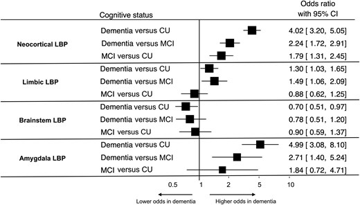
Odds of each subtype of LBP by cognitive status. CU = cognitively unimpaired; LBP = Lewy body pathology; MCI = mild cognitive impairment.
Limbic LBP showed modest increases in dementia compared with CU (OR 1.30, 95% CI 1.03–1.65, P = 0.027) and MCI (OR 1.49, 95% CI 1.06–2.08, P = 0.021), but these associations did not remain significant after Bonferroni correction. Limbic LBP did not differ between MCI and CU (OR 0.88, 95% CI 0.62–1.25, P = 0.462). Brainstem LBP was not associated with cognitive status. Although brainstem LBP was less frequent in dementia than CU (OR 0.70, 95% CI 0.51–0.97, P = 0.030), this did not survive correction for multiple comparisons. Amygdala-predominant LBP was more frequent in dementia than CU (OR 4.99, 95% CI 3.08–8.11, P < 0.001) and MCI (OR 2.71, 95% CI 1.45–5.42, P = 0.023); only the comparison with CU remained significant after Bonferroni correction. Amygdala-predominant LBP did not differ between MCI and CU (OR 1.84, 95% CI 0.72–4.71, P = 0.20).
Association between LBP and co-pathologies
Co-pathologies across all participants with each anatomical subtype of LBP are described in Table 3. Severe burden of NFTs (Braak NFT stages V–VI) was more frequent in neocortical LBP (42.2%) compared with limbic- (33.7%) and brainstem-predominant LBP (16.1%). Similarly, frequent CERAD neuritic plaques were more common in neocortical (38.8%) than limbic- (30.6%) or brainstem-predominant LBP (20.9%). Advanced Thal Aβ phases (4–5) were present in 57.1% of neocortical, 46.3% of limbic- and 34.2% of brainstem-predominant LBP cases.
Proportion with each staging of co-pathology across participants separated by anatomical distribution of LBP
| Staging of co-pathology | All participants (N = 7309) | Anatomical distribution of LBP | P-value | ||||
|---|---|---|---|---|---|---|---|
| Neocortical (N = 606) | Limbic-predominant (N = 409) | Brainstem-predominant (N = 244) | Amygdala-predominant (N = 137) | Olfactory only (N = 41) | |||
| Braak NFT staging, % (n)a | |||||||
| 0–II | 75.4% (5497) | 11.3% (68) | 17.6% (72) | 28.5% (69) | 8.9% (12) | 10.0% (4) | <0.001 |
| III–IV | 46.4% (280) | 48.7% (199) | 55.4% (134) | 17.8% (24) | 25.0% (10) | <0.001 | |
| V–VI | 24.6% (1758) | 42.2% (255) | 33.7% (138) | 16.1% (39) | 73.3% (99) | 65.0% (26) | <0.001 |
| CERAD neuritic amyloid plaque stages, % (n) | |||||||
| None | 52.8% (3854) | 18.2% (110) | 27.4% (112) | 38.5% (94) | 5.1% (7) | 17.1% (7) | <0.001 |
| Sparse | 10.6% (64) | 14.9% (61) | 16.8% (41) | 10.9% (15) | 9.8% (4) | 0.073 | |
| Moderate | 26.3% (1795) | 32.3% (196) | 27.1% (111) | 23.8% (58) | 38.0% (52) | 26.8% (11) | 0.017 |
| Frequent | 22.7% (1655) | 38.8% (236) | 30.6% (125) | 20.9% (51) | 46.0% (63) | 46.3% (19) | <0.001 |
| Thal Aβ phase, % (n)b | |||||||
| 0 | 23.8% (1666) | 11.1% (66) | 13.2% (53) | 21.7% (52) | 1.5% (2) | 7.9% (3) | <0.001 |
| 1–2 | 32.5% (2274) | 11.9% (71) | 23.4% (94) | 21.7% (52) | 8.1% (11) | 10.5% (4) | <0.001 |
| 3 | 19.9% (119) | 17.2% (69) | 22.5% (54) | 9.6% (13) | 13.2% (5) | 0.019 | |
| 4–5 | 43.6% (3050) | 57.1% (341) | 46.3% (186) | 34.2% (82) | 80.9% (110) | 68.4% (26) | <0.001 |
| ADNC, % (n)c | |||||||
| Mild or absent | 51.2% (3450) | 25.7% (153) | 38.9% (157) | 52.3% (125) | 11.3% (6) | – | <0.001 |
| Moderate or severe | 48.8% (3289) | 74.3% (442) | 61.1% (247) | 47.7% (114) | 88.7% (47) | – | <0.001 |
| LATE-NC (TDP-43) stages, % (n)d | |||||||
| Stage 0 | 66.7% (5226) | 40.4% (241) | 52.9% (211) | 63.7% (151) | 43.1% (59) | 26.3% (10) | <0.001 |
| Stage 1 | 17.3% (103) | 17.8% (71) | 13.9% (33) | 23.4% (32) | 23.7% (9) | 0.17 | |
| Stage 2–3 | 23.3% (1578) | 42.3% (252) | 29.3% (117) | 22.4% (53) | 33.6% (46) | 50.0% (19) | <0.001 |
Differences in proportions across groups were assessed in X2 tests with significant in bold at the 5% level.
Aβ = amyloid-β; ADNC = Alzheimer’s disease neuropathological change; APOE = Apolipoprotein E; CERAD = Consortium to Establish a Registry for Alzheimer's Disease; NFT = neurofibrillary tangle; LATE-NC = Limbic-predominant age-related-43 encephalopathy neuropathological change.
LBP = Lewy body pathology; NFT = neurofibrillary tangle; CERAD = Consortium to Establish a Registry for Alzheimer's Disease; ADNC = Alzheimer’s disease neuropathological change; LATE-NC = limbic-predominant age-related TDP-43 encephalopathy neuropathological change.
aNumber missing across each subgroup: neocortical n = 3; limbic n = 2; brainstem n = 2; amygdala n = 2 olfactory n = 1.
bNumber missing across each subgroup: neocortical n = 9; limbic n = 7; brainstem n = 2; amygdala n = 1; olfactory n = 3.
cNumber missing in each subgroup: neocortical n = 11, limbic n = 5, brainstem n = 5, amygdala n = 84.
dNumber missing across each subgroup: neocortical n = 10; limbic n = 10; brainstem n = 7; amygdala n = 1; olfactory n = 3.
Fewer cohorts staged amygdala-predominant or olfactory-only LBP, as reflected by the smaller sample size. However, the prevalence of ADNC co-pathology in these groups was markedly higher than in the whole cohort: among amygdala-predominant cases, 73.3% had severe NFTs, 46.0% had frequent CERAD plaques and 80.9% had Thal Aβ phase 4–5.
ADNC and LBP
Among participants without LBP, ADNC was present in 45.0% (n = 2471). In those with LBP, the prevalence varied by anatomical subtype of LBP: it was highest in neocortical LBP (74.3%, n = 442), followed by limbic-predominant LBP (61.1%, n = 247) and brainstem-predominant LBP (47.7%, n = 114). In three cohorts reporting ADNC in amygdala-predominant LBP, ADNC was observed in 88.7% (n = 47) of cases.
Mixed-effects logistic regression models with cohort included as a random intercept and adjusted for the overall sex distribution and mean age of the cohort showed that neocortical LBP was associated with more than 2-fold higher odds of ADNC compared with individuals without neocortical LBP (OR 2.30, 95% CI 1.88–2.81, P < 0.001) (Fig. 3). Limbic-predominant LBP was also significantly associated with ADNC, though to a lesser extent (OR 1.40, 95% CI 1.13–1.74, P = 0.002). However, brainstem-predominant LBP was not significantly associated with ADNC (OR 0.88, 95% CI 0.67–1.17, P = 0.386). Amygdala-predominant LBP showed a strong association with ADNC in the three cohorts reporting this subtype, albeit with wider confidence intervals due to smaller numbers (OR 12.74, 95% CI 5.30–30.58, P < 0.001). Between-cohort variance was modest (intraclass correlation coefficient 0.028, 95% CI 0.010–0.079).
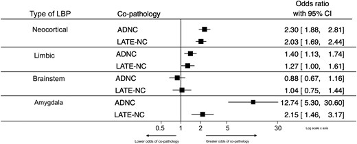
Odds of ADNC and LATE-NC co-pathology in each subtype of LBP. ADNC = Alzheimer’s disease neuropathological change; LATE-NC = limbic-predominant age-related TDP-43 encephalopathy neuropathological change; LBP = Lewy body pathology; TDP = transactive response DNA-binding protein.
To explore variability in the distribution of Braak NFT co-pathology across studies, a random effects meta-analysis was conducted for each regional subgroup of LBP shown in Supplementary Figs 5–8. As expected, there was substantial heterogeneity in the prevalence of NFT across groups but the pooled, weighted estimate generated by random effects models was similar to the prevalence estimated from total numbers.
LATE-NC and LBP
LATE-NC was also most frequent in neocortical LBP (42.3%), followed by limbic (29.3%) and brainstem LBP (22.4%). In mixed-effects logistic regression models adjusted for age and sex and including cohort as a random intercept shown in Fig. 3, the odds of LATE-NC were increased in individuals with neocortical LBP (OR 2.03, 95% CI 1.69–2.44, P < 0.001) and amygdala-predominant LBP (OR 2.15, 95% CI 1.46–3.18, P < 0.001). Limbic-predominant LBP was associated with an increase in odds of LATE-NC (OR 1.27, 95% CI 1.00–1.61, P = 0.047) but this did not meet the statistical significance threshold after Bonferroni correction. Brainstem-predominant LBP was not associated with LATE-NC (OR 1.04, 95% CI 0.75–1.44, P = 0.831).
Cognitive status and co-pathology
Co-pathology in LBP stratified by cognitive status and sex distribution was available in three cohorts (BAS-USP n = 2075, ROS-MAP n = 2087, UKy n = 217). Across all anatomical subtypes of LBP, co-existing ADNC and LATE-NC were significantly more frequent in participants with dementia (Table 4).
Co-pathology stratified by cognitive status across each anatomical subtype of LBP
| ADNC present (N = 2020), % (n) | LATE-NC present, % (n) | P ADNC | P LATE | ||
|---|---|---|---|---|---|
| Neocortical LBP (N = 368)a | Dementia (n = 249) | 82.9% (204) | 47.4% (118) | <0.001 | 0.003 |
| MCI (n = 56) | 56.4% (31) | 33.9% (19) | |||
| CU (n = 63) | 48.4% (30) | 25.4% (16) | |||
| Limbic LBP (N = 234)b | Dementia (n = 124) | 81.8% (99) | 45.2% (56) | <0.001 | <0.001 |
| MCI (n = 40) | 56.4% (22) | 25.0% (10) | |||
| CU (n = 70) | 37.1% (26) | 11.4% (8) | |||
| Brainstem LBP (N = 107)c | Dementia (n = 31) | 51.6% (16) | 38.7% (12) | 0.020 | 0.001 |
| MCI (n = 19) | 41.2% (7) | 26.3% (5) | |||
| CU (n = 57) | 22.8% (13) | 5.5% (3) | |||
| Amygdala LBP (N = 51)d | Dementia (n = 40) | 100% (39) | 52.5% (21) | <0.001 | 0.033 |
| MCI (n = 4) | 75% (3) | 0 | |||
| CU (n = 7) | 33% (2) | 14.3% (1) | |||
| No LBP (N = 3621)e | Dementia (n = 1092) | 68.9% (752) | 36.2% (366) | <0.001 | <0.001 |
| MCI (n = 620) | 49.5% (307) | 24.1% (146) | |||
| CU (n = 1909) | 23.8% (454) | 7.2% (126) |
Comparisons across groups conducted with X2 tests.
ADNC = Alzheimer’s disease neuropathological change; LATE-NC = limbic-predominant age-related TDP-43 encephalopathy neuropathological change; LBP = Lewy body pathology, MCI = mild cognitive impairment; CU = cognitively unimpaired; reduced numbers reported for some subgroups across the cohorts.
aNeocortical LBP, ADNC reported in: dementia n = 246, MCI n = 55, CU n = 62.
bLimbic LBP, ADNC reported in: dementia n = 121, MCI n = 39.
cBrainstem LBP, ADNC reported in MCI n = 17, LATE-NC reported in CU n = 55.
dAmygdala LBP reported in ADNC dementia n = 39, CU n = 6.
eNo LBP reported in LATE-NC dementia n = 1012, MCI n = 605, CU n = 1758.
In multinomial logistic regression adjusted for the cohort and individual sex of participants with neocortical LBP, comorbid ADNC was associated with an increased relative risk of dementia compared with CU participants [RRR = 5.53 (95% CI 2.98–10.2), P < 0.001] and with dementia versus MCI [RRR = 3.92 (95% CI 2.06–7.47), P < 0.001]. There was no significant difference in risk between MCI and CU [RRR = 1.41 (95% CI 0.66–2.99), P = 0.372]. Similarly, co-morbid LATE-NC in neocortical LBP was associated with an increased relative risk of dementia versus CU [RRR = 3.00 (95% CI 1.59–5.68), P = 0.001], but not versus MCI [RRR = 1.73 (95% CI 0.93–3.21), P = 0.085] or between MCI and CU [RRR = 1.74 (95% CI 0.77–3.93), P = 0.182].
Similarly, in participants with limbic LBP, comorbid ADNC was associated with an increased relative risk of dementia versus CU [RRR = 6.81 (95% CI 3.41–13.6), P < 0.001] and MCI [RRR = 3.74 (95% CI 1.69–8.23), P = 0.001] but there was no significant difference in risk between MCI and CU with comorbid ADNC [RRR = 1.82 (95% CI 0.79–4.18), P = 0.157]. Co-morbid LATE-NC in limbic LBP was also associated with an increased relative risk of dementia versus CU [RRR = 6.81 (95% CI 3.41–13.6), P < 0.001] and versus MCI [RRR = 2.76 (95% CI 1.22–6.24), P = 0.015], but not with MCI versus CU participants.
In participants with brainstem LBP, co-morbid ADNC was not associated with increased risk of dementia or MCI. Co-morbid LATE-NC was associated with greater relative risk of dementia compared with CU [RRR = 8.03 (95% CI 1.94–33.2), P = 0.004], but there was no significant difference in risk between dementia and MCI or between MCI and CU. Multinomial regression could not be performed for the amygdala-predominant LBP group because of small sample size; however, it is notable that all participants with dementia and amygdala-predominant LBP had co-morbid ADNC (Fisher’s exact P < 0.001).
Association with demographic and genetic factors
Age of death
The mean age at death was positively associated with the frequency of neocortical LBP (r = 0.79, P = 0.01), but no significant correlations were observed between age at death and prevalence of limbic-, brainstem- or amygdala-predominant LBP (Supplementary Fig. 9). Consistent with this, meta-regression showed that age at death was significantly associated with the proportion of neocortical LBP [β = 0.01 (0.002–0.01), P = 0.001] but not with LBP in other regions [limbic β = 0.002 (−0.002–0.005), P = 0.44; brainstem β = 0.002 (−0.001–0.005), P = 0.15; amygdala β = 0.001 (−0.002–0.003), P = 0.37].
APOE ɛ4 genotype status
There was no correlation between the cohort percentage with APOE ɛ4 positivity and the proportion of the reported subtypes of LBP as shown in Supplementary Fig. 10.
Epidemiological factors
There was no evidence of geographic variation in regional LBP prevalence between US and non-US cohorts. Neither test of subgroup differences [neocortical Q(between)(1) = 0.60, P = 0.44, limbic: Q(between)(1) = 0.00, P = 1.00, brainstem Q(between)(1) = 0.19, P = 0.67, amygdala Q(between)(1) = 0.38, P = 0.54] nor age-adjusted meta-regression [neocortical β = 0.00 (−0.06–0.06), P = 0.81; limbic β = −0.19 (−0.09–0.05), P = 0.58; brainstem β = −0.03 (−0.07–0.014), P = 0.18; amygdala β = 0.02 (−0.02–0.06), P = 0.40] showed any association. Similarly, LBP prevalence did not differ between population-based and community-based cohorts in any brain region [neocortical Q(between)(1) = 0.70, P = 0.40, limbic: Q(between)(1) = 0.00, P = 1.00, brainstem: Q(between)(1) = 0.00, P = 0.99, amygdala: Q(between)(1) = 1.67, P = 0.20) and age-adjusted meta-regression demonstrated no association between study design and LBP subtype prevalence [neocortical β = −0.006 (−0.056–0.04), P = 0.81; limbic β = 0.008 (−0.05–0.07), P = 0.81; brainstem β = 0.01 (−0.032–0.055), P = 0.61; amygdala β = −0.01 (−0.05–0.03), P = 0.61].
Sex
The frequency of neocortical and limbic LBP, each summed across cohorts, did not differ by sex (Table 5). The proportion of each type of LBP was evaluated in meta-analyses, the pooled frequency is reported in Table 5 and forest plots are shown in Supplementary Figs 11–14. In univariate analyses brainstem-predominant LBP was significantly more common in men (X2 = 6.34, P = 0.012) and amygdala-predominant LBP was significantly more common in women (X2 = 9.82, P = 0.002). All other neurodegenerative pathologies staged in the cohorts (amyloid plaque severity stages, Braak NFT stages and LATE-NC stages) were significantly more common in women than men as illustrated in Table 5.
Prevalence of each subtype of LBP and other neurodegenerative pathologies stratified across men and women
| Pathologic features | Men | Women | P-value | |||
|---|---|---|---|---|---|---|
| Total number across cohorts (N = 2995) | Meta-analysis proportion (95% CI) | Total number across cohorts (N = 4314) | Meta-analysis proportion (95% CI) | |||
| LBP subtype, n (%) | Neocortical | 230 (7.7%) | 9% (5%–13%) | 376 (8.7%) | 8% (5%–11%) | 0.11 |
| Limbic | 162 (5.4%) | 7% (4%–10%) | 247 (5.7%) | 6% (4%–9%) | 0.56 | |
| Brainstem | 119 (4.0%) | 5% (3–6%) | 125 (2.9%) | 4% (2%–6%) | 0.012 | |
| Amygdalaa | 45 (2.1%) | 3% (1–5%) | 92 (3.6%) | 4% (2–6%) | 0.002 | |
| Other neurodegenerative pathologies | Total across cohorts, n (%) | Total across cohorts, n (%) | |
|---|---|---|---|
| Thal Aβ phases 4 or 5b | 913 (36.5%) | 1978 (51.7%) | <0.001 |
| Braak NFT stages V or VIc | 504 (19.1%) | 1186 (29.7%) | <0.001 |
| CERAD neuritic plaque stages moderate or frequentd | 1058 (40%) | 2210 (55.3%) | 0.003 |
| LATE-NC stages 2 or 3e | 424 (17.6%) | 1004 (26.9%) | <0.001 |
Difference in proportions across sex were evaluated in χ2 tests, significant results highlighted in bold if P < 0.05.
CI = confidence interval; LBP = Lewy body pathology; NFT = neurofibrillary tangle; CERAD = Consortium to Establish a Registry for Alzheimer's Disease; LATE-NC = limbic-predominant age-related TDP-43 encephalopathy neuropathological change.
Number of men and women participants each neurodegenerative pathology was reported in:
aAmygdala-predominant LBP: men, n = 2189, women n = 2570.
bThal phase men n = 2504, women n = 3823.
cBraak NFT staging men n = 2637, women n = 3987.
dCERAD staging, men n = 2645, women n = 3998.
eLATE-NC staging, men n = 2413, women = 3728.
In mixed-effects logistic regression models adjusted for age and including cohort as a random intercept, the odds of neocortical and limbic LBP did not differ by sex (Supplementary Fig. 15; neocortical: female versus male OR 0.89, 95% CI 0.74–1.06, P = 0.184; limbic: OR 0.87, 95% CI 0.71–1.07, P = 0.197). In contrast, brainstem-predominant LBP was more frequent in men (female versus male OR 0.64, 95% CI 0.49–0.84, P = 0.001) while amygdala-predominant LBP was more frequent in women than men (female versus male OR 1.53, 95% CI 1.06–2.22, P = 0.023), but this association did not remain significant after Bonferroni correction. Together, these findings suggest sex-related differences in LBP vary across regional distribution.
Subgroup analysis for ADNC co-pathology in LBP by sex
Subgroup analysis of three cohorts assessed variation in ADNC co-pathology across regional subtypes of LBP by sex. In these cohorts, a higher proportion of women with neocortical and limbic LBP tended to have ADNC than men but the difference was not statistically significant (Supplementary Table 3).
Concordance with clinical diagnosis and core clinical features
Three of the nine cohorts (Duke/UNC ADRC, UCI90, UKy-ARDC) reported clinical diagnoses made during life. There were 29 clinical diagnoses of DLB and 11 diagnoses of PDD across the cohorts (Duke n = 5; UK n = 13; UCI90 n = 22). Across participants with a clinical diagnosis of DLB, 41.3% had neocortical LBP, 7.9% had limbic-predominant LBP and 17.2% had brainstem-predominant LBP and 34.5% had no LBP (Supplementary Table 4). In participants with PDD, 45.5% had neocortical LBP, 36.4% had limbic LBP, 9.1% had brainstem-predominant LBP and 9.1% had no LBP. Limbic LBP was more common in PDD than DLB but there were no other significant differences in neuropathology across the clinical groups.
Three cohorts (UKy-ADRC, Duke/UNC-ADRC and BAS-USP) included additional clinical information for a subgroup of participants with LBP. In participants with neocortical LBP, 38.0% (n = 30) reported hallucinations, 60.3% (n = 44) reported parkinsonism, 25.0% (n = 9) reported cognitive fluctuations and 33.3% (n = 26) reported sleep disturbance. Core clinical features were less frequently reported in limbic-, brainstem- and amygdala-predominant LBP than in neocortical LBP shown in Supplementary Fig. 16.
Discussion
In this large, international, aggregated sample of 7309 autopsied research participants across nine population- and community-based cohorts, over a quarter of participants had LBP (neocortical 8%; limbic-predominant 7%; brainstem-predominant 5%; amygdala-predominant 4%; olfactory-only 4%). Among participants with dementia, neocortical LBP was most frequent (15%), followed by limbic-predominant (8%), amygdala-predominant (7%) and brainstem-predominant LBP (4%). However, the prevalence of clinically diagnosed LBD in dementia populations has previously been estimated as 5%.4 This conflicts with the current post-mortem study, suggesting significant underdiagnosis in clinical populations, with the majority of individuals with neocortical or limbic LBP not receiving a diagnosis of LBD. Among those with MCI, 8% had neocortical LBP and 4% had limbic LBP, providing a relatively population-generalizable estimate of LBP in MCI populations that exceed prior clinical estimates of prodromal DLB in MCI.82
Additionally, the odds of neocortical LBP were more than four times greater in participants with dementia compared with CU individuals and more than 2-fold higher than those with MCI.17 Limbic-predominant LBP was also more common in dementia, although the odds of dementia in this LBP subgroup were lower than neocortical LBP. The presence of co-morbid ADNC and LATE-NC in neocortical and limbic LBP further increased the odds of dementia, highlighting the additive impact of each co-pathology on cognitive decline. In contrast, the prevalence of brainstem-predominant LBP was stable across cognitive groups and showed no association with dementia, supporting an aetiological role for neocortical and limbic, but not brainstem, LBP in cognitive decline. Furthermore, although earlier reports indicate up to 25% of older people without dementia have LBP without clinical manifestations,17,83 only 11% of CU participants in the current study had LBP (neocortical 3%, limbic- 4%, brainstem- 3% and amygdala-predominant LBP 1%) suggesting prior studies may have overestimated incidental LBP. Although the prevalence of amygdala-predominant and olfactory-only LBP was higher in dementia groups, this most likely reflects the high burden of concomitant ADNC co-pathology observed in these populations. Indeed, in sub-analyses, all participants with amygdala-predominant LBP and dementia had co-morbid ADNC, indicating that amygdala LBP alone is not a driver of dementia.
The neuropathological heterogeneity of LBP is well established, with ‘pure’ disease relatively uncommon.35,84,85 In the current study, co-pathology was frequent, particularly in neocortical LBP, where 74% of participants had co-morbid substantial ADNC and 42.3% had stage 2 or 3 LATE-NC—higher than prior estimates not stratifying by distribution of LBP.34 These findings suggest that synergistic interactions between ADNC, LATE-NC and LBP may be particularly important in neocortical LBP.86,87 The odds of ADNC and LATE-NC were also markedly elevated in amygdala-predominant LBP, where severe NFT and diffuse amyloid pathology were common, supporting earlier reports of a link between amygdala involvement and both ADNC and LATE-NC.51,88,89 In contrast, the odds of ADNC co-pathology were not increased in brainstem-predominant LBP. Recent evidence suggests two possible trajectories of LBP progression: a caudo-rostral ‘body-first’ pathway and an amygdala or olfactory initiated ‘brain-first’ pathway.52,53 The high prevalence of ADNC reported in amygdala-predominant relative to brainstem-predominant LBP suggests that comorbid ADNC may be more characteristic of the ‘brain-first’ variant, contributing to clinical heterogeneity in LBD. There may also be protective factors inherent to brainstem-predominant LBP that limit the spread of multiple pathologies to supratentorial regions.
Previous studies have reported varying results on the association between age and prevalence of LBP, with some reporting stable prevalence across the ageing spectrum (or even decreased prevalence in persons living beyond 90 years of age), while others describe increasing prevalence with age.8,25 In the present study, cohort-level increased age was associated with increased prevalence of neocortical, but not other distributions of LBP which may help explain inconsistencies across earlier reports not differentiating by anatomical subtype of LBP.
Although clinical cohorts of DLB and PDD typically report higher prevalence in men,28,34 in the current study we found no sex differences in the prevalence of neocortical or limbic LBP—the subtypes most associated with dementia—suggesting that women with LBP may be underdiagnosed in these clinical cohorts. Indeed, prior studies suggest men more often exhibit parkinsonism and rapid eye movement (REM) sleep behavioural disorder, clinical features that facilitate diagnosis and increase representation in specialty clinics,36,90 while women often present with fewer core features, contributing to higher rates of misdiagnosis and diagnostic delay.29 Co-pathology may also influence presentation: pure neocortical LBP is more common in men, whereas women more often have concomitant ADNC in mixed disease presentations which are also associated with fewer core features and higher rates of misdiagnosis.19,27,32,91-94
In the current study, other neurodegenerative pathologies were more common in women, however, analysis of ADNC across LBP subtypes by sex was limited by practical factors to a subgroup of three cohorts and did not reach statistical significance, likely due to low statistical power.
However, other sex-related differences in the regional distributions of LBP were observed with higher odds of brainstem-predominant LBP in men and higher odds of amygdala-predominant LBP in women. These findings suggest that men and women may be differentially affected by pathological mechanisms, with brainstem-associated ‘body-first’ subtypes more common in men and amygdala-onset ‘brain-first’ subtypes more common in women.95 The differences in the prevalence of ADNC across amygdala and brainstem-predominant distributions, with ADNC also more common in women than men suggests there may be common factors driving these pathways. Such divergence may contribute to sex-specific patterns of clinical manifestation and underdiagnosis. Recognition of these differences, alongside increased use of biomarkers, will be required to improve diagnostic accuracy and reduce sex disparities in identifying LBD in the clinical setting.
Clinicopathological concordance for DLB was quite poor with almost a third of participants who were diagnosed with DLB lacking LBP at autopsy, in line with previous studies.96 There were no significant differences in the neuropathological burden associated with clinical diagnoses of DLB and PDD but this sub-analysis was likely underpowered statistically. Neocortical LBP was the subtype with the highest percentage of each core clinical features of DLB but there was heterogeneity across clinical symptoms. Furthermore, variability in diagnostic performance across cohorts highlights the need for harmonization of diagnostic protocols and greater integration of biomarkers to improve detection in clinical populations.4,36,37,97
Strengths and limitations
Strengths of this study include the aggregation of data from nine high-quality community-based autopsy studies to explore the prevalence of LBP in the largest sample to date with neuropathologic workup also differentiated by LBP anatomical distribution. The population- and community-based design of the included cohorts reduces the circularity inherent in many clinicopathological studies allowing less biased assessment of the relationship between pathology and clinical parameters, and avoiding selection of more severe and specific clinical phenotypes. While none of the included cohorts were recruited from dementia- or movement disorder-specific clinical settings, their human population representativeness varied. For example, ROS-MAP specifically recruited religious clergy, while BAS-USP included a younger, more diverse population with a higher proportion of CU individuals at death. The biological and cultural variation in age, geography, cognition and ethnoracial parameters likely contributed to heterogeneity across studies but despite methodological differences, the aggregate APOE ɛ4 prevalence in this study (27%) was close to population-based estimates,98,99 suggesting the cohorts were broadly unselected. This study thus was able to discern significant clinical-biological phenomena that would have been impossible to detect using smaller cohorts.
However, several important caveats should be considered. Neuropathological protocols and assessment methods varied across cohorts. One cohort staged LBP using Braak PD criteria, which were approximated to McKeith staging for this analysis and may have led to misclassification in some cases. Cohorts also used different α-synuclein antibodies (Supplementary Table 1); however, prior studies suggest that staging reliability is not dependent on antibody choice,17 and all sites used state-of-the-art diagnostic methods. Selection bias related to consent to autopsy is another limitation, with variation in the proportion of the recruited population who were neuropathologically examined across cohorts. Sensitivity analyses performed in some cohorts suggest minimal impact of selection bias but these data were not collected by all cohorts. Another concern related to the current study was that most cohorts reported only summary data, precluding individual-level regression analyses. There was also notable between-study heterogeneity, including differences in cognitive assessments and dementia classification. While differences in study design and geographic location were explored, and no significant differences in LBP prevalence were observed between population-based and community-based cohorts or between US and non-US cohorts, global geographic variation could not be fully assessed due to the predominance of US cohorts (6/9), with only two European and one Brazilian cohort included. Except for the BAS cohort, participants were predominantly White and highly educated, limiting generalizability to more diverse populations and restricting analysis of the impact of ethnicity and social or structural determinants of health. Future studies should aim to investigate LBP prevalence in post-mortem populations with greater ethnic, social and educational diversity. Additionally, associations between APOE ɛ4 allele positivity and LBP could not be examined due to the absence of participant-level joint distributions. Some sub-analyses were underpowered, and for most cohorts, clinical information beyond cognitive status was unavailable for participants with LBP, with motor symptoms not systematically reported. Finally, post-mortem data represent a single time point and do not capture the dynamic evolution of neuropathology across the lifespan; in vivo biomarkers have yet to be routinely incorporated into large, longitudinal, community-based studies.
Conclusion
This study establishes robust estimates of LBP prevalence across anatomical subtypes, stratified by cognition, sex and co-pathologies. As expected, neocortical LBP pathology had the strongest association with dementia with frequent co-morbid ADNC and LATE-NC, each contributing additively to dementia risk. However, there was also marked clinical and neuropathological heterogeneity of LBP, with brainstem-predominant LBP more frequent in men without increased ADNC while amygdala-predominant LBP was more common in women and strongly associated with ADNC. Thus, distinct anatomical distributions of LBP were associated with different demographic and neuropathological signatures. Improving recognition of sex-specific and mixed disease presentations, with wider implementation of biomarkers to support diagnoses, will be critical in reducing underdiagnosis of LBP and advancing precision medicine for clinical trials and for routine clinical management.
Data availability
The data in this study are collated from nine international cohorts. The data that support the findings of this study are available from the corresponding author, upon reasonable request.
Acknowledgements
We are deeply grateful to the research participants and their families without whom this work would be impossible. We are also grateful to the many clinicians, research investigators and staff who have made this study possible and are stewards of this data. The BLSA is supported by the National Institute on Aging’s (NIA’s) Intramural Research Program. The views expressed in this paper do not represent the views of the National Institute of Aging or National Institute of Health. The Adult Changes in Thought (ACT) study is also supported the NIA/NIH. You can learn more about ACT at: https://actagingstudy.org/.
Funding
The data collection in each of the autopsy cohorts included in this study was funded as described below. Funders did not have any role in the writing of this manuscript or the decision to submit it for publication. Authors were not precluded from accessing data from the study and all authors accept responsibility for publication. This research was supported, in part, by the Intramural Research Program of the National Institutes of Health (NIH). The contributions of the NIH author(s) are considered Works of the United States Government. The findings and conclusions presented in this paper are those of the author(s) and do not necessarily reflect the views of the NIH or the U.S. Department of Health and Human Services. L.L.G. is funded by a The National Institute for Health and Care Research (NIHR) Clinical Lectureship. The Biobank for Aging Studies was funded by Fundacao de Apoio a Pesquisa do Estado de São Paulo (FAPESP: 2020/14339-3 and 2024/03917-7), Alzheimer’s Association (AARG-20-678884 and 24CBIDR-1185483). R.E.P.L. is funded by Alzheimer’s Association (AARGD-22-972378). J.A. was supported by Brains for Dementia research, a joint venture between Alzheimer’s Society and Alzheimer’s Research UK and by the The National Institute for Health and Care Research (NIHR) Newcastle Biomedical Research Centre awarded to the Newcastle upon Tyne Hospitals NHS Foundation Trust and Newcastle University. The John Hopkins study is funded by ARDC (NIH grant P30AG066507). S.-H.J.W. is funded by NIH grant P30 AG072958. The UCI 90+ study is funded by several grants R01AG021055, UF1AG057707, P30AG066519. APOE genotyping by the National Centralized Repository for Alzheimer’s Disease and Related Dementias (NCRAD), which receives government support under a cooperative agreement grant (U24AG021886) awarded by the National Institute on Aging (NIA), were used in this study. The ACT study was funded by the National Institute on Aging (U19AG066567). Data collection for this work was additionally supported, in part, by prior funding from the National Institute on Aging (U01AG006781). G.G.K. Holds Rossy Chair for PSP research at UHN. The University of Washington BioRepository and Integrated Neuropathology (BRaIN) Laboratory and Precision Neuropathology Core are supported by the National Institutes of Health (NIH) through the UW Alzheimer’s Disease Research Center (P30AG066509), the Adult Changes in Thought (ACT) study (U19AG066567 and R01AG060942), the BRAIN Initiative Cell Atlas Network (UM1 MH130981 and UM1MH134812), the Seattle Alzheimer’s Disease Brain Cell Atlas (U19AG060909), cooperative agreements (U24AG072458; U24NS133949; U24NS133945; U24NS135651; U01NS137500; and U01NS137484), the US Department of Defense (DoD W81XWH-21-S-TBIPH2). C.D.K. receives additional support by the Allen Institute for Brain Science, and as a Weill Neurohub Investigator and the Nancy and Buster Alvord Endowed Chair in Neuropathology. The Vantaa 85+ Study is supported by the Academy of Finland (341007), HUS Diagnostic Center and Helsinki University Hospital competitive research fund (TYH2020231, TYH2022316), Medicinska Understödsföreningen Livoch Hälsa rf, Finska Läkaresällskapet, and Konung Gustaf Voch Drottning Victorias Stiftelse. The ROSMAP study is funded by multiple grants R01AG017917; P30AG072975; P30AG1016; R01AG015819. P.T.N. is funded by NIH grants (P01 AG078116, P30 AG072946, and U24 NS133945).
Competing interests
K.A.W. is an Associate Editor for Alzheimer’s & Dementia: The Journal of the Alzheimer’s Association, Alzheimer’s & Dementia: Translational Research and Clinical Interventions (TRCI), and on the Editorial Board of Annals of Clinical and Translational Neurology. K.A.W. is on the Board of Directors of the National Academy of Neuropsychology. K.A.W. has given unpaid presentations and seminars on behalf of SomaLogic. D.A. has received research support and/or honoraria from Astra-Zeneca, H. Lundbeck, Novartis Pharmaceuticals, Evonik, Roche Diagnostics, GE Health, Sanofi, and served as paid consultant for H. Lundbeck, Eisai, Heptares, Eli Lilly, Enterin, Acadia, EIP Pharma, Biogen, and Takeda, Eli Lilly, Eisai, BioArctic. L.T.G. reported personal fees from Guidepoint (consulting), Otsuka (educational event), Medscape and Weill Neurohub outside the submitted work and she is part of the Medical and Scientific Advisory Group from the Alzheimer Association. G.A.J. has received research support from Alnylam, Cognition Therapeutics, Eisai, Novo Nordisk. The other authors report no competing interests.
Supplementary material
Supplementary material is available at Brain online.


