


 https://orcid.org/0000-0002-3325-7641

Abstract
Alzheimer's disease patients frequently experience seizures, sleep disturbances and other forms of neural network dysfunction that accelerate cognitive decline. Although tau-lowering therapies may alleviate these features, they also risk disrupting essential physiological functions of tau, leading to motor impairments. We hypothesized that targeted mutations within tau's proline-rich domain—a region critical for binding SH3-containing proteins implicated in seizures and excitotoxicity—could selectively disrupt pathological interactions while preserving normal cognition and behaviour.
To test this, we generated two tau knock-in mouse lines: AxxA6, carrying proline-to-alanine mutations in the sixth PxxP motif; and R221A, containing an arginine-to-alanine substitution at residue 221. Tau protein interactions were evaluated using proximity ligation assays in cultured hippocampal neurons and co-immunoprecipitation mass spectrometry of cortical lysates. To model epilepsy, Kv1.1 heterozygous knockout mice were crossed with tau knock-in mice. Mice underwent 24 h cortical EEG recordings. Seizure susceptibility was assessed following intraperitoneal kainic acid (25 mg/kg). Hippocampal slice electrophysiology was used to measure epileptic bursting after picrotoxin/4-AP application. Comprehensive motor and cognitive testing were performed in AxxA6 and R221A lines at young and older ages.
Both variants reduced tau's binding to the SH3-containing proteins BIN1, PLCγ1 and p85α/PI3K, with AxxA6 specifically decreasing Fyn interaction (P < 0.0001). Co-immunoprecipitation mass spectrometry revealed variant-specific alterations in tau interactomes, including increased synaptotagmin 5 binding in both lines (P < 0.05). AxxA6 knock-in mice displayed unique resistance to kainic acid-induced seizures. AxxA6 knock-in also reduced epileptic spike rates in Kv1.1−/− mice (P = 0.02), along with improved beta power during REM (P < 0.05), and rescued sleep disruptions (P < 0.002). Both AxxA6 and R221A prevented the increase in epileptiform bursting in Kv1.1−/− hippocampal slices after picrotoxin/4-AP (P < 0.05) and improved survival in Kv1.1−/− mice. Motor function, cognition and body weight were preserved in both lines across ageing (3–7 and 14–18 months), in contrast to age-related weight gain and motor deficits in tau knockout mice.
These findings demonstrate that precision targeting of tau's sixth PxxP motif can selectively disrupt pathological protein interactions while preserving physiological function, offering a promising therapeutic strategy to mitigate tau-driven neuronal and network dysfunction without compromising cognitive or motor health.
Introduction
Alzheimer's disease (AD), the most prevalent form of dementia, presents an increasingly critical public health challenge as global populations age.1 While traditionally viewed through the lens of cognitive decline and amyloid pathology, mounting evidence reveals epileptic activity as a significant component of AD pathophysiology.2 Recent studies indicate that patients with AD exhibit seizures or subclinical epileptiform activity, leading to accelerated cognitive decline and disease progression.3–6 This bidirectional relationship between AD and epilepsy suggests shared pathogenic mechanisms that could offer novel therapeutic opportunities.5
Tau plays a central role in both AD pathogenesis and epileptic activity. While tau's classical function involves microtubule assembly, emerging evidence highlights its crucial role in regulating neuronal excitability through interactions with various signalling proteins.7 Of particular significance are tau's interactions with SH3-containing proteins, including phospholipase Cγ1 (PLCγ1), the p85α regulatory subunit of phosphatidylinositol 3-kinase (PI3K) and the tyrosine kinase Fyn.8 PLCγ1 mediates epileptogenesis through regulation of neuronal excitability,9,10 while p85α/PI3K signalling modulates excitotoxicity through downstream effectors such as GSK3β.11,12 Fyn's interaction with tau affects NMDA receptor function and subsequent excitotoxicity.13 Many of these interactions are mediated by tau's proline-rich domain (PRD).
Current therapeutic strategies targeting tau primarily focus on reducing total tau levels or preventing its aggregation.14 However, these approaches present significant limitations, including potential disruption of tau's physiological functions and associated adverse effects. Moreover, broad tau reduction may unintentionally impact multiple cellular pathways. This underscores the need for more precise interventions that target specific tau interactions while preserving its essential functions.15
In this study, we present a precision approach using two tau variants, AxxA6 (P-to-A substitutions in the sixth PxxP motif of tau's PRD) and R221A (R-to-A substitution at position 221), which specifically disrupt tau's interactions with SH3-containing proteins without reducing total tau levels. These variants offer unique opportunities to investigate the role of specific tau interactions in AD and epilepsy pathogenesis, potentially leading to more targeted therapeutic strategies, including the development of small molecule inhibitors or gene therapy approaches.
To investigate the therapeutic potential of these tau variants, we employed comprehensive behavioural, electrophysiological, seizure and sleep analyses in the tau variant mouse models. Our approach includes the Kv1.1 knockout epilepsy model, chosen for its well-characterized epileptic phenotype and relevance to human diseases featuring network hyperexcitability.16,17 This combination allows us to examine how modulating specific tau interactions affects epileptic phenotypes relevant to AD.
Additionally, we investigated sleep disturbances—a critical but often overlooked aspect of AD that may connect epileptiform activity with cognitive decline. Sleep disruption in AD patients has been increasingly recognized as an important factor, potentially contributing to both cognitive decline and disease progression.18 Our study uniquely positions sleep analysis alongside epileptiform activity measurements, providing insights into how tau-mediated signalling might influence these interconnected phenomena. We also investigated how genetic perturbations in tau's PRD affect its interactome.
We hypothesized that selective disruption of tau's interactions with SH3-containing proteins through these variants will ameliorate epileptic activity and associated sleep changes while avoiding the drawbacks of broader tau-reduction approaches. This work not only advances our understanding of tau's role in neuronal excitability but also presents a therapeutic strategy that could lead to more effective treatments for both AD and epilepsy. The translational potential of this approach lies in its specificity, potentially allowing for the development of targeted interventions that modify discrete tau interactions while preserving its essential functions.
Materials and methods
Animals
All mice were derived from the C57BL/6J background strain. We generated two distinct tau (Mapt) knock-in point mutations, referred to in this study as AxxA6 and R221A knock-ins. AxxA6 knock-in mice have homozygous knock-in mutations resulting in Pro→Ala amino acid substitutions in the sixth proline-rich motif in mouse tau, corresponding to amino acids 216–219 in human 2N4R tau. R221A knock-in mice have homozygous knock-in mutations resulting in an Arg→Ala amino acid substitution at the position corresponding to amino acid 221 in human 2N4R tau (Fig. 1). To incorporate the epilepsy model into the tau variant models, Kv1.1 (Kcna1) heterozygous knockout (KO) C57BL/6J mice were crossed with AxxA6 and R221A knock-in groups to create two hybrid models: Kv1.1_AxxA6 and Kv1.1_R221A. Additionally, Tau KO mice19 were crossed with Kv1.1 heterozygous KO to create a Kv1.1_Tau model. In all models, mice with the wild-type allele were used as controls and knock-in/knockout were experimental. We refer to the genotypes of mutant models as follows: mutant+/+ (mutant homozygous knock-in); mutant+/wt (mutant heterozygous knock-in); mutantwt/wt (endogenous tau); Tau−/− (Tau KO); Tau+/+ (endogenous tau); Kv1.1−/− (Kv1.1 KO); and Kv1.1+/+ (wild-type Kv1.1). All genotypes within each cohort were included and randomly ordered for all in vivo experiments. For ex vivo electrophysiology, genotypes were randomly ordered, and for cell cultures and assays, all genotypes per cohort were included at every step (see Supplementary material for additional details).
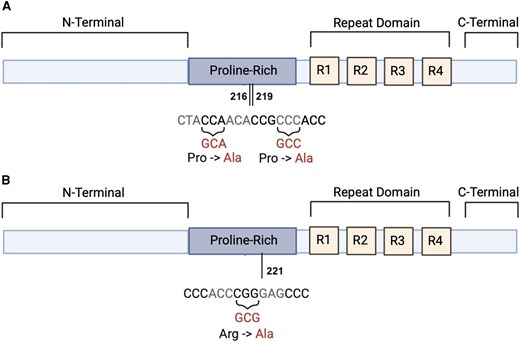
AxxA6 and R221A tau variants. Red letters indicate amino acid substitutions in (A) the 0N4R AxxA6 mutant tau sequence and (B) the 0N4R R221A mutant tau sequence. AxxA6 tau has proline-to-alanine substitutions at amino acid positions corresponding to 216 and 219 in human 2N4R tau (A). R221A tau has an arginine-to-alanine substitution at the position corresponding to 221 in human 2N4R tau (B).
Primary neuronal cultures
Primary neuronal cultures were established from postnatal Day 0 pups, as previously described20 (Supplementary material). Briefly, hippocampal tissue from littermate pups was dissected, enzymatically digested with papain and processed into single cells. Neurons were then plated on poly-L-lysine and laminin-coated dishes in Neurobasal medium with supplements. Glial proliferation was inhibited using 5-fluoro-2′-deoxyuridine. Cultures were maintained at 37°C with 5% CO2 and half-medium changes on Day 7, before they were used for proximity ligation assay and immunohistochemistry.
Proximity ligation assay and immunohistochemistry
The proximity ligation assay (PLA) process, as previously described,21 detects interactions between proteins within 40 nm or less and can be used to quantify the degree of interaction (Supplementary material). Briefly, after 14 days, cultured hippocampal neurons were fixed, permeabilized and blocked. Cells were incubated overnight with primary antibodies [Tau (1:400, Agilent, A0024), Fyn (1:1000, Abcam, ab1881), PI3K (1:100, Millipore, 05-512, clone AB6), PLCγ-1 (1:100, Millipore, 05-163), BIN1 (1:200, Sigma-Aldrich, 05-449, clone 99D)]. PLA was performed using Duolink probes (1 h incubation) with ligation and amplification steps (Anti-Rabbit PLUS and Anti-Mouse MINUS secondary antibodies at 37°C). Samples were counterstained using 1:500 Alexa Fluor 488-conjugated secondary antibody against rabbit IgG (for Dako Tau) or mouse IgG (for Tau-1) and imaged. For each protein–protein interaction, three independent neuronal cultures prepared from different animals were analysed. Within each culture, PLA signal intensities were quantified from multiple individual neurons, whose measurements were pooled across the three cultures for statistical comparison between genotypes (pooled sample size per genotype ranged from 94–101 for AxxA6 and 94–108 for R221A).
Imaging and analysis
Images were acquired at ×40 magnification using epifluorescence deconvolution (Nikon) or confocal Stimulated Emission Depletion microscopes (Leica). Investigators were blinded to genotype during analysis. Signal was quantified using ImageJ2 from 4–5 neuronal regions per image after background subtraction. PLA signal intensity was normalized to tau immunofluorescence levels for each region.
Western blotting
Mice between the ages of 20–26 weeks were anaesthetized using isoflurane. After confirming the depth of anaesthesia, mice were transcardially perfused with PBS and brains were extracted and stored at −80°C. Samples were collected from homozygous, heterozygous and wild-type mice from AxxA6 (n = 18), R221A (n = 18) and Tau KO (n = 6) lines. Hemibrains were sliced (450 µm), with slices collected in cold dissection buffer composed of PBS with 1:100 of each of Halt protease inhibitor cocktail and Halt phosphatase inhibitor cocktail (ThermoFisher). Membranes were probed with rabbit anti-tau (1:10 000, EP2456Y, Abcam) and mouse anti-actin (1:5,000, mAbGEa, Invitrogen) overnight, then with fluorescent secondary antibodies (1:5000, Alexa Fluor plus, ThermoFisher). Signals were detected using the iBright imaging system (Supplementary material).
Behaviour
The behavioural assessments were performed according to University of Minnesota protocols (#2003-37959A; Supplementary material). Young (aged 2.4–7.4 months) and old (aged 14–19 months) mice of AxxA6 and R221A models underwent behaviour tests [AxxA6: n = 12 (young), 6–12 (old); R221A: n = 7–14 (young), 9–26 (old)]. These mice were not used for any other experiments. Behavioural tests included: elevated plus maze (anxiety—open arm time, 5 min exploration), open field (locomotion and anxiety—distance/centre time, 30 min exploration), rotarod (motor coordination—fall latency over 3 days, four trials/day), Y-maze (working memory—spontaneous alternation, 5 min exploration/session), Barnes maze (spatial learning—escape latency/target quadrant time, explored over 4 days, four trials/day) and pole test (motor function—descent time, with a pole 50 cm high and 1 cm wide).
Kainic acid-induced seizures
Male and female mice of AxxA6 (n = 11–14) and R221A (n = 10–18) models, aged 15–20 weeks were challenged with a single intraperitoneal dose of kainic acid (25 mg/kg) and monitored by video for 3 h. The latency, severity and duration of seizures on video recordings were scored based on a validated scoring system. We modified a seven seizure stage classification22 as 0 = no abnormality and 8 = death (Supplementary material).
EEG surgeries and recordings
EEG surgeries were performed on double mutant models, with epidural screw electrodes inserted over motor (1 mm anterior, ±1 mm lateral) and parietal cortices (2 mm posterior, ±2 mm lateral to bregma), referenced to right motor cortex, for spike, sleep and spectral power analyses (Supplementary material).
Acute hippocampal slice electrophysiology
CA3 pyramidal neurons are known to display heightened excitability in the absence of Kv1.1.23 To test the effect of mutation on hyperexcitability, picrotoxin/4-amino-pyridine (4-AP)-induced CA3 bursts were measured in mice (4–7 weeks) with genotypes Kv1.1+/+_AxxA6 (or _R221A)wt/wt, Kv1.1+/+_AxxA6 (or _R221A)+/+, Kv1.1−/−_AxxA6 (or _R221A)wt/wt and Kv1.1−/−_AxxA6 (or _R221A)+/+. Transversal hippocampal slices were prepared using a vibratome, with 350 μm thickness (Supplementary material). Slices were then transferred to artificial CSF (ACSF), maintained at 32°C and awaited recording. The recorded signal was amplified by a MultiClamp 700B instrument, digitized by a Digidata 1550B instrument and collected using Clampex software (Molecular Devices). Slices were recorded for 30 min, perfused with normal ACSF for an initial 5 min, followed by ACSF containing picrotoxin (50 μM) and 4-AP (200 μM) for the remaining duration. The signal from 25 to 30 min was analysed. Data analysis was performed using Clampfit.
Co-immunoprecipitation and mass spectrometry-based proteomics
To investigate how genetic perturbations in tau's PRD affect its interactome, cortical samples were subjected to tau co-immunoprecipitation (Co-IP) followed by mass spectrometry-based proteomics, with tau as the only predefined interaction target (Supplementary material).
Data analysis
See Supplementary material for details on data analysis of epileptic spikes, sleep, spectral power and mass spectrometry. All data analyses were performed under blinded conditions.
Statistics
Sample sizes were determined a priori using power analyses based on pilot studies. Calculations targeted around 80% power at α = 0.05 to detect effect sizes observed in preliminary data. We used the Shapiro–Wilk test to determine if the data were normally distributed. For parametric data, we used Student's t-test and ANOVA for two- and three-group comparisons, respectively. Welch's correction was applied if the standard deviations were unequal. Tukey's multiple comparisons test was used for multiple comparisons of groups. For non-parametric data, we used the Mann–Whitney test and Kruskal–Wallis test for two- and three-group comparisons, respectively. Dunn's multiple comparisons test was used for multiple comparisons of groups that used non-parametric tests. The robust regression and outlier removal (ROUT) test, with Q = 2%, was used to identify outliers, where Q = 10% was used for highly skewed spike rate data. Individual neurons were considered as independent biological units for statistical analysis, as described previously.24,25 For spectral analysis, we performed repeated measures mixed model tests, where the post hoc comparisons have examined genotype differences within each frequency band. For behavioural tests with measurements across multiple days, to evaluate within-genotype changes over time, the post hoc comparisons were used to examine the changes across days within each genotype. Samples with video acquisition failure or poor video quality were excluded from analysis. Seizure stage latency data were analysed using two-way ANOVA with Holm–Šidák's multiple comparisons test. Analysis was performed using GraphPad Prism v10.2. For mass spectrometry analysis, the relative abundances of peptides and their P-values were provided by Proteome Discoverer 2.4 (ThermoFisher Scientific). Gene Ontology (GO) analysis was conducted through the GO knowledgebase.26 Fisher's exact tests were conducted to generate raw P-values and corresponding false discovery rates (FDRs). Fold enrichment values were generated based on the number of proteins in our dataset compared to the number of all known Mus musculus proteins identified in the specific category listed.27–29
Results
Tau expression is unaffected in AxxA6 and R221A models
We measured the expression of tau in AxxA6 and R221A models using western blotting. Mice were aged 19–28 weeks. We measured tau and actin levels of mutantwt/wt, mutant+/wt, mutant+/+, Tau−/− and Tau+/+. The ratio of tau to actin was analysed. The ratios are normalized with respect to the average ratio of respective mutantwt/wt mice. For both AxxA6 and R221A models, we found a significant difference between the ratios at the group level (n = 6 for all groups except n = 3 for each of Tau−/− and Tau+/+; total n = 48; AxxA6: F = 25.20, P < 0.0001; R221A: F = 13.91, P < 0.0001; ANOVA). However, at the individual comparison level, all the genotypes had similar tau expression, except for Tau−/−, which had lower expression than the rest of its group (P < 0.0001, AxxA6; P ≤ 0.0003, R221A; Tukey multiple comparison test; Supplementary Fig. 2). These results show that both tau mutations did not alter tau expression.
AxxA6 and R221A models decrease tau's interactions with SH3-containing proteins
We then examined how these tau variants affect tau's interactions with specific SH3-containing proteins using PLA. We found that, in cultured hippocampal neurons, both AxxA6 and R221 variants reduced tau's interactions with the SH3-containing proteins: PLCγ1 (P < 0.0001 for both variants), the p85α subunit of PI3K (P < 0.0001 for both variants) and BIN1 (AxxA6: P < 0.001; R221A: P < 0.0001). Additionally, the AxxA6 variant had reduced tau’s interaction with Fyn (P < 0.0001) (Fig. 2).
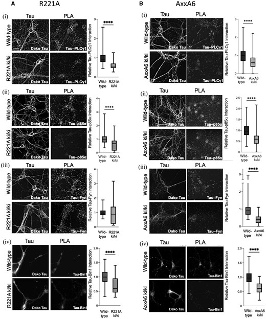
Proximity ligation assay results for tau binding to SH3-containing proteins in the AxxA6 and R221A line. (A) R221A homozygous knock-in (ki) and wild-type proximity ligation assay results were analysed for: PLCγ1 (ki/ki, n = 98, median = 0.5658; +/+, n = 104, median = 0.8922; ****P < 0.0001, Mann–Whitney U = 1192), p85α/PI3K (ki/ki, n = 94, median = 0.6659; +/+, n = 97, median = 0.9630; ****P < 0.0001, Mann–Whitney U = 2243), Fyn (ki/ki, n = 97, median = 0.8859; +/+, n = 98, median = 0.9608; P = 0.27, Mann–Whitney U = 4318) and BIN1 (ki/ki, n = 108, median = 0.5890; +/+, n = 100, median = 0.9960; ****P < 0.0001, Mann–Whitney U = 2239). (B) AxxA6 homozygous knock-in and wild-type proximity ligation assay results were analysed for: PLCγ1 (ki/ki, n = 94, median = 0.5880; +/+, n = 94, median = 0.9845; ****P < 0.0001, Mann–Whitney U = 2164), p85α/PI3K (ki/ki, n = 94, median = 0.5987; +/+, n = 96, median = 0.9642; ****P < 0.0001, Mann–Whitney U = 1492), Fyn (ki/ki, n = 98, median = 0.3853; +/+, n = 99, median = 0.8991; ****P < 0.0001, Mann–Whitney U = 778) and BIN1 (ki/ki, n = 100, median = 0.6103; +/+, n = 101, median = 0.9722; ****P < 0.0001, Mann–Whitney U = 1017). Micrographs in i–iii were obtained on an epifluorescence deconvolution microscope system; micrographs in iv were obtained on a confocal Stimulated Emission Depletion microscope system. Scale bar in A(i) = 20 µm. Graphs show min to max on box and whisker plots. *P < 0.05, **P < 0.01, ***P < 0.001, ****P < 0.0001.
Behaviour is unaffected in AxxA6 and R221A models
Behaviour tests were performed on young [aged 2.4–7.4 months, n = 17–24 (AxxA6), 16–26 (R221A)] and old [aged 14–19 months, n = 20–23 (AxxA6), 18–49 (R221A)] mice. We analysed performance differences between mutantwt/wt and mutant+/+ within each age group. Among the AxxA6 variants (Fig. 3), both age groups spent similar amounts of time in the open arms of the elevated plus maze, with no difference between AxxA6wt/wt and AxxA6+/+. They also travelled similar total distances. In the open field test, young and old mice spent similar amounts of time in the centre and travelled similar total distances. In the rotarod test, for both ages, the latency to fall times were similar between genotypes. All genotypes of all ages continued to improve their latency times on Day 2 and Day 3 with respect to Day 1, except older AxxA6+/+ mice, whose performance plateaued after Day 2. In the Y-maze spontaneous alternation test, for both ages, the short-term memory was similar between genotypes. In the Barnes maze, for both ages, the escape latency was similar between genotypes. All genotypes of all ages continued to improve their escape latency times on subsequent days. All genotypes of all ages spent similar times in the target quadrant. In the pole test, in old mice, the latency to descend the pole was similar between genotypes. These results show that the AxxA6 mutation did not affect behaviour. Similarly, the R221A mutation did not notably affect behaviour, except for an increased anxiety-like response in older mice (Supplementary Fig. 3).
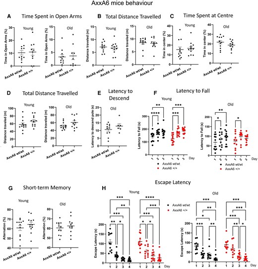
Behaviour is unaffected in AxxA6 mice, in both young (aged 2.4–7.4 months) and old (14–19 months) mice. In the elevated plus maze, with respect to their control genotypes, (A) both ages spent similar times in the open arms (as %), with no difference between AxxA6wt/wt and AxxA6+/+ (young mice, n = 12wt/wt, 12+/+, mean % = 12.7wt/wt, 24.9+/+, U = 52, P = 0.84); old mice, n = 11wt/wt, 9+/+, mean % = 4.5wt/wt, 8.7+/+, U = 45, P = 0.76; Mann–Whitney test) and (B) they travelled similar total distances (in metres) (young mice, mean = 7.0wt/wt, 6.6+/+, U = 62, P = 0.58; old mice, mean = 8.5wt/wt, 8.2+/+, U = 53, P = 0.44; Mann–Whitney test). In the open field test, with respect to their control genotypes, both ages performed similarly for (C) time spent at the centre (as %) (young mice, n = 12wt/wt, 12+/+, mean % = 15.4wt/wt, 16.7+/+, t = 0.39, P = 0.69; old mice, n = 12wt/wt, 11+/+, mean % = 21.7wt/wt, 18.7+/+, t = 1.32, P = 0.19; unpaired t-test) and (D) total distance travelled (in metres) (young mice, mean = 57.7wt/wt, 66.8+/+, t = 1.54, P = 0.13; old mice, mean = 51.5wt/wt, 60.5+/+, t = 1.72, P = 0.09; unpaired t-test). (E) In the pole test, in old mice, the latency to descend the pole (in seconds) was similar between genotypes (n = 11wt/wt, 6+/+, mean = 10.6wt/wt, 12.8+/+, t = 1.13, P = 0.27; unpaired t-test). (F) In the rotarod test, for both ages, the latency to fall times (in seconds) was similar between genotypes [young mice, n = 12wt/wt, 12+/+, F(1,22) = 0.034, P = 0.85; old mice, n = 12wt/wt, 11+/+, F(1,22) = 0.03408, P = 0.85; two-way repeated measure ANOVA]. All genotypes of all ages continued to improve their latency times on Days 2 and 3 with respect to Day 1, except older AxxA6+/+ mice, whose performance plateaued after Day 2 (P < 0.05, Šídák's multiple comparisons test). (G) In the Y-maze spontaneous alternation test, for both ages, short-term memory (as %) was similar between genotypes (young mice, n = 12wt/wt, 12+/+, mean % = 60.8wt/wt, 64.4+/+, t = 1.00, P = 0.32; old mice, n = 12wt/wt, 11+/+, mean % = 60.4wt/wt, 62.5+/+, t = 0.66, P = 0.51; unpaired t-test). (H) In the Barnes maze, for both ages, the escape latency was similar between genotypes [young mice, n = 12wt/wt, 12+/+, F(1,22) = 3.792, P = 0.06); old mice, n = 12wt/wt, 11+/+, F(1,21) = 0.9536, P = 0.33; two-way repeated measure ANOVA]. All genotypes of all ages continued to improve their escape latency times on subsequent days (P < 0.05, Šídák's multiple comparisons test). Tukey's multiple comparisons test/unpaired t-test. n = 12 (young), 6–12 (old). Error bars represent standard error of the mean. *P < 0.05, **P < 0.01, ***P < 0.001, ****P < 0.0001. wt = wild-type.
AxxA6 and R221A mice were of similar weights between genotypes at ages 3–5 months and 18–20 months (Supplementary Fig. 4).
AxxA6 reduces seizure susceptibility
After intraperitoneal kainic acid infusion (25 mg/kg), AxxA6+/wt (n = 13) and AxxA6+/+ (n = 14) variants exhibited significantly increased latency to seizure stages compared to AxxA6wt/wt (n = 11) [P < 0.0001+/wt and P < 0.05+/+, Holm–Šídák test/two-way ANOVA; Fig. 4A(i)]. In contrast, R221A+/wt (n = 18) and R221A+/+ (n = 14) variants showed no significant difference in seizure latency compared to R221Awt/wt [n = 10; Fig. 4A(ii)]. Not all mice reached Stage 8 after kainic acid treatment. These results show that AxxA6 is protective against seizure induction, delaying seizure onset.
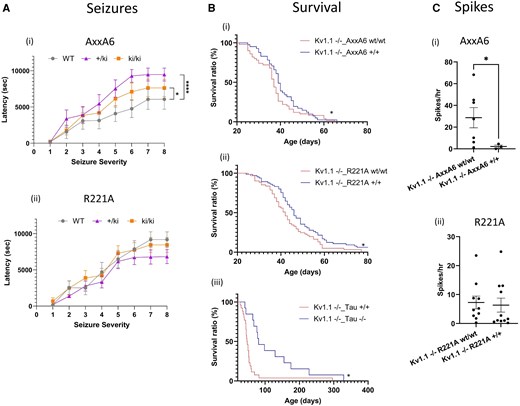
AxxA6 tau knock-in exhibits unique resistance to kainic acid-induced seizures and reduces epileptic spikes in the Kv1.1 knockout (KO) epilepsy model. [A(i and ii)] Graphs display latency to seizure stages in the (i) AxxA6 and (ii) R221A knock-in genotypes, compared to their wild-type (wt) mice, after intraperitoneal administration of kainic acid (25 mg/kg). Seizure stage was determined from video recordings. AxxA6: n = 11 wt/wt; n = 14 +/+; n = 13 +/wt; R221A: n = 10 wt/wt; n = 14 +/+; n = 18 +/wt; ages 15–20 weeks. *P < 0.05, ****P < 0.0001 (two-way ANOVA with Holm-Šidák's multiple comparison test). [B(i–iii)] AxxA6 and R221A improve early survival outcomes, as shown by survival curves for double mutants (i) Kv1.1_AxxA6, n = 41–50, (ii) Kv1.1_R221A, n = 61–65 and (iii) Kv1.1_Tau, n = 13–26. *P < 0.05. Gehan–Breslow–Wilcoxon test. With respect to the median lifespan of the Kv1.1−/−_mutant wild-type control, all mutant and tau models significantly improved the early mortality rates (P < 0.05, chi-square test). [C(i and ii)] AxxA6 reduced intracranial EEG Kv1.1 KO-induced epileptic spikes in mice, whereas R221A had no effect, as revealed by the spike rates (per hour) for (i) AxxA6 (showing reduced spike rate in AxxA6+/+, *P < 0.05, unpaired t-test) and (ii) R221A (showing similar spike rates between genotypes, Mann–Whitney test). Error bars represent the standard error of the mean. *P < 0.05, **P < 0.01, ***P < 0.001, ****P < 0.0001.
AxxA6 and R221A improve early survival outcomes
We examined whether mutations could increase survival times in Kv1.1 KO mice, which are otherwise known to die prematurely.23 The median lifespan of Kv1.1−/−_AxxA6+/+ (n = 41, median 39 days) mice was slightly more than Kv1.1−/−_AxxA6wt/wt (n = 50, median 37 days; P = 0.046, Gehan–Breslow–Wilcoxon test). We further observed that, with respect to the median lifespan of Kv1.1−/−_AxxA6wt/wt, Kv1.1−/−_AxxA6+/+ significantly reduced the early mortality rates [56% of Kv1.1−/−_AxxA6wt/wt, 34.1% of Kv1.1−/−_AxxA6+/+; χ2 = 4.329, degrees of freedom (df) = 1, P = 0.037, chi-square test], representing a 39.3% relative risk reduction due to the mutation [Fig. 4B(i)]. Similarly, the median lifespan of Kv1.1−/−_R221A+/+ (n = 61, median 46 days) mice was slightly more than Kv1.1−/−_R221Awt/wt (n = 65, median 42 days; P = 0.017, Gehan–Breslow–Wilcoxon test). With respect to the median lifespan of Kv1.1−/−_R221Awt/wt, Kv1.1−/−_R221A+/+ significantly reduced the early mortality rates (54.1% of Kv1.1−/−_AxxA6wt/wt, 34.4% of Kv1.1−/−_AxxA6+/+; χ2 = 4.463, df = 1, P = 0.034, chi-square test), representing a 34.6% relative risk reduction due to the mutation [Fig. 4B(ii)]. The median lifespans of Kv1.1−/−_Tau+/+ (n = 26) and Kv1.1−/−_Tau−/− (n = 13) mice were 47.5 and 81 days, respectively [P = 0.001, Gehan–Breslow–Wilcoxon test; Fig. 4B(iii)]. With respect to the median lifespan of Kv1.1−/−_Tau+/+, Kv1.1−/−_Tau−/− significantly reduced the early mortality rates (50.0% of Kv1.1−/−_Tau+/+, 15.4% of Kv1.1−/−_Tau−/−; χ2 = 4.388, df = 1, P = 0.036, chi-square test), representing a 69.2% relative risk reduction due to the knockout. These results suggest that both AxxA6 and R221A variants decrease the mortality rate.
AxxA6 reduces intracranial EEG Kv1.1 KO-induced epileptic spikes in mice, whereas R221A has no effect
Epileptic spike rates were studied in AxxA6 and R221A hybrid models. Spikes were rarely found in mice of wild-type genotype, and therefore we could not test our hypothesis on the benefits of tau mutations. We hence used double mutants for this spike study: Kv1.1_AxxA6 and Kv1.1_R221A. Among the mice with the Kv1.1+/+ allele, no spikes were found in 83.3% of all AxxA6 genotypes (n = 12), 54.1% of all R221A genotypes (n = 24) and 85.7% of all tau genotypes (n = 14). Among Kv1.1_AxxA6 mice, those with Kv1.1−/−_AxxA6wt/wt had a high spike rate (n = 7, mean 28.69 spikes/h) and mice with Kv1.1−/−_AxxA6+/+ had a significantly reduced spike rate [n = 3 (1 outlier), mean 2.16 spikes/h, t = 2.82, P = 0.02, unpaired t-test with Welch's correction; Fig. 4C(i)]. In Kv1.1_R221A mice, the spike rate in Kv1.1−/−_R221Awt/wt [7.25 spikes/h, n = 12 (2 outliers)] was similar to Kv1.1−/−_R221A+/+ [6.34 spikes/h, n = 12 (1 outlier), P = 0.38, Mann–Whitney test; Fig. 4C(ii)]. These results suggest that AxxA6 rescues pathological spikes.
AxxA6 rescues Kv1.1 KO-induced abnormal network activity during REM, whereas R221A amplifies gamma activity
We analysed the spectral power for the hybrid models for each sleep state: Awake, non-REM (NREM) and REM for Kv1.1_AxxA6 [n = 6, 5, 6, 4 (1 outlier) for +/+_wt/wt, +/+_+/+, −/−_wt/wt, −/−_+/+, respectively] and Kv1.1_R221A [n = 17 (4 outliers), 7, 12, 12 for +/+_wt/wt, +/+_+/+, −/−_wt/wt, −/−_+/+, respectively; Fig. 5 and Supplementary Fig. 5]. For spectral power and sleep analysis, animals with video acquisition failure or poor video quality were excluded from analysis (AxxA6: n = 1+/+_wt/wt, 1−/−_wt/wt). As Kv1.1 KO mice were reported to have hemispheric asymmetry in spontaneous spreading depolarization events,30 with 74% of events occurring unilaterally, we analysed each hemisphere separately to account for potential hemispheric differences that could be masked by combined analysis. For the awake state spectral power of Kv1.1_AxxA6 hybrid mice, there was significant interaction between the frequency band and the genotype factor for the left parietal cortex (LPC) [F(12,80) = 2.352, P = 0.01, mixed model analysis], but not for the right parietal cortex (RPC) [F(12,80) = 1.023, P = 0.43, mixed model analysis]. Multiple comparisons revealed no differences among all the pairs during this awake state (P > 0.05, Holm–Šídák test; Fig. 5A). During NREM, in both the LPC and RPC, there was significant interaction between the frequency band and genotypes [LPC: F(12,80) = 2.960, P = 0.001; RPC: F(12,75) = 3.546, P = 0.0003; mixed model analysis]. In pairwise multiple comparisons, Kv1.1−/− had increased beta and gamma power in both the LPC and RPC (beta: Kv1.1−/−_AxxA6wt/wt versus Kv1.1+/+_AxxA6wt/wt, LPC: P = 0.004; RPC: P = 0.003; gamma: Kv1.1−/−_AxxA6wt/wt and Kv1.1−/−_AxxA6+/+ versus Kv1.1+/+_AxxA6wt/wt, P < 0.05 both sides; Holm–Šídák test; Fig. 5B). During REM, in both the LPC and RPC, there was significant interaction between the frequency band and the genotype factor [LPC: F(12,80) = 5.091, P < 0.0001; RPC: F(12,80) = 1.965, P = 0.03; mixed model analysis]. In pairwise multiple comparisons, Kv1.1−/− had increased beta power in both the LPC and RPC (Kv1.1−/−_AxxA6wt/wt versus Kv1.1+/+_AxxA6wt/wt, P = 0.01, both sides; Holm–Šídák test). Interestingly, AxxA6+/+ rescued the Kv1.1 KO-induced increased beta activity, as shown by decreased beta power in Kv1.1−/−_AxxA6+/+ when compared to Kv1.1−/−_AxxA6wt/wt mice (LPC: P = 0.005, RPC: P = 0.01, Holm–Šídák test). Moreover, Kv1.1−/−_AxxA6+/+ had higher theta power than Kv1.1−/−_AxxA6wt/wt mice in the LPC (P = 0.008, Holm–Šídák test), but not with Kv1.1+/+_AxxA6wt/wt (P > 0.05, Holm–Šídák test), indicating healthy compensation in theta power, due to a reduced beta, which is important during sleep (Fig. 5C). These results show that AxxA6 rescues pathological beta activity during REM sleep. On the other hand, R221A partially (only LPC) amplified pathological beta and gamma activity during REM sleep (Supplementary Fig. 5).
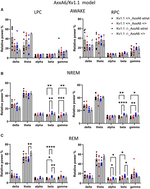
AxxA6 rescues Kv1.1 KO-induced abnormal network activity during REM, but not during NREM. We analysed spectral power for the AxxA6 for each sleep state (A) Awake, (B) NREM and (C) REM for Kv1.1_AxxA6 (n = 6, 5, 6 and 3 for +/+_wt/wt, +/+_+/+, −/−_wt/wt and −/−_+/+, respectively). In both the LPC and RPC, Kv1.1−/− showed increased beta and gamma in NREM, and beta power in REM. AxxA6+/+ rescued the Kv1.1 KO-induced increased beta activity, as shown by decreased beta power in Kv1.1−/−_AxxA6+/+ when compared to Kv1.1−/−_AxxA6wt/wt mice (LPC: P = 0.005, RPC: P = 0.01, Holm-Šídák test). Additionally, Kv1.1−/−_AxxA6+/+ exhibited higher theta power than Kv1.1−/−_AxxA6wt/wt mice in the LPC. Repeated measure mixed model. Error bars represent the standard error of the mean. *P < 0.05, **P < 0.01, ***P < 0.001, ****P < 0.0001. LPC = left parietal cortex; KO = knockout; NREM = non-REM; RPC = right parietal cortex; wt = wild-type.
AxxA6 has no effect on sleep duration, but R221A amplifies Kv1.1 KO-induced abnormal sleep
We analysed wake, NREM, REM and total sleep (NREM + REM) durations in the same Kv1.1_AxxA6 and Kv1.1_R221A mice previously used for network activity measurements (Supplementary Fig. 6). There was a significant overall difference between genotypes of the Kv1.1_AxxA6 model in awake duration [W = 5.224 (3.000, 7.692), P = 0.02, Welch/ANOVA], but there were no differences between genotypes at the multiple comparison level (P > 0.05, Dunnett's T3 multiple comparisons test). Kv1.1_AxxA6 also had similar durations of NREM, REM and total sleep (P > 0.05, Welch/ANOVA). Genotypes of the Kv1.1_R221A hybrid showed differences in awake duration [W = 14.79 (3.000, 20.17), P < 0.0001, Welch/ANOVA], in which Kv1.1−/−-containing groups resulted in increased awake time compared to Kv1.1+/+-containing groups (P < 0.008, Dunnett's T3), where R221A+/+ did not contribute to the increase (P > 0.05, Dunnett's T3). Similarly, in NREM, the genotypes showed a difference [W = 8.230 (3.000, 20.43), P < 0.0001, Welch/ANOVA], in which Kv1.1−/−_R221Awt/wt and Kv1.1−/−_R221A+/+ mice had reduced NREM duration compared to Kv1.1+/+_R221Awt/wt mice (P < 0.003, Dunnett's T3). For REM duration, the genotypes showed a difference [W = 4.896 (3.000, 21.46), P = 0.0096, Welch/ANOVA], in which Kv1.1−/−_R221A+/+ mice had reduced NREM duration compared to Kv1.1+/+_R221Awt/wt mice (P = 0.005, Dunnett's T3). For total sleep duration, the genotypes showed a difference [W = 10.60 (3.000, 20.38), P = 0.0096, Welch/ANOVA], where Kv1.1−/−_R221Awt/wt and Kv1.1−/−_R221A+/+ mice had reduced total sleep duration compared to Kv1.1+/+_R221Awt/wt mice (P < 0.001, Dunnett's T3). These results show that the hyperexcitable Kv1.1 model has reduced sleep duration and mutant models could not rescue this.
AxxA6 rescues Kv1.1 KO-induced sleep fragmentation, whereas R221A has no effect
We analysed the number of transitions (transition count) from one sleep state to another for the double mutant models (Fig. 6). The Kv1.1_AxxA6 hybrid model showed significant differences between its genotypes (P = 0.01, two-way ANOVA). In multiple comparison tests, Kv1.1−/−_AxxA6wt/wt mice had increased transitions from Awake to NREM (P = 0.002, Tukey's multiple comparisons test) and NREM to Awake (P = 0.02, Tukey's test) compared to Kv1.1+/+_AxxA6wt/wt mice. Interestingly, Kv1.1−/−_AxxA6+/+ mice had reduced transitions from Awake to NREM (P < 0.0001, Tukey's test) and NREM to Awake (P = 0.002, Tukey's test) when compared to Kv1.1−/−_AxxA6wt/wt mice. Additionally, all transition counts of Kv1.1−/−_AxxA6+/+ mice were similar to Kv1.1+/+_AxxA6wt/wt mice (all P > 0.48, Tukey's test). In the Kv1.1_R221A hybrid model, differences between its genotypes were marginally significant (P = 0.0501, two-way ANOVA). In multiple comparison tests, both Kv1.1−/−_R221Awt/wt and Kv1.1−/−_R221A+/+ mice had increased transitions from Awake to NREM (P = 0.0005 and P < 0.0001, respectively, Tukey's tests) and NREM to Awake (P = 0.0002 and P < 0.0001, respectively, Tukey's tests) compared to Kv1.1+/+_R221Awt/wt mice. These results show that AxxA6 reduces Kv1.1 KO-induced sleep fragmentation.
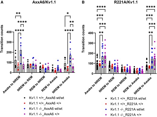
AxxA6 rescues Kv1.1 KO-induced sleep fragmentation. We analysed the number of transitions (transition count) from one sleep state to another for the models (A) Kv1.1_AxxA6 and (B) Kv1.1_R221A. (A) Kv1.1−/−_AxxA6wt/wt mice showed increased transitions from Awake to NREM (P = 0.002, Tukey's multiple comparisons test) and NREM to Awake (P = 0.02, Tukey's test) compared to Kv1.1+/+_AxxA6wt/wt mice. Kv1.1−/−_AxxA6+/+ mice exhibited fewer transitions from Awake to NREM (P < 0.0001, Tukey's test) and NREM to Awake (P = 0.002, Tukey's test) when compared to Kv1.1−/−_AxxA6wt/wt mice. All transition counts for Kv1.1−/−_AxxA6+/+ mice were similar to Kv1.1+/+_AxxA6wt/wt mice (all P > 0.48, Tukey's test). (B) Both Kv1.1−/−_R221Awt/wt and Kv1.1−/−_R221A+/+ mice exhibited increased transitions from Awake to NREM (P = 0.0005 and P < 0.0001, respectively, Tukey's tests) and NREM to Awake (P = 0.0002 and P < 0.0001, respectively, Tukey's tests) compared to Kv1.1+/+_R221Awt/wt mice. Error bars represent the standard error of the mean. *P < 0.05, **P < 0.01, ***P < 0.001, ****P < 0.0001. KO = knockout; NREM = non-REM; wt = wild-type.
AxxA6 and R221A mutations reduce Kv1.1 KO-induced ex vivo CA3 hyperexcitability
To determine the effect of tau mutation on Kv1.1 KO-induced local circuit hyperexcitability, we measured the CA3 bursts in Kv1.1_AxxA6 (slices per genotype: 9–10, mice per genotype: 3–4) and Kv1.1_R221A (slices per genotype: 8–21, mice per genotype: 3–4) mice (Fig. 7). We calculated the burst frequency (Hz), defined as total number of bursts per recording period (5 min). For Kv1.1_AxxA6 mutants, there was a significant difference between genotypes in burst frequency (Kruskal–Wallis score = 12.96, P = 0.004, Kruskal–Wallis test). Kv1.1 KO increased the burst frequency almost by double in Kv1.1−/−_AxxA6wt/wt (mean 0.159 Hz) when compared with Kv1.1+/+_AxxA6wt/wt (mean 0.081 Hz; P = 0.04, Dunn's multiple comparisons/Kruskal–Wallis test). Interestingly, Kv1.1−/−_AxxA6+/+ (mean 0.082 Hz) fully reversed the burst frequency to the levels of wild-type slices (Kv1.1−/−_AxxA6+/+ versus Kv1.1−/−_AxxA6wt/wt, P = 0.047, Dunn's multiple comparisons/Kruskal–Wallis test; Fig. 7C). For Kv1.1_R221A mutants, there was a significant difference between genotypes in burst frequency (Kruskal–Wallis score = 9.816, P = 0.02, Kruskal–Wallis test). Kv1.1 KO increased the burst frequency by 1.48-fold in Kv1.1−/−_R221Awt/wt (mean 0.47 Hz) when compared with Kv1.1+/+_R221Awt/wt (mean 0.32 Hz; P = 0.026, Dunn's multiple comparisons/Kruskal–Wallis test). Interestingly, Kv1.1−/−_R221A+/+ (mean 0.30 Hz) fully reversed the burst frequency to the levels of wild-type slices (Kv1.1−/−_R221A+/+ versus Kv1.1−/−_R221Awt/wt, P = 0.05, Dunn's multiple comparisons/Kruskal–Wallis test; Fig. 7D). These results demonstrate that both mutants have significant capability to rescue local network hyperactivity in CA3, and potentially in other hippocampal regions.
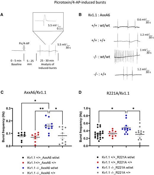
AxxA6 and R221A mutations reduce Kv1.1 KO-induced ex vivo CA3 hyperexcitability. (A) Hippocampal slices were initially recorded in normal ACSF for 5 min, then perfused with ACSF containing picrotoxin (50 μM) and 4-amino-pyridine (200 μM). After allowing 20 min for drug equilibration, induced bursts were measured for a period of 5 min at 25–30 min. A representative signal of CA3 activity is shown. (B) Representative signals from all genotypes of the Kv1.1_AxxA6 double mutation. We measured the CA3 bursts in (C) Kv1.1_AxxA6 (slices per mouse: 9–10, mice: 3–4) and (D) Kv1.1_R221A (slices per mouse: 9–10, mice: 3–4). We calculated the burst frequency (Hz), defined as the total number of bursts per recording period (5 min). (C) Kv1.1 KO increased the burst frequency almost two-fold in Kv1.1−/−_AxxA6wt/wt (mean 0.159 Hz) when compared with Kv1.1+/+_AxxA6wt/wt (mean 0.081 Hz) (P = 0.04, Dunn's multiple comparisons/Kruskal–Wallis test). Kv1.1−/−_AxxA6+/+ (mean 0.082 Hz) reversed the burst frequency to wild-type levels (Kv1.1−/−_AxxA6+/+ versus Kv1.1−/−_AxxA6wt/wt, P = 0.047, Dunn's multiple comparisons/Kruskal–Wallis test). (D) Kv1.1 KO increased the burst frequency by 1.48 times in Kv1.1−/−_R221Awt/wt (mean 0.47 Hz) when compared with Kv1.1+/+_R221Awt/wt (mean 0.32 Hz) (P = 0.026, Dunn's multiple comparisons/Kruskal–Wallis test). Kv1.1−/−_R221A+/+ (mean 0.30 Hz) reversed the burst frequency to wild-type levels (Kv1.1−/−_R221A+/+ versus Kv1.1−/−_R221Awt/wt, P = 0.05, Dunn's multiple comparisons/Kruskal–Wallis test). Error bars represent the standard error of the mean. *P < 0.05, **P < 0.01. ACSF = artificial CSF; KO = knockout; NREM = non-REM; wt = wild-type.
AxxA6 enhances tau protein interactions; R221A reduces them
Co-IP mass spectrometry on cortical samples revealed opposing effects of AxxA6 and R221A mutations [n = 12 mice/line, n = 4 mice per genotype (wt/wt, wt/ki and ki/ki); Supplementary Fig. 7 for criteria]. Genotype expression levels were normalized to the corresponding wild-type genotype within each line. The AxxA6 mutation (ki/ki and +/ki combined) altered tau's interaction with 67 distinct proteins (P < 0.05), with the majority of interactions increased [94% had an abundance ratio (AR) > 1]. In contrast, R221A mutation (ki/ki and +/ki combined) altered tau's interaction with 10 distinct proteins (P < 0.05), with the majority of interactions decreased (90% had an AR < 1). Among the homozygous genotypes of both lines, synaptotagmin 5 was the only protein that was affected, showing increased interaction in both genotypes (Fig. 8).
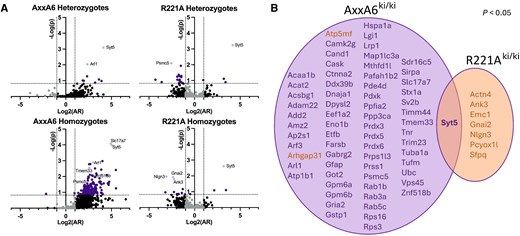
Co-immunoprecipitation mass spectrometry results revealed that tau variants have different effects on tau protein interactions, with both variants showing increased binding to SYT5. (A) Volcano plots derived from the log2[abundance ratio(AR)] and −log(P-value) for the proteins observed in the R221A and AxxA6 knock-in (ki) lines relative to wild-type; grey = P > 0.15 and AR < 2-fold changes; black = P > 0.15 and AR ≥ 2-fold changes; dark purple = P < 0.15; light purple = protein with P < 0.05. (B) Venn diagram of significant co-immunoprecipitation/mass spectrometry results in AxxA6 and R221A homozygotes (all proteins P < 0.05; purple font indicates AR > 1, orange font indicates AR < 1).
Next, we examined how proteins differed between genotypes by listing proteins that were significant (P < 0.05) in both genotypes within their line. Three proteins were identified for each of AxxA6 and R221A: Arl1, Gpm6b and Syt5 in AxxA6; and Nlgn3, Pcyox1l and Syt5 in R221A (Supplementary Fig. 8). The AR and P-values of these proteins are AxxA6ki/ki: Arl1 (8.0, 0.001), Gpm6b (10.1, 0.006), Syt5 (32.8, 0.0001); AxxA6wt/ki: Arl1 (5.1, 0.009), Gpm6b (0.01, <0.0001), Syt5 (17.4, 0.0008); R221Aki/ki: Nlgn3 (0.1, 0.01), Pcyox1l (0.2, <0.0001), Syt5 (9.0, 0.002); and R221Awt/ki: Nlgn3 (0.01, <0.0001), Pcyox1l (0.3, 0.01), Syt5 (18.1, 0.0005).
Synaptic proteins are affected by AxxA6 and R221A
When P < 0.15 was considered, 170, 11, 18 and 23 proteins were identified for AxxA6ki/ki, AxxA6wt/ki, R221Aki/ki and R221Awt/ki, respectively. GO analysis of Co-IP proteins revealed distinct enrichment patterns (Supplementary Fig. 9 and Supplementary Tables 1 and 2). AxxA6 mutations enriched synaptic, vesicular and SNARE complex proteins, suggesting tau relocalization to synapses, dendrites and the cell body. R221A enriched axonal and myelin proteins, indicating axonal membrane localization. Both variants influence neuronal excitation, with subcellular associations reflecting their specific amino acid substitutions.
AxxA6 tau alters biological processes linked to hyperexcitability and neurodegeneration
GO analysis of AxxA6ki/ki proteins (P < 0.05) uniquely revealed enrichment in autophagy, endocytosis, energy metabolism, redox homeostasis and synaptic vesicle recycling (Supplementary Fig. 9 and Supplementary Tables 1 and 2), indicating homozygous AxxA6 profoundly alters tau functionality beyond other genotypes.
Discussion
In this study, we demonstrated that distinct modifications within tau's PRD differentially modulate seizure susceptibility and sleep architecture and alter tau protein interactions with signalling molecules. Although both AxxA6 and R221A tau showed reduced binding to several SH3-containing proteins, only AxxA6 tau exhibited decreased interaction with Fyn (Fig. 2). Notably, AxxA6 tau conferred protection against kainic acid-induced seizures and reduced epileptic spikes in Kv1.1 KO mice (Fig. 4), underscoring the functional relevance of tau–Fyn interactions in seizure susceptibility. The apparent longer latencies in AxxA6 heterozygotes compared to homozygotes may suggest a non-linear gene dosage effect. However, homozygotes provide clearer mechanistic interpretation without confounding wild-type tau. Co-IP mass spectrometry of cortical lysates from AxxA6 and R221A +/ki and ki/ki mice revealed distinct tau interactomes relative to wild-type tau. The AxxA6 mutation broadly increased tau protein interactions, while the R221A mutation led to an overall reduction (Fig. 8). We observed minimal overlap between the specific proteins significantly altered by each mutation, indicating that tau protein interactions are highly sensitive to PRD sequence composition. These findings have broad implications for therapeutic strategies targeting tau's PRD and its interactions with cellular signalling pathways.
Molecular basis for precision tau targeting over broad reduction strategies
Our findings with the AxxA6 variant provide compelling evidence for precision tau targeting approaches over broadly eliminating tau. The AxxA6 mutations selectively disrupt tau–SH3 domain interactions while preserving microtubule-binding regions, demonstrating that targeted interventions at specific protein–protein interfaces have potential to yield therapeutic benefits equivalent to complete tau knockout effects. Recent structural studies reveal that tau functions as an ‘adaptable sticky surface’ with multiple proline-rich (PxxP) motifs spanning 85 residues, providing functional redundancy that enables selective disruption without complete functional loss.31,32 The AxxA6 variant specifically targets the sixth PxxP motif (positions 216–219), which mediates critical interactions with Fyn kinase, PLCγ1 and PI3K p85α—key components of pathological signalling cascades rather than physiological tau functions.32,33 This precision approach contrasts sharply with broad tau reduction strategies like antisense oligonucleotides (MAPTRx), which eliminate all tau species and may compromise essential cellular functions.14 The success of structure-based precision targeting aligns with recent clinical failures of N-terminal tau antibodies (semorinemab, tilavonemab) compared to promising results from MTBR-targeted approaches (E2814, bepranemab),34 supporting a paradigm shift towards domain-specific therapeutic intervention.15,35
AxxA6-mediated rescue of hyperexcitability
The rescue of spike rate, reduction of seizure stage onset and, to some degree, survival in Kv1.1−/− mice by the AxxA6 variant illuminate critical molecular mechanisms underlying tau-mediated hyperexcitability. AxxA6 disrupts tau interactions with PLCγ1, p85α/PI3K, BIN1 and Fyn.8,36 Disrupting the tau–Fyn interaction could prevent the formation of pathological NMDA receptor signalling complexes that drive hyperexcitability and excitotoxicity.8 Wild-type tau facilitates Fyn localization to postsynapses where Fyn phosphorylates GluN2B Y1472, stabilizing NMDAR–PSD95 complexes and promoting synaptic NMDA receptor function.37,38 The AxxA6 mutations could eliminate this scaffolding function, effectively decoupling tau from excitotoxic signalling pathways while maintaining tau's structural roles in microtubule binding. This selective disruption may contribute to the variant's ability to normalize CA3 burst frequency from pathological levels (0.52 Hz in Kv1.1−/− mice) to wild-type ranges (0.17 Hz), demonstrating mechanistic specificity rather than broad cellular dysfunction. Tau KO mice showed the greatest survival benefit in our study, as anticipated from prior work.23 The superior protective effect of tau deletion likely reflects complete elimination of tau-mediated excitatory and excitotoxic signalling, including Fyn kinase, whereas AxxA6 reduces but does not eliminate tau's interactions with Fyn and other binding partners (Fig. 2B). The preserved survival benefits of AxxA6, although modest compared to Tau KO, avoids potential adverse effects of complete tau loss39,40 and supports the precision targeting strategy of tau–SH3 interactions. We also showed that the AxxA6 mice displayed intact motor function, measured by the pole test, whereas Tau KO mice of similar age displayed impaired performance in the pole test.41 Additionally, AxxA6 also exhibited normal weight at both 3–5 months and 18–20 months of age, in contrast to the weight gain observed in 12-month-old Tau KO BL6 mice.41 Overall, these findings support the non-deleterious effect of AxxA6.
Sleep disruption as a biomarker and mechanistic driver of neurodegeneration
Our demonstration that AxxA6 normalizes REM beta oscillations and reduces sleep fragmentation provides crucial evidence for bidirectional tau–sleep interactions as both a biomarker and therapeutic target. Sleep architecture disruption represents an early measurable consequence of tau pathology that precedes cognitive symptoms, offering a non-invasive monitoring approach for therapeutic intervention.42,43 The rescue of pathological beta activity (13–35 Hz) during REM sleep by the AxxA6 mutation suggests that tau-mediated protein interactions are critical for maintaining healthy sleep architecture, with normalization of REM oscillatory patterns serving as a functional readout of therapeutic efficacy. Sleep fragmentation accelerates neurodegeneration through: (i) AQP4-mediated glymphatic dysfunction impairing protein clearance; (ii) NADPH oxidase-2 pathways promoting neuroinflammation; and (iii) hyperexcitability during sleep states increasing seizure susceptibility.44,45 By partially normalizing sleep parameters, AxxA6 could potentially slow disease progression through mechanisms beyond direct tau targeting.
Tau PRD demonstrates versatile responses to sequence modifications with distinct interactomes
We illustrated that distinct modifications within tau's PRD can have an extensive modulating effect. Although both AxxA6 and R221A tau showed reduced binding to the SH3-containing proteins BIN1, PLCγ1 and p85α/PI3K, only AxxA6 tau exhibited decreased interaction with Fyn. These disrupted interactions are notable, as all four proteins are implicated in epilepsy susceptibility7,9,46,47 and dementia.22,48 Tau-bound Fyn, when recruited to PSD95–NMDAR complexes, alters NMDAR trafficking, a mechanism linked to epilepsy.49 Fyn also phosphorylates the NR2B subunit of NMDARs, promoting p85α/PI3K-NR2B binding,36 which is further enhanced under ischaemic conditions.36 NMDAR signalling is additionally modulated by PLCγ binding to both NR2A and NR2B subunits.36,50 Beyond their roles in NMDAR function, tau activation of PLCγ and interactions with p85α/PI3K have further implications in epileptogenesis.7,51,52 Similarly, BIN1 has been shown to regulate network excitability through its tau-dependent interaction with L-type voltage-gated calcium channels.47 Tau–BIN1 binding is implicated in both AD and late-onset epilepsy, suggesting broader relevance to disease-associated network dysfunction.53 Together, these findings suggest that tau–Fyn interactions play a central role in seizure susceptibility, as only AxxA6 tau conferred protection against kainic acid-induced seizures; however, interactions with other SH3-containing proteins, such as BIN1, PLCγ1 and p85α/PI3K, may also contribute to hyperexcitability through cell type-specific mechanisms that warrant further investigation.
Clinical translation potential and biomarker development
Our results support clinical translation of precision tau targeting approaches, particularly given the failure of broad tau reduction strategies in human trials.54 Sleep-based biomarkers offer additional advantages for clinical monitoring, as sleep architecture parameters can be measured non-invasively and respond dynamically to therapeutic intervention.55 The bidirectional relationship between sleep disruption and neurodegeneration creates opportunities for early detection and intervention before extensive neuronal loss occurs.56 The finding that the MAPT-A152T variant, a genetic risk factor for tau-related neurodegenerative disorders, promotes neuronal hyperactivity through Fyn–NMDAR signalling in human induced pluripotent stem cell-derived neurons57 provides a translatable human cellular platform to evaluate whether disruption of tau–Fyn binding can normalize this hyperexcitability. The intact behavioural performance even in aged mice supports the conclusion that the mutation does not cause cognitive decline or behavioural deficits, suggesting it is well-tolerated across different life stages. Small molecule inhibitors or stapled peptides that specifically target the PxxP-binding groove of relevant SH3 domains could competitively block their interaction with tau's 216–219 motif while sparing other SH3-mediated interactions.
Limitations
This study presents methodological constraints. Mouse models do not fully recapitulate human AD tau-related pathophysiology and epilepsy, limiting translational extension. The Kv1.1 epilepsy model was selected to isolate tau's effects on neuronal hyperexcitability. Sample sizes and durations of recordings were not sufficient to easily track spontaneous seizures. The divergent effects between AxxA6 and R221A variants need further mechanistic explanation. Furthermore, longitudinal studies may be necessary to fully understand the functional role of tau's PRD. For detection techniques, Co-IP preferentially detects proteins with high abundance or stable interactions, so we are likely capturing only a subset of proteins with the strongest tau binding. To complement this, we selectively used PLA to assess weaker interactions with specific SH3-containing proteins. However, PLA is also limited in the number of proteins that can be practically evaluated. Finally, this study was conducted in mice, which express only 4R tau, whereas humans express both 3R and 4R isoforms.
Conclusion
This study demonstrates that precision targeting of specific tau protein interactions offers therapeutic advantages over broad tau reduction strategies. The AxxA6 variant, which selectively disrupts tau–SH3 domain interactions while preserving essential tau domains, successfully reduced pathological hyperexcitability in both local CA3 and regional parietal cortical networks, reduced susceptibility to seizures and epileptic spikes, improved survival, normalized sleep fragmentation and rescued beta activity during REM in epilepsy models, all without affecting baseline behaviour. In contrast, R221A showed limited benefits, thereby highlighting the specificity requirements for effective tau-targeted interventions. These findings also support structure-based therapeutics that can modulate discrete tau interactions rather than protein elimination, potentially offering safer and more effective treatments for AD and related tauopathies.
Future directions
Future research can focus on elucidating the molecular mechanisms underlying the differential effects of AxxA6 versus R221A through more detailed protein interaction mapping, cell-type specific expression and assays, cognitive assays in models exhibiting hyperexcitability and downstream signalling analysis. Long-term studies are needed to assess the sustained therapeutic benefits and potential adverse effects over full lifespans. Development of small molecule inhibitors targeting specific tau–SH3 interactions based on AxxA6 structural insights represents a critical translational step.
Data availability
The data that support the findings of this study are available from the corresponding author, upon reasonable request.
Acknowledgements
We thank the ES Cell Targeting Core at UCSF and Transgenic Gene Targeting Core at the Gladstone Institutes for their outstanding assistance in developing the tau-variant knock-in mouse lines using CRISPR/Cas9. We also thank the University Imaging Centers at the University of Minnesota, and Advanced Light Microscopy and Spectroscopy Laboratory at UCLA for assistance with cell imaging. We thank Dr Jeffrey Noebels for generously sharing the Kv1.1 knockout mice. We thank Dr Lindsay Lueptow for assistance in mouse behaviour tests and Dr Stanley Thayer for assistance in electrophysiology experiments. Finally, we thank Dr Istvan Mody and Dr Xiaofei Wei for their invaluable support and guidance in setting up the hippocampal slice electrophysiology. The thumbnail image for the online table of contents was created with Microsoft Copilot (GPT 5).
Funding
This study was supported by National Institutes of Health grants R01 AG073377 to V.J. and R01 AG058820, R01 AG075955 and RF1 NS033310 to K.V., the Fineberg Foundation and an anonymous gift.
Competing interests
The authors report no competing interests.
Supplementary material
Supplementary material is available at Brain online.


