


Abstract
A hexanucleotide (GGGGCC) repeat expansion in the C9orf72 gene represents the most frequent genetic cause of amyotrophic lateral sclerosis (ALS) and frontotemporal dementia (FTD), resulting in reduced C9orf72 mRNA and protein expression. C9orf72 is highly expressed in the cerebellum, and growing evidence implicates C9orf72-associated cerebellar pathology across neurodegenerative disorders, including ALS/FTD, yet the pathogenic mechanisms remain unresolved.
Here, we demonstrate that in vivo C9orf72 loss of function leads to cerebellar atrophy, loss of GABAergic interneurons and depletion of Purkinje and granule cells. Additionally, we demonstrate that these cerebellar anomalies precede motor defects. Single-cell transcriptomics of the C9orf72 zebrafish brain revealed the downregulation of a purine biosynthetic gene, paics, in Purkinje cells.
Furthermore, we demonstrate the reduced expression of PAICS in human post-mortem cerebellar sections and induced pluripotent stem cell-derived motor neurons from C9orf72 and sporadic ALS patients. Knockout of paics in zebrafish recapitulates cerebellar neuronal loss, neuromuscular junction disruption, motor impairment and widespread DNA damage and repair defects, including suppression of key DNA repair pathways.
Restoring paics expression in C9orf72 zebrafish resolves DNA damage and preserves Purkinje cells and granule cells, revealing PAICS as a crucial mediator of cerebellar degeneration and a promising therapeutic avenue for C9orf72-associated ALS and FTD.
Introduction
Amyotrophic lateral sclerosis (ALS) is a progressive neurodegenerative disorder characterized by the selective loss of motor neurons that leads to muscular atrophy, paralysis and ultimately death within 2–5 years of the clinical onset of symptoms.1-3 The hexanucleotide repeat expansion (HRE) of GGGGCC in the first intronic region of the C9orf72 gene represents the most common genetic cause of ALS and frontotemporal dementia (FTD).4-6 The HRE in the C9orf72 gene accounts for ∼40% of familial and 7% of sporadic cases of ALS.7 HRE drives the pathogenesis of the disease through three distinct mechanisms: (i) the formation of toxic nuclear RNA foci through bidirectional transcription of the expanded repeats8-10; (ii) the accumulation of dipeptide repeat proteins generated by repeat-associated non-AUG (RAN) translation11-13; and (iii) haploinsufficiency resulting in C9orf72 protein loss-of-function (LOF).5,6,14 Although gain-of-function mechanisms are well reported and studied,15-19 emerging evidence indicates that C9orf72 haploinsufficiency contributes significantly to the pathogenesis of ALS.14,20-22
Although motor neuron degeneration remains the primary hallmark of ALS, neuroimaging studies have revealed broader neurodegeneration affecting multiple brain regions.23,24 Consequently, the cerebellum, which plays a crucial role in motor coordination, has emerged as a crucial brain area that shows pathological changes in ALS.25-29 Functional MRI, voxel-based morphometry and diffusion-tensor imaging have revealed both functional and structural cerebellar changes in patients with ALS, supporting the characterization of ALS as a multi-system neurodegenerative disorder.26-28,30,31 Nonetheless, the molecular mechanisms behind these changes remain poorly understood. C9orf72 is highly expressed in the cerebellum.6,32 Additionally, C9orf72 RNA foci and dipeptide repeat proteins are reported to be present extensively in the cerebellum.29 Neuroimaging studies in ALS patients carrying C9orf72 repeat expansions display significant cerebellar atrophy.33-35 Notably, the characteristics of the HRE in the C9orf72 gene are correlated with clinical and pathological aspects of the disease in the cerebellum.36,37 Impairment of cerebellar-specific tasks in C9orf72 patients has been reported.38 Moreover, reduced levels of C9orf72 transcript variants32,39,40 and C9orf72 protein in the cerebellum have been reported in C9orf72 repeat expansion carriers compared with healthy controls.41,42 Interestingly, there is a significant association between survival and expression level of the C9orf72 transcript in the cerebellum.40 A recent study has demonstrated focal cerebellar degeneration and disruptions in cerebro-cerebellar connectivity in patients with ALS, which might contribute to both motor and non-motor symptoms of the disease.35 Emerging evidence suggests abnormalities in inhibitory GABAergic neurons, particularly the loss of Purkinje cells (PCs), and synapses in ALS.43,44 Given the role of the cerebellum in motor coordination and the evidence of its involvement in ALS pathology, studying cerebellar changes in ALS is crucial.
Given that the precise role and mechanism of cerebellar change in ALS remain elusive, we address this gap by investigating the impact of C9orf72 LOF on cerebellar pathology using the zebrafish as a model organism. In this study, we used a stable zebrafish transgenic line with c9orf72 LOF (C9-mir), which exhibits the characteristic hallmarks of ALS,20 to study the structural and functional changes in the C9-mir zebrafish brain, focusing particularly on the cerebellum. The findings demonstrated global atrophy and the loss of GABAergic neurons in C9-mir zebrafish brain and showed cerebellar loss of PCs and granule cells (GCs) preceding motor defects. Notably, our single-cell transcriptomic analysis revealed significant negative enrichment of the purine-containing compound biosynthetic process in PCs, with marked downregulation of the paics (phosphoribosylaminoimidazole carboxylase/phosphoribosylaminoimidazole succinocarboxamide synthetase) gene. Importantly, we validated the clinical relevance of these findings by demonstrating significant downregulation of PAICS in the cerebellum and induced pluripotent stem cell (iPSC)-derived motor neurons from both sporadic and C9orf72-HRE-carrying ALS patients, suggesting a conserved pathogenic mechanism. Furthermore, paics downregulation was sufficient to cause PC and GC loss and DNA damage in C9-mir zebrafish. Altogether, our findings strongly demonstrate that paics is a key mediator of DNA damage pathology in C9orf72-associated ALS, underscoring the potential role of purine metabolism imbalance in promoting DNA damage and repair failure and fostering disease progression.
Materials and methods
Ethics statement
Adult and larval zebrafish (Danio rerio) were maintained and experimental procedures performed in compliance with the Canadian Council on Animal Care guidelines and approved by the INRS-LNBE ethics committee. All procedures with human material were approved by the Ethical Committees in Human Research (protocols CER-INRS 2026-659/25-851 for INRS, DURCAN_iPSC/2019–5374 and Institutional Review Board study number A03-M19-22A for McGill University).
Zebrafish
Experimental model maintenance and handling
Wild-type (WT) and C9-mir adult zebrafish (Danio rerio) were maintained at 28°C under a 12 h–12 h light–dark cycle, as previously described.45 The C9orf72 zebrafish model (C9-mir) was generated using a micro RNA-based gene-silencing approach.20 Embryos were raised at 28.5°C and were collected and staged as previously described.46 The following transgenic zebrafish lines were used for imaging studies: Tg(dlx5a/6a:GFP) and Tg(vglut2:RFP).
Generation of paics F0 knockout zebrafish
Zebrafish paics F0 knockout (KO) embryos were generated using the CRISPR-Cas9 system.47-49 The target sequences for guide RNAs (gRNAs) were chosen using CHOPCHOP and Integrated DNA Technologies design tools. Three gRNAs targeting three different exons of the paics gene were selected based on the tool-based predictions, with higher priority given to those with fewer off-target effects. The gRNAs targeting the paics gene were assembled by annealing each of the three CRISPR RNA (crRNA) (Integrated DNA Technologies) to the Alt-R CRISPR-Cas9 trans-activating crRNA (tracrRNA) (Integrated DNA Technologies, Cat. No. 1072532). Each gRNA was then mixed with Cas9 protein (Alt-R S.p. Cas9 Nuclease V3, Integrated DNA Technologies, Cat. No. 1081058) to form a Cas9/gRNA ribonucleoprotein. All three ribonucleoproteins were pooled (cocktail mix of 6 μl), and 1 nl of the mixture was injected into one-cell-stage embryos. High-resolution melting analysis was performed for genotyping the paics mutant zebrafish. The sequences of all primer sets are available upon request.
Zebrafish immunostaining and confocal imaging
Zebrafish larvae/sections were fixed in 4% paraformaldehyde (PFA) in PBS at 4°C overnight. The next day, the fish were rinsed three times in a solution of 0.1% Tween-20 in PBS (PBS-Tween) for 15 min at room temperature (RT). Larvae were then rinsed twice with PBS-DT [1% bovine serum albumin (BSA), 1% Triton X-100 and 1% dimethyl sulfoxide (DMSO)] for 10 min each time. Antigen retrieval was performed for zebrafish brain sections at 85°C with 10 mM sodium citrate buffer for 20 min. Larvae/sections were then incubated in a blocking solution [5% normal goat serum (NGS), 1% BSA, 1% DMSO and 1% Triton X-100 in PBS] for 1 h. Primary antibodies [anti-pvalb7 (1:200; Millipore Sigma, Cat. No. MAB1572), anti-neurod150 (gift from M. Hibi; 1:500) and anti-γ-H2AX (1:500; GeneTex, Cat. No. GTX127342)] were added to the blocking solution and incubated overnight at 4°C. The following day, after three 15 min-washes in PBS-Tween, larvae were incubated with a secondary antibody diluted in blocking solution [goat anti-mouse Alexa Fluor 488 (Invitrogen, Cat. No. A10680) or goat anti-rabbit Alexa Fluor 568 (Invitrogen)] at 4°C overnight. The next day, the fish were rinsed three times with PBS-Tween at RT, for 15 min each time. The larvae were mounted the next day in 0.5% low-melting point agarose (Invitrogen, Cat. No. 16520-100) or fluoromount-G (Invitrogen, Cat. No. 00-4958-02) and imaged using a Zeiss LSM 780 confocal microscope.
Neuromuscular junction staining and analysis
Zebrafish larvae at 5 days postfertilization (dpf) were fixed overnight at 4°C in 4% paraformaldehyde in PBS. After three 15 min rinses in PBS-Tween at RT, larvae were permeabilized in a collagenase solution (1 mg/ml in PBS; Sigma-Aldrich, Cat. No. C0130) for 2.5 h at RT on a rotator. After three 15 min rinses with PBS-Tween, larvae were blocked for 1 h at RT in a blocking solution (1% BSA, 1% DMSO, 1% Triton X-100 and 2% NGS in PBS). Larvae were incubated for 30 min in α-BTX (tetramethylrhodamine-conjugated α-bungarotoxin; 10 mg/ml in 0.1% PBS-Tween; ThermoFisher, Cat. No. T1175) to label postsynaptic acetylcholine receptors. Then, the primary antibody SV2 (1:200; Developmental Studies Hybridoma Bank) was applied overnight at 4°C in blocking solution. After three 15 min PBS-Tween rinses, larvae were incubated for 4 h at RT in Alexa Fluor 488 goat anti-mouse secondary antibody (1:1000; Invitrogen, Cat. No. A10680) in blocking buffer on a rotator. Larvae were rinsed three times in PBS-Tween and mounted in fluoromount-G (Invitrogen, Cat. No. 00-4958-02). Confocal imaging was performed using a Zeiss LSM 780 microscope. The analysis was performed via NMJ Analyser using the default parameter of zebrafish NMJ.51
Single-cell RNA sequencing
Library preparation and sequencing
Single-cell RNA sequencing (scRNA-seq) libraries were prepared from dissociated adult C9-mir and WT zebrafish brains using standard protocols for cell capture, complementary DNA synthesis and library preparation, followed by sequencing on an Illumina NovaSeq 6000 platform (for detailed methodology, see the Supplementary material).
Data analysis
Single-cell RNAseq data were processed and analysed using Seurat (v.5) and DoubletFinder in R for rigorous quality control, doublet exclusion, dimensional reduction and cell type annotation. Subsequent differential gene expression and gene set enrichment analysis (GSEA) were performed to identify transcriptomic alterations and dysregulated pathways in specific cerebellar populations, particularly PCs and GCs. Detailed bioinformatics pipelines, parameters and filtering thresholds are provided in the Supplementary material.
Induced pluripotent stem cell-derived motor neurons
Cell lines and reprogramming
Peripheral blood mononuclear cells (PBMCs) were obtained through the C-BIG Repository at the Montreal Neurological Institute (The Neuro, McGill University). Three cells lines were used, which were derived from a patient carrying a C9orf72-HRE and a patient with sporadic ALS, reprogrammed from PBMCs of a 66-year-old male and a 65-year-old female, respectively, and a previously published and characterized control cell line, AIW002-02,52 reprogrammed from PBMCs of a 37-year-old Caucasian male. Differentiation of iPSCs into motor neurons was performed using a previously published protocol.53 The iPSC-derived motor neurons were cultured and collected for two independent studies, including this one. The detailed methodology is described by Lescouzères et al.54 Briefly, iPSCs were initially induced into neuroepithelial progenitors, which were next specified into motor neuron progenitor cells over 6 days. The motor neuron progenitor cells were subsequently replated for terminal differentiation into mature motor neurons. These were cultured for 14 and 28 days post-plating to capture two complementary maturation stages, representing early post-mitotic motor neurons with initial axon extension and mature motor neurons, respectively. Differentiation efficiency was confirmed by immunofluorescence and qPCR.54 Expression of the mature motor neuron markers HB9 (MNX1; Supplementary Fig. 1), ISL1 and CHAT was strongly upregulated in mature motor neurons, whereas it was nearly absent in the iPSCs and motor neuron progenitor cells.54
Human post-mortem tissues
Cerebellum samples
Human cerebellum samples were acquired from the Target ALS Multicenter Human Postmortem Tissue Core. Fixed, paraffin-embedded sections of cerebellum were obtained from three patients with C9orf72 ALS, a sporadic case and three non-neurological controls. Samples were from a mixture of males and females.
Extended experimental procedures
Detailed protocols regarding zebrafish in vitro transcription, morphological and behavioural assessments, histological analyses (haematoxylin and eosin, TUNEL and immunostaining) and molecular assays (qPCR and western blotting) are provided in the Supplementary material. Additionally, comprehensive methodologies for immunostaining, western blotting and qPCR of iPSC-derived motor neurons, in addition to the processing and immunostaining of human post-mortem cerebellar tissues, are described in the Supplementary material.
Statistical analysis
Significance was determined using Student’s t-test or one-way ANOVA followed by a multiple comparisons test. Graphs were plotted using Graphpad PRISM or custom-made visualization scripts in R. Significance is indicated as P < 0.05, P < 0.01 or P < 0.001; N represents the number of experimental repeats, and n represents the number of fish. For the iPSC-derived motor neurons, each experiment was performed using technical replicates generated by independent differentiations. Data are presented as the mean ± standard error of the mean.
Results
Brain atrophy and loss of GABAergic neurons in C9orf72 loss-of-function zebrafish model
We recently showed that a zebrafish C9orf72 LOF (C9-mir) model at 6 days postfertilization (6 dpf) recapitulates ALS phenotypes, including motor deficits, motor neuron loss, muscle atrophy and TDP-43 pathology.20 Capitalizing on the strength of this in vivo ALS model, we sought to investigate the effect of C9orf72 LOF on brain architecture. We performed histological studies of brain sections from C9-mir zebrafish at the larval stage (6 dpf) and the adult stage (12 months postfertilization). Haematoxylin and eosin staining of transverse sections at 6 dpf showed a significant reduction in the size of the C9-mir brain along the whole rostrocaudal axis (Fig. 1A and B). Interestingly, we observed weaker haematoxylin staining in the cerebellum of C9-mir zebrafish brain compared with controls, suggesting cellular depletion in the region (Fig. 1A). This phenotype persisted in adulthood, as demonstrated by a sagittal section of the adult C9-mir zebrafish brain showing a significant reduction in the area of the cerebellum (Fig. 1C and D). Taken together, these findings are in line with clinical observations in ALS-FTD patients, where neuroimaging studies have documented progressive brain atrophy extending beyond motor regions.23,24,55,56
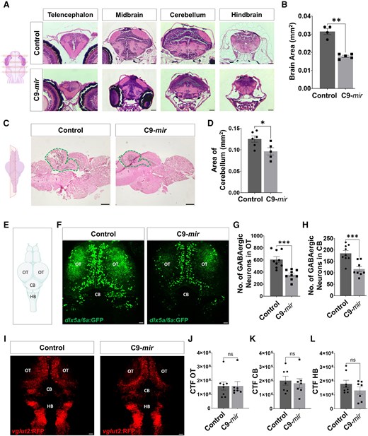
C9orf72 loss-of-function causes early brain atrophy and loss of GABAergic neurons in zebrafish. (A) Haematoxylin and eosin staining of the telencephalon, midbrain, cerebellum and hindbrain sections of control and C9-mir zebrafish at 6 days postfertilization (dpf). Green dashed line indicates the cerebellum. Scale bar = 10 µm. (B) Quantitative analysis demonstrated a significant decrease in the brain area of C9-mir compared with control zebrafish brain at 6 dpf. N = 4 or 5 biological replicates. (C) Haematoxylin and eosin staining on a sagittal section of the adult control and C9-mir zebrafish brain. Green dashed line indicates the cerebellum. Scale bar = 100 µm. (D) Quantitative analysis demonstrated a significant decrease in cerebellar area in C9-mir compared with control adult zebrafish. N = 5 or 6 biological replicates. (E) Schematic representation of the zebrafish brain. (F) Representative images of GABAergic neurons (dlx5a/6a:GFP) in control versus C9-mir zebrafish brain at 5 dpf. Scale bar = 20 µm. (G) Quantitative analysis demonstrated a significant decrease in the number of GABAergic neurons (dlx5a/6a:GFP) in C9-mir compared with control optic tectum at 5 dpf, n = 9 samples. (H) Quantitative analysis demonstrated a significant decrease in the number of GABAergic neurons (dlx5a/6a:GFP) in C9-mir compared with control cerebellum at 5 dpf, n = 8 or 9 samples. (I) Representative images of glutamatergic neurons (vglut2:RFP) in control versus C9-mir zebrafish brain at 5 dpf. Scale bar = 20 µm. (J) Quantitative analysis demonstrated no significant changes in the corrected total fluorescence (CTF) of glutamatergic neurons (vglut2:RFP) in C9-mir compared with control optic tectum at 5 dpf, n = 7 or 8 samples. (K) Quantitative analysis demonstrated no significant changes in the CTF of glutamatergic neurons (vglut2:RFP) in C9-mir compared with control cerebellum at 5 dpf, n = 7 or 8 samples. (L) Quantitative analysis demonstrated no significant changes in the CTF of glutamatergic neurons (vglut2:RFP) in C9-mir compared with control hindbrain at 5 dpf, n = 7 or 8 samples. Data are mean ± standard error of the mean. Student’s t-test; *P < 0.05, **P < 0.01, ***P < 0.001, ****P < 0.0001; ns = non-significant. CB = cerebellum; HB = hindbrain; OT = optic tectum. Figure partially created in BioRender. Patten, K. (2026) https://BioRender.com/a32owax.
Given the brain size and cerebellar abnormalities in C9-mir zebrafish, we next conducted an analysis of the brain neuronal networks. We observed a significant reduction in the number of GABAergic neurons in the C9-mir zebrafish brain at 5 dpf (Fig. 1E and F), particularly in the optic tectum (Fig. 1F and G) and cerebellum (Fig. 1F and H). These findings align with the emerging clinical evidence of GABAergic system dysfunction in ALS patients.57-63 We did not observe any changes in the glutamatergic neurons in the C9-mir zebrafish brain at 5 dpf (Fig. 1I–L) or 6 dpf (Supplementary Fig. 1B–E). These data suggest a potential imbalance between excitatory and inhibitory neural networks in the zebrafish C9-mir ALS model.
Loss of Purkinje cells and granule cells in the cerebellum precedes motor defects in C9-mir zebrafish
To complement our previous observations of cerebellar abnormalities in C9-mir zebrafish and explore cerebellar cell populations further, using immunostaining, we assessed two main cerebellar neuronal cell types: parvalbumin7 (pvalb7)-positive PCs and neurod1-positive GCs, both integral to cerebellar function.
At 5 dpf, we observed a significant reduction in the numbers of PCs and GCs within the cerebellum of C9-mir zebrafish (Fig. 2A–C). This neuronal loss persisted into adulthood, as depicted by the sagittal sections of the adult C9-mir zebrafish cerebellum, which continued to show a marked decrease in the number of PCs (Fig. 2D and E). The cerebellum plays a central role in the coordination of motor functions, such as swimming behaviour, in larval zebrafish.64,65 We thus investigated the temporal relationship between cerebellar pathology and motor dysfunction in the C9-mir zebrafish using the automated Noldus Ethovision XT behaviour monitoring system. At 5 dpf, the C9-mir zebrafish exhibited no significant motor impairments, with both distance travelled and velocity comparable to control fish (Fig. 2F–H). However, by 6 dpf, the C9-mir zebrafish displayed significant motor deficits, characterized by reduced distance travelled and decreased velocity (Fig. 2I–K). These findings indicate that loss of PCs and GCs in C9-mir zebrafish cerebellum precedes motor dysfunction, thus emphasizing the importance of early cerebellar involvement in the pathogenesis of ALS.
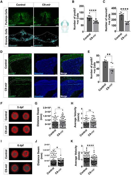
Purkinje and granule cell loss precedes the onset of motor impairment in C9-mir zebrafish. (A) Immunostaining of Purkinje cells (pvalb7) and granule cells (neurod1) in control and C9-mir zebrafish cerebellum at 5 days postfertilization (dpf). Scale bar = 10 µm for Purkinje cells and 20 µm for granule cells. (B) Quantitative analysis demonstrated a significant decrease in the number of Purkinje cells (pvalb7) in C9-mir zebrafish compared with control zebrafish cerebellum at 5 dpf, n = 14–18. (C) Quantitative analysis demonstrated a significant decrease in the number of granule cells (neurod1) in C9-mir zebrafish compared with control zebrafish cerebellum at 5 dpf, n = 10 or 11. (D) Sagittal section of adult zebrafish cerebellum in control and C9-mir zebrafish. White dashed lines indicate the cell bodies of Purkinje cells. Scale bar = 50 µm. (E) Quantitative analysis demonstrated a significant decrease in the number of Purkinje cells (pvalb7) in adult C9-mir zebrafish compared with control zebrafish section of cerebellum, n = 6. (F) Representative swimming tracks of control and C9-mir zebrafish at 5 dpf. (G and H) The C9-mir zebrafish (n = 46) did not show any significant changes in swimming distance (G) and average velocity (H) compared with control zebrafish (n = 47–48) at 5 dpf. (I) Representative swimming tracks of control and C9-mir zebrafish at 6 dpf. (J and K) The C9-mir zebrafish (n = 62–69) displayed a significant decrease in swimming distance (J) and average velocity (K) compared with control zebrafish (n = 71–77) at 6 dpf. All data are represented as the mean ± standard error of the mean. Statistical significance was calculated by Student’s t-test. ns = non-significant; *P < 0.05, **P < 0.01, ***P < 0.001, ****P < 0.0001. Figure partially created in BioRender. Patten, K. (2026) https://BioRender.com/9maheqa.
Dysregulated purine-containing compound biosynthesis process in C9-mir Purkinje cells
To elucidate the mechanistic link between the loss of PCs and GCs and the pathogenesis of ALS, we performed scRNA-seq on the adult C9-mir and WT control zebrafish brain tissue. The data were processed using the Seurat standard pipeline for data filtering and clustering to determine cell type identity based on genetic markers of zebrafish brain cell populations.60,61 We identified 15 major cell clusters within the zebrafish brain (Fig. 3A), each characterized by unique genetic markers (Fig. 3B). We used the zebrafish cell landscape,66 zebrahub,67,68 the single-cell transcriptome atlas for zebrafish69-71 and the Max Planck zebrafish brain (mapzebrain) atlas72 as major reference databases of genetic markers for cell annotations. Given that PCs did not appear in the 15 major cell clusters, we specifically isolated a cluster of PCs using carbonic anhydrase 8 (ca8) as a genetic marker.73 Furthermore, we cross-referenced the top 10 differentially expressed genes (DEGs) in each of the identified cell clusters against publicly available ALS studies.74-78 Notably, this comparison revealed that a substantial number of these genes have previously been associated with ALS in the literature (Supplementary Table 1), providing strong validation for our data.
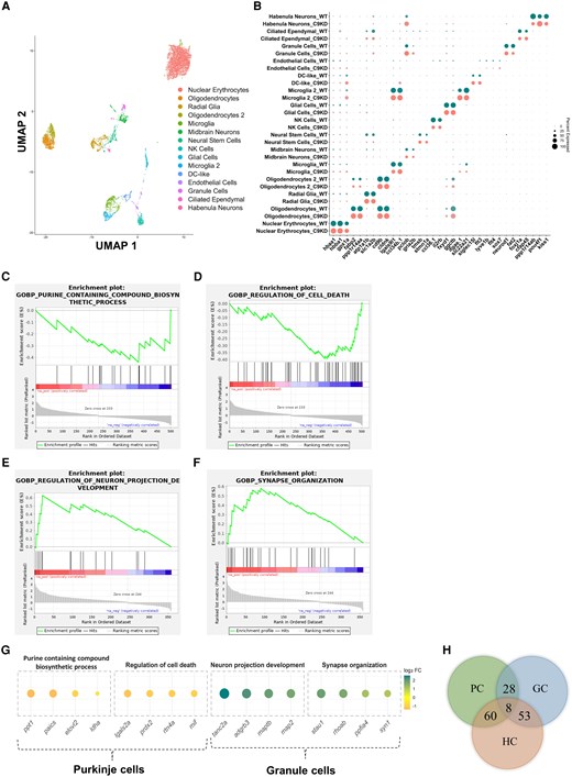
Comprehensive analysis of single-cell RNA sequencing and differential gene expression in adult wild-type and C9-mir zebrafish brain. (A) Uniform manifold approximation and projection (UMAP) of single-cell RNA sequencing profiles from adult wild-type (WT) and C9-mir zebrafish brains, representing 15 cell clusters. (B) Dot plot representing two or three marker genes for each cell cluster in the UMAP. (C–F) Enrichment plots from gene set enrichment analysis representing top enriched biological processes from Purkinje cells (PCs) and granule cells (GCs) in C9-mir zebrafish cerebellum. (G) Dot plot representing the top differentially expressed genes (DEGs) from the enriched biological processes in PCs and GCs. (H) Venn diagram illustrating the overlap of DEGs between human cerebellar tissue from C9orf72-HRE amyotrophic lateral sclerosis patients and C9-mir zebrafish cerebellar cell types. Right: Fifty-three shared DEGs between human cerebellum (HC) and GCs. Left: Sixty shared DEGs between HC and PCs.
Next, we performed a GSEA on the DEGs in PCs and GCs, respectively, to identify dysregulated biological processes in C9-mir zebrafish. GSEA offers the advantage of assessing gene expression data at the level of gene sets, making it particularly capable of detecting slight, coordinated changes in biological pathways that otherwise could be missed when focusing solely on individual genes.79,80 GSEA on the DEGs in PCs revealed significant negative enrichment in pathways related to the nucleoside biosynthetic process (Supplementary Fig. 2A), purine-containing compound biosynthetic process (Fig. 3C and Supplementary Fig. 2A) and regulation and negative regulation of cell death in the C9-mir PCs (Fig. 3D and Supplementary Fig. 2A). This suggests a downregulation of genes involved in purine nucleotide synthesis and cell survival mechanisms, most notably paics and rtn4a (Fig. 3G and Supplementary Table 2), potentially compromising neuronal function and viability. Additionally, pathways related to neuron projection development (Fig. 3E and Supplementary Fig. 2B), synapse organization (Fig. 3F and Supplementary Fig. 2B) and autophagy (Supplementary Fig. 2B) were found to be dysregulated in the C9-mir GCs, indicating alterations in genes essential for neuronal connectivity, synaptic integrity and cellular maintenance (Fig. 3G and Supplementary Table 2), which might contribute to cerebellar dysfunction.
To further validate the translational relevance of our findings, we compared our single-cell transcriptomic data from C9-mir zebrafish with published human cerebellar transcriptome data from C9orf72-HRE-carrying ALS patients.81 This cross-species analysis revealed remarkable overlap in DEGs. Specifically, 53 DEGs were shared among human cerebellar tissue and GCs in our zebrafish dataset, and 60 DEGs were shared among human cerebellar tissue and PCs (Fig. 3H). These overlapping genes, particularly in cerebellar cell types, represent robust clinical validation of our zebrafish model as a reliable system for studying ALS-related molecular mechanisms.
Conserved PAICS downregulation in C9-mir zebrafish and C9orf72-ALS patient samples
Recent studies have underlined the importance of purine metabolism in neurodegenerative diseases, including ALS.82,83 Our single-cell RNA data revealed significant downregulation of the paics gene, which encodes an enzyme essential for de novo purine biosynthesis, in PCs of the C9-mir zebrafish cerebellum (Fig. 4A and Supplementary Fig. 3A). Consistent with the transcriptomic changes, the level of Paics protein was also significantly reduced in C9-mir zebrafish, as determined by western blot analysis (Fig. 4B and C). To explore whether PAICS might interact physically with C9orf72, we modelled a PAICS–C9orf72 complex using AlphaFold 384 (DeepMind). The individual protein structures were predicted with high confidence, but a low interface predicted template modelling score (ipTM = 0.32) and diffuse inter-chain predicted aligned error indicated that AlphaFold does not support a stable and well-defined PAICS–C9orf72 complex (Supplementary Fig. 3B). These in silico results from AlphaFold 3, therefore, argue against a strong direct interaction and hint at an indirect mechanism linking PAICS to C9orf72. Further analysis of paics expression across 14 distinct cell types in the C9-mir zebrafish brain revealed widespread downregulation in 9 of these cell types (Supplementary Fig. 3C).
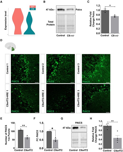
PAICS expression is downregulated in C9-mir zebrafish, human post-mortem cerebellum sections and induced pluripotent stem cell-derived motor neurons from patients with amyotrophic lateral sclerosis. (A) Violin plot representing the expression level of paics gene in C9-mir versus wild-type (WT) Purkinje cells. (B) Western blot depicting the level of Paics protein in control and C9-mir zebrafish at 5 days postfertilization (dpf). (C) Quantification of the Paics protein level in control and C9-mir zebrafish (n = 3) at 5 dpf. (D) Immunofluorescence staining of PAICS protein in post-mortem cerebellar sections from control and C9orf72-HRE ALS patients. N = 3. Scale bar = 30 μm. (E) Quantitative analysis demonstrated a significant decrease in the number of PAICS+ puncta in C9orf72-HRE cerebellum sections compared with controls (N = 3). (F) Quantitative PCR (qPCR) analysis of PAICS mRNA in iPSC-derived motor neurons (MNs) from control and C9orf72-HRE ALS patients at 3 weeks post-differentiation. (N′ = 3). (G) Western blot depicting the level of PAICS protein in iPSC-derived MNs from control and C9orf72-HRE ALS patients at 1 week post-differentiation. (H) Quantification of the PAICS protein level in iPSC-derived MNs from control and C9orf72-HRE ALS patients at 1 week post-differentiation (N′ = 4). All data are represented as the mean ± standard error of the mean. Statistical significance was calculated by Student’s one-tailed t-test (with Welch’s correction for unequal variances). *P < 0.05, **P < 0.01, ***P < 0.001, ****P < 0.0001. ALS = amyotrophic lateral sclerosis; iPSC = induced pluripotent stem cell; n = zebrafish sample size; N = number of human donors from whom post-mortem cerebellar tissue was obtained; N′ = number of donors from whom iPSC-derived MN lines were generated. Figure partially created in BioRender. Patten, K. (2026) https://BioRender.com/h0zh8h0.
The zebrafish genome encodes a single paics orthologue that shares a high degree of similarity with human PAICS (71.3% nucleotide and 76.3% amino acid identity). According to the Human Protein Atlas, PAICS is expressed in all tissues, including highly in the brain, particularly the cerebral cortex and cerebellum. To validate the translational significance of Paics downregulation observed in our C9-mir zebrafish model, we next examined the PAICS expression in post-mortem cerebellar tissue from three controls and three C9orf72-HRE ALS patients. Hoechst staining confirmed the characteristic cerebellar foliation in these samples (Supplementary Fig. 3D). In line with our zebrafish data, we observed a significant reduction in PAICS expression in the cerebellum of C9orf72-HRE ALS patients compared with controls (Fig. 4D and E). Notably, the PAICS signal appeared enriched in cells with a morphology and perisomatic distribution consistent with Bergmann glia surrounding Purkinje neuron cell bodies (Supplementary Fig. 3E). Additionally, the examination of cerebellar tissue from one sporadic ALS patient revealed reduced PAICS expression relative to control (Supplementary Fig. 3F), thereby raising the possibility that PAICS downregulation is not restricted to C9orf72-ALS.
Given that we observed a significant loss of GCs in the C9-mir zebrafish cerebellum, we next sought to examine the GC population in post-mortem human cerebellar tissue. Mirroring the changes observed in the C9-mir zebrafish cerebellum, we observed a significant reduction in the GABA-AR α1 signal, which marks GCs in the cerebellum from three C9orf72-HRE ALS patients compared with three controls (Supplementary Fig. 4A and B). Examination of cerebellar tissue from one sporadic ALS patient similarly showed a reduction in GABA-AR α1 signal relative to the control (Supplementary Fig. 4C).
As indicated by our single-cell RNA data, that paics downregulation was not restricted to the cerebellum but occurred across 9 of 14 identified cell types in the zebrafish brain (Supplementary Fig. 3C). Moreover, we observed a global reduction in the expression of the Paics protein in C9-mir zebrafish (Fig. 4B and C). Given this widespread downregulation and the fact that motor neuron loss is a primary hallmark of ALS, we next examined PAICS expression in iPSC-derived motor neurons from C9orf72-HRE carriers and sporadic ALS patients. To this end, we generated iPSC-derived motor neurons from C9orf72-HRE carriers, sporadic ALS patients and controls using a sequential differentiation protocol (Supplementary Fig. 4D). Quantitative PCR (qPCR) and western blot analysis of iPSC-derived motor neurons from C9orf72-HRE patients revealed a significant reduction of PAICS mRNA (Fig. 4F) and protein levels (Fig. 4G and H), respectively, compared with controls. Immunostaining confirmed PAICS expression in motor neurons, but no major difference was observed in the cellular location of PAICS in control versus C9orf72-HRE iPSC-derived motor neuron lines (Supplementary Fig. 4E). Additionally, we also observed reduced PAICS mRNA expression in iPSC-derived motor neurons from patients with sporadic ALS relative to controls (Supplementary Fig. 4F). Altogether, these findings indicate that downregulation of PAICS expression is a conserved feature across zebrafish, post-mortem cerebellum and patient-derived motor neurons.
paics knockout in zebrafish leads to motor dysfunction
PAICS is a bifunctional enzyme that catalyses sequential steps in the de novo purine biosynthesis pathway: the conversion of 5-aminoimidazole ribonucleotide to 5′-phosphoribosyl-4-carboxy-5-aminoimidazole and the subsequent transformation of 5′-phosphoribosyl-4-carboxy-5-aminoimidazole into 5-aminoimidazole-4-(N-succinylcarboxamide) ribonucleotide.85 These reactions are important in the synthesis of purine nucleotides, the basic components of DNA and RNA, and play a critical role in cellular energy metabolism. Dysregulation in the function of PAICS has been implicated in several pathological conditions, including neurodevelopmental disorders,86 highlighting its importance in maintaining cellular homeostasis.
To investigate the functional consequences of paics downregulation, we generated a paics F0 CRISPR KO zebrafish line. Zebrafish paics F0 KO embryos were generated using the CRISPR-Cas9 system.49 We targeted three gRNA sites at exons 3, 5 and 6 of the zebrafish paics gene to disrupt its function (Supplementary Fig. 5A). This approach facilitated the generation of a biallelic zygotic KO directly in the injected embryos of the F0 generation.47,48 A high-resolution melting assay87 revealed that the gRNAs were highly efficient in creating indels in injected embryos (Supplementary Fig. 5B).
We observed that paics F0 KO fish displayed a significant reduction in head length (Fig. 5A) and head area (Fig. 5A) compared with WT control zebrafish at 5 dpf. Motor behavioural analysis further revealed that paics F0 KO zebrafish exhibited notable motor deficits at 5 dpf, with a significant reduction in the distance travelled and velocity of the paics F0 KO zebrafish compared with WT control zebrafish at 5 dpf (Fig. 5B). These results were consistent with the motor phenotype observed in our C9-mir zebrafish model (Fig. 2I–K). To investigate the impact of paics KO on neuromuscular function further, we examined the neuromuscular junction (NMJ) integrity in the larval paics F0 KO and control fish. Immunostaining and the analysis of NMJ morphology by NMJ analyser51 revealed a significant reduction in the number of presynaptic (SV2a) and postsynaptic (α-BTX) puncta in paics F0 KO zebrafish compared with WT zebrafish (Fig. 5C–E). Additionally, we observed a significant reduction in the co-localization of presynaptic (SV2a) puncta with postsynaptic (α-BTX) puncta, in addition to a decrease in the co-localization of postsynaptic (α-BTX) puncta with presynaptic (SV2a) puncta in paics F0 KO zebrafish compared with WT zebrafish (Fig. 5C and F). These impaired motor behaviours and NMJ defects in larval paics F0 KO zebrafish are consistent with findings that we and others have reported in several other zebrafish models of ALS.20,88-93
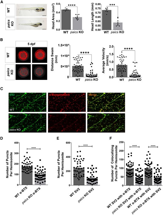
Morphological, locomotor and neuromuscular junction defects are induced by paics knockout in zebrafish. (A) Morphological comparison of willd-type (WT) and paics F0 knockout (KO) zebrafish at 5 days postfertilization (dpf). Quantitative analysis includes measurements of head length and head area in both WT (N = 8) and paics F0 KO (N = 8 or 9) zebrafish. Scale bar = 1 mm. (B) Representative swimming tracks of WT and paics F0 KO zebrafish at 5 dpf. The paics F0 KO zebrafish (n = 46) displayed a significant decrease in swimming distance and average velocity compared with WT zebrafish (n = 46) at 5 dpf. (C) Co-immunostaining of zebrafish neuromuscular junctions with presynaptic (SV2a; green) and postsynaptic (α-bungarotoxin; red) markers at 5 dpf. Scale bar = 20 µm. (D) Quantitative analysis demonstrated a significant decrease in the number of postsynaptic (α-BTX) puncta in paics F0 KO zebrafish compared with WT zebrafish. (E) Quantitative analysis demonstrated a significant decrease in the number of presynaptic (SV2a) puncta in paics F0 KO zebrafish compared with WT zebrafish. (F) Co-localization analysis revealed a significant decrease in the co-localization of presynaptic (SV2a) puncta with postsynaptic (α-BTX) puncta in paics F0 KO zebrafish compared with WT zebrafish. Likewise, co-localization analysis of postsynaptic (α-BTX) puncta with presynaptic (SV2a) puncta revealed a marked reduction in co-localizing puncta in paics F0 KO zebrafish compared with WT zebrafish. n = 42–52. All data are represented as the mean ± standard error of the mean. Statistical significance was calculated by Student’s t-test. ns = non-significant; *P < 0.05, **P < 0.01, ***P < 0.001, ****P < 0.0001. α-BTX = α-bungarotoxin; n = number of hemisomites.
Elevated DNA damage results in GABAergic and Purkinje cell loss in paics knockout zebrafish cerebellum
We next sought for any alterations in the brain and cerebellar structures in paics F0 KO zebrafish. Analysis of GABAergic and glutamatergic neuronal networks in the paics F0 KO revealed a significant reduction in the number of GABAergic neurons in the optic tectum and the cerebellum of the paics F0 KO zebrafish brain at 5 dpf (Fig. 6A–C), which parallels our observations in the C9-mir zebrafish brain. Furthermore, we observed a reduction in the number of glutamatergic neurons only in the optic tectum, but no significant changes were observed in the cerebellum and the hindbrain of the paics F0 KO zebrafish brain at 5 dpf (Fig. 6A and D–F). Consistent with our previous observations, we found a significant reduction in the number of pvalb7+ PCs in the cerebellum of paics F0 KO zebrafish larvae at 5 dpf (Fig. 6G and H), precisely phenocopying our observations of PCs loss in C9-mir zebrafish.
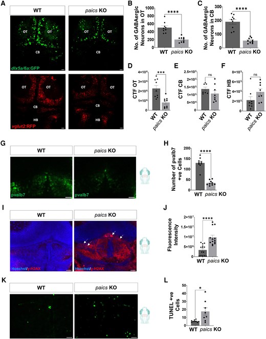
GABAergic and Purkinje cell loss and increased DNA damage in zebrafish cerebellum after paics knockout. (A) Representative images of GABAergic neurons (dlx5a/6a:GFP) and glutamatergic neurons (vglut2:RFP) in wild-type (WT) versus paics F0 knockout (KO) zebrafish brain at 5 days postfertilization (dpf). Scale bar = 20 µm. (B) Quantitative analysis demonstrated a significant decrease in the number of GABAergic neurons (dlx5a/6a:GFP) in paics F0 KO zebrafish compared with WT zebrafish optic tectum at 5 dpf, n = 9. (C) Quantitative analysis demonstrated a significant decrease in the number of GABAergic neurons (dlx5a/6a:GFP) in paics F0 KO zebrafish compared with WT zebrafish cerebellum at 5 dpf, n = 9. (D) Quantitative analysis demonstrated significant decrease in the corrected total fluorescence of glutamatergic neurons (vglut2:RFP) in paics F0 KO zebrafish compared with WT zebrafish optic tectum at 5 dpf, n = 9. (E) Quantitative analysis demonstrated no significant changes in the corrected total fluorescence of glutamatergic neurons (vglut2:RFP) in paics F0 KO zebrafish compared with WT zebrafish cerebellum at 5 dpf, n = 7 or 8. (F) Quantitative analysis demonstrated no significant changes in the corrected total fluorescence of glutamatergic neurons (vglut2:RFP) in paics F0 KO zebrafish compared with WT zebrafish hindbrain at 5 dpf, n = 8. (G) Immunostaining of Purkinje cells (pvalb7+) in WT and paics F0 KO zebrafish brain at 5 dpf. Scale bar = 10 µm. (H) Quantitative analysis demonstrated a significant decrease in the number of Purkinje cells (pvalb7+) in paics F0 KO zebrafish compared with WT zebrafish cerebellum at 5 dpf, n = 9. (I) Immunolabelling of γ-H2AX in WT and paics KO zebrafish cerebellum at 5 dpf. White arrows represent DNA damage foci, and dashed white arrows represent DNA damage apoptotic rings. Scale bar = 20 µm. (J) Quantitative analysis revealed significant upregulation in γ-H2AX signal in paics F0 KO zebrafish cerebellum at 5 dpf, n = 13 or 14. (K) Representation of TUNEL-positive cells in WT and paics KO zebrafish cerebellum at 5 dpf. Scale bar = 20 µm. (L) Quantitative analysis revealed a significant increase in the number of apoptotic cells in paics F0 KO zebrafish versus WT zebrafish cerebellum at 5 dpf, n = 8 or 9. All data are represented as the mean ± standard error of the mean. Statistical significance was calculated by Student’s t-test. ns = non-significant; *P < 0.05, **P < 0.01, ***P < 0.001, ****P < 0.0001. CB = cerebellum; HB = hindbrain; OT = optic tectum. Figure partially created in BioRender. Patten, K. (2026) https://BioRender.com/ndmvh57.
The human PAICS gene has been implicated in regulating DNA damage and repair (DDR).94,95 Accumulation of DNA damage has been reported in ALS96-98 and other movement disorders, such as Parkinson’s disease99,100 and cerebellar ataxia.101 To explore this possibility, we investigated whether paics loss alone could trigger DNA damage in our KO model. In the paics KO zebrafish, we found that the γ-H2AX signal was significantly enhanced in the cerebellum at 5 dpf, supported by an increase in γ-H2AX foci and rings, a marker of double-strand breaks, reflecting increased DNA damage (Fig. 6I and J). At this stage, this corresponded to an enhanced number of TUNEL-positive cells, showing increased apoptosis within the cerebellum of the paics KO zebrafish (Fig. 6K and L). These findings suggest that paics is a critical mediator of DNA damage that results in cerebellar degeneration.
Loss of paics suppresses key DNA repair pathways in paics knockout and C9-mir zebrafish
To unravel the molecular underpinnings of the DNA damage in the paics KO zebrafish, we carried out qPCR analysis for 18 genes associated with DDR pathways.102-106 Transcriptional profiling of these genes revealed a marked upregulation of DNA damage sensors, including atm, atr, tp53 and and rad17, in paics KO zebrafish, indicating an activated DNA damage response (Fig. 7A). However, genes involved in DNA repair processes were significantly downregulated. Specifically, we observed reduced expression of genes critical for base excision repair (ogg1 and apex1), nucleotide excision repair (ercc1 and ddb2) and mismatch repair (mlh1, msh2 and msh6) (Fig. 7A). We also found reduced expression of regulatory kinases (chek1 and chek2) that are critical for coordinating DDR signalling, which suggests compromised checkpoint activation (Fig. 7A). Checkpoint kinase dysregulation, as shown by reduced chek1 and chek2 expression in the paics KO zebrafish, might further exacerbate ALS pathology by impairing cell cycle arrest and apoptosis.
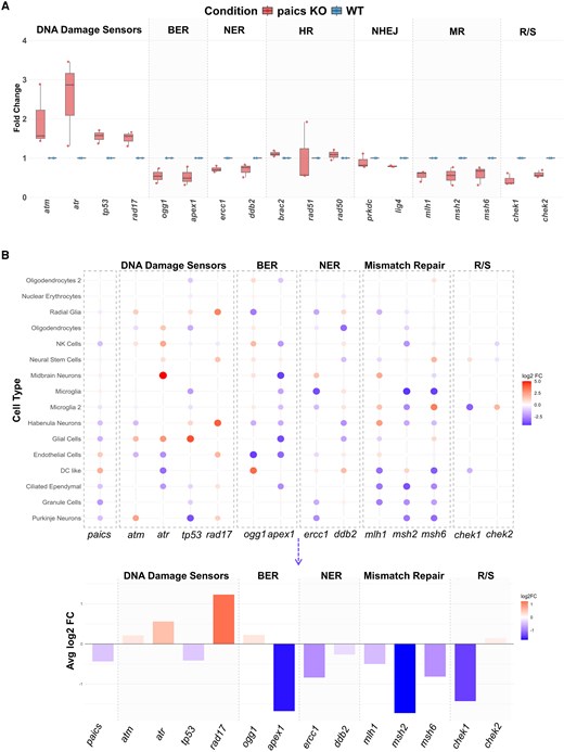
Suppression of DNA repair pathways in paics F0 knockout and C9-mir zebrafish. (A) Box plot illustrating the relative mRNA expression levels of DNA damage response and repair (DDR) pathway genes in wild-type (WT) and paics F0 knockout (KO) zebrafish. The x-axis represents genes categorized by functional pathway [DNA damage sensors, base excision repair (BER), nucleotide excision repair (NER), homologous recombination (HR), non-homologous end joining (NHEJ), mismatch repair (MR) and regulatory/signalling (R/S)], while the y-axis denotes the fold change (FC) relative to WT. Data are derived from qPCR analysis (N = 3 biological replicates). Each box plot displays the median (centre line), interquartile range (box) and individual data points (dots). Red and blue colours denote paics KO and WT conditions, respectively. (B) Dot plot and bar graph illustrating cell type-specific and averaged mRNA expression changes of DDR genes in C9-mir zebrafish brain. The dot plot (top) displays the log2FC of gene expression across 16 cell types (y-axis), categorized by functional pathway (DNA damage sensors, BER, NER, MR and R/S). Each circle represents the log2FC for a specific gene (x-axis) in a cell type, with colour intensity indicating magnitude (blue, downregulated; red, upregulated). The bar graph (bottom) shows the average log2FC across all cell types for each gene. Data were derived from single-cell RNA sequencing analysis. All data are represented as the mean ± standard error of the mean.
To resolve cell type-specific contributions, we analysed the expression of genes involved in DDR across distinct cell types in our single-cell data of C9-mir zebrafish brain. The data revealed heterogeneous expression patterns, yet the overall aggregate analysis (average expression across all cell types) aligned with our initial qPCR results in paics KO zebrafish. DDR sensors (atm, atr and rad17) showed elevated average expression, although tp53 was an exception, displaying a slight decrease in the expression (Fig. 7B). For base excision repair pathways, apex1 expression was markedly reduced, whereas the expression of ogg1 exhibited a modest but inconsistent upregulation in selected cell types (Fig. 7B). In nucleotide excision repair and mismatch repair pathways, the expression of the genes was predominantly downregulated across cell types, with average expression levels of ercc1, ddb2, mlh1, msh2 and and msh6 showing substantial reductions (Fig. 7B). Regulatory kinases exhibited divergent trends, whereby chek1 expression was greatly decreased, whereas chek2 showed a subtle increase in average expression (Fig. 7B).
This convergence of DDR defects across C9-mir and paics KO zebrafish further underscores paics as a critical mediator of DNA damage pathology in C9orf72-associated ALS. The upregulation of DDR sensors and the persistent presence of unresolved DNA damage, as observed through γ-H2AX accumulation, aligns with findings from ALS patient-derived tissues.96,97,107,108 Additionally, impaired base excision repair (BER) genes (ogg1 and apex1) contribute to oxidative DNA damage, a hallmark of ALS.98 ,109,110 Reduced expression of mismatch repair (MMR) genes (mlh1, msh2 and msh6) compromise genomic integrity as slippage in repetitive sequences, mirroring microsatellite instability, another characteristic hallmark of C9orf72-associated ALS.111,112 Critically, the loss of paics phenocopies C9orf72-associated ALS at the genomic level, positioning purine metabolism as a linchpin connecting nucleotide pool imbalance, DDR failure and ALS progression.
paics overexpression rescues Purkinje and granule cell loss and DNA damage response in C9-mir zebrafish
Given the critical role of paics in maintaining cerebellar neuronal integrity and DNA repair, we examined whether restoring the expression of paics could rescue the cerebellar deficits observed in C9-mir zebrafish. We cloned the WT zebrafish paics coding sequence into the pCS2+ plasmid and, from that, we generated the capped WT paics mRNA (paicsWT) using in vitro transcription and injected it into C9-mir zebrafish embryos at the one-cell stage (Supplementary Fig. 6A). Immunostaining for pvalb7 and neurod1 revealed a significant reduction in the number of PCs and GCs in the cerebellum of C9-mir larvae compared with controls at 5 dpf, which was consistent with our previous observations (Fig. 8A–C). Remarkably, overexpression of paicsWT mRNA into C9-mir zebrafish resulted in a robust rescue of the number of PCs and GCs to levels comparable to control larvae (Fig. 8A–C).
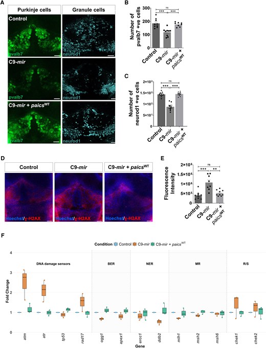
Injection of paics mRNA rescues Purkinje and granule cell loss and DNA damage response in C9-mir zebrafish. (A) Immunostaining of Purkinje cells (pvalb7+) and granule cells (neurod1+) in control, C9-mir and C9-mir + paicsWT zebrafish cerebellum at 5 days postfertilization (dpf). Scale bar = 20 µm. (B) Quantitative analysis demonstrated a significant decrease in the number of Purkinje cells (pvalb7+) in C9-mir zebrafish compared with control zebrafish cerebellum at 5 dpf. Injection of paics mRNA into C9-mir zebrafish significantly restored Purkinje cell numbers, reaching levels comparable to those of control larvae, n = 8 or 9. (C) Quantitative analysis demonstrated a significant decrease in the number of granule cells (neurod1+) in C9-mir zebrafish compared with control zebrafish cerebellum at 5 dpf. Injection of paics mRNA into C9-mir zebrafish significantly restored granule cell numbers, reaching levels comparable to those of control larvae, n = 7 or 8. (D) Immunolabelling of γ-H2AX to detect DNA damage in control, C9-mir and C9-mir + paicsWT zebrafish cerebellum at 5 dpf. Scale bar = 20 µm. (E) Quantitative analysis demonstrated a significant upregulation in γ-H2AX signal in C9-mir zebrafish compared with control zebrafish cerebellum at 5 dpf. Injection of paics mRNA into C9-mir zebrafish significantly mitigated the DNA damage, reaching levels comparable to those of control larvae, n = 8 or 9. (F) Box plot illustrating the relative mRNA expression levels of DNA damage response genes in control, C9-mir and C9-mir + paicsWT zebrafish. The x-axis represents genes categorized by functional pathway [DNA damage sensors, base excision repair (BER), nucleotide excision repair (NER), mismatch repair (MR) and regulatory/signalling (R/S)], while the y-axis denotes the fold change relative to controls. Data are derived from qPCR analysis (N = 3 biological replicates). Each box plot displays the median (centre line), interquartile range (box) and individual data points (dots). Blue, orange and teal denote control, C9-mir and C9-mir + paicsWT conditions, respectively. All data are represented as the mean ± standard error of the mean. Statistical significance was calculated by one-way ANOVA followed by Dunnett’s T3 multiple comparisons test. ns = non-significant; *P < 0.05, **P < 0.01, ***P < 0.001, ****P < 0.0001. WT = wild-type.
Given that the downregulation of paics was associated with increased DNA damage in paics KO zebrafish, we next assessed whether overexpression of paicsWT mRNA could also mitigate DNA damage in C9-mir larvae. Immunostaining for γ-H2AX, a marker of DNA double-strand breaks, revealed extensive DNA damage in the cerebellum of C9-mir zebrafish at 5 dpf (Fig. 8D and E). However, paicsWT mRNA-injected C9-mir larvae exhibited a significant reduction in the γ-H2AX signal, with levels comparable to control fish (Fig. 8D and E).
Given that loss of paics in both paics KO and C9-mir zebrafish was associated with activation of DNA damage sensors and suppression of multiple DNA repair pathways, we next asked whether the restoration of paics expression via mRNA injection could reverse transcriptional dysregulation of DDR genes in the C9-mir zebrafish. We therefore measured the expression of the same panel of 13 DDR genes involved in DNA damage sensing and repair. In C9-mir larvae, we observed the characteristic pattern of marked upregulation of DDR sensors (atm, atr and rad17) together with reduced expression of base excision repair (ogg1 and apex1), nucleotide excision repair (ercc1 and ddb2) and mismatch repair (mlh1 and msh2) at 6 dpf (Fig. 8F). Strikingly, we observed the upregulation of checkpoint regulators (chek1 and chek2) in the C9-mir zebrafish at 6 dpf (Fig. 8F). The injection of paicsWT mRNA to restore paics expression in C9-mir zebrafish largely normalized the DDR profile by reducing the elevated DNA damage sensor transcripts and restoring the expression of DNA repair and checkpoint genes towards control levels (Fig. 8F).
Together, these findings demonstrate that restoration of paics expression is sufficient to rescue both PC and GC loss and DDR pathways in C9-mir zebrafish, positioning paics as a critical mediator of C9orf72-associated cerebellar pathology in ALS.
paics overexpression ameliorates motor deficits in C9-mir zebrafish larvae
Having established that paics overexpression can rescue cerebellar cellular populations and DDR pathways in C9-mir zebrafish, we next sought to test whether motor function can also be rescued. Consistent with our previous observations, C9-mir zebrafish exhibited notable motor deficits at 6 dpf, with a significant reduction in the distance travelled and average velocity of C9-mir zebrafish compared with control zebrafish at 6 dpf (Supplementary Fig. 6B and C). Remarkably, injection of paicsWT mRNA in C9-mir zebrafish showed a significant rescue of swimming distance and average velocity, reaching values comparable to those of control larvae (Supplementary Fig. 6B and C). These data indicate that paics overexpression not only corrects cerebellar degeneration and DNA damage phenotypes but also confers a robust functional rescue at the level of motor behaviour.
Discussion
In this study, we demonstrate that the C9orf72 LOF in zebrafish initiates a cascade of neurodegenerative events that extends beyond motor neuron pathology. Our study provides new insights into the early cerebellar pathology resulting from C9orf72 LOF, revealing key mechanistic links between paics, a gene involved in de novo purine biosynthesis and DDR failure.
We show that C9orf72 LOF in zebrafish leads to global brain atrophy, selective reduction in GABAergic neurons and loss of PCs and GCs prior to motor dysfunction. These findings align with clinical neuroimaging studies of cerebellar atrophy and cerebro-cerebellar connectivity changes in C9orf72-ALS patients.23,24,35 Notably, our work extends earlier findings of cerebellar RNA foci and dipeptide repeat protein deposition in ALS29 to demonstrate that C9orf72 haploinsufficiency is sufficient to cause cerebellar vulnerability independently.
A crucial mechanistic insight from our study is the identification of disrupted purine metabolism as a factor contributing to cerebellar pathology in ALS. Through the single-cell transcriptomics and GSEA, we observed a disruption in the purine-containing compound biosynthetic process in the PCs of C9-mir zebrafish, characterized by downregulation of the paics gene, which encodes an essential enzyme in the de novo purine biosynthesis pathway. Crucially, this transcriptional reduction translates to the proteomic level, in that we observed a significant downregulation of Paics protein in the C9-mir zebrafish. Interestingly, a recent KEGG pathway analysis in a separate study94 identified ALS as one of the top-enriched pathways among 4039 genes co-expressed with PAICS in non-small cell lung cancer. Our paics KO study, which recapitulated the cerebellar pathology and motor defects of the C9-mir zebrafish, also highlights the link between metabolic imbalance and the extensive dysregulation of DDR. The upregulation of DNA damage sensors and the downregulation of key DNA repair genes, especially the genes involved in the mismatch repair pathway, suggest that disrupted purine homeostasis can exacerbate genomic instability, thus propagating neurodegeneration in ALS.
Importantly, the translational relevance of our findings is supported by the convergence of molecular and cellular pathology in human ALS patient tissues. We observed a significant downregulation of PAICS protein in the post-mortem cerebellar sections from C9orf72-HRE carriers compared with controls. Crucially, this molecular defect was accompanied by a marked reduction in the GABA-AR α1, which is a marker for GCs. This demonstrates that the loss of cerebellar GCs we identified in the C9-mir zebrafish is a clinically conserved feature with human ALS cerebellar pathology. We also observed reduced PAICS expression and GC signal in a sporadic ALS case, suggesting that this cerebellar pathology might extend beyond C9orf72-linked ALS.
Given that motor neuron loss is a primary hallmark of ALS, and our observation of global downregulation of Paics protein in C9-mir zebrafish, we next investigated whether this purine biosynthesis defect extends to motor neurons. We found that PAICS is significantly downregulated at the transcriptomic level in iPSC-derived motor neurons from both sporadic and C9orf72 ALS patients, with further proteomic analysis confirming reduced PAICS protein levels in C9orf72 iPSC-derived motor neurons. Collectively, our data indicate that PAICS deficiency is a broad, systemic consequence of C9orf72 LOF rather than a defect restricted to a single cell type. To validate the role of paics functionally, we restored the expression of paics in C9-mir zebrafish via mRNA injection. Remarkably, paics overexpression rescued the PC and GC loss and significantly reduced DNA damage and restored DDR pathways in the C9-mir zebrafish, to levels comparable to controls. Moreover, the rescue of cerebellar phenotypes resulted in a robust functional rescue of motor behaviour in the C9-mir zebrafish. These rescue experiments directly demonstrate that paics downregulation is a key driver of cerebellar neuronal vulnerability and DNA damage downstream of c9orf72 LOF.
Our data propose a model wherein C9orf72 LOF disrupts purine biosynthesis via paics downregulation, leading to the depletion of nucleotides required for DNA repair. This creates a self-perpetuating cycle of unresolved DNA damage (evidenced by γ-H2AX accumulation), repair pathway suppression and neurodegeneration. These defects mirror genomic instability in ALS patients and suggest that C9orf72 haploinsufficiency converges on DDR failure. Our results make the case for a broader model of ALS pathogenesis that integrates both motor and non-motor systems and combines metabolic defects with genomic instability. Furthermore, this metabolic restriction offers a plausible explanation for the persistence of neurodegeneration in our model, despite the innate regenerative capacity of the zebrafish CNS. Successful regeneration requires rapid proliferation of progenitor cells, a process heavily dependent on available nucleotide pools for DNA replication. We postulate that Paics downregulation creates a metabolic bottleneck, wherein the depletion of purine substrates limits the clonal expansion required for tissue repair. Additionally, the observed upregulation of DNA damage checkpoints (atm and atr) is likely to trigger cell cycle arrest, effectively locking progenitor cells in a non-proliferative state and preventing compensatory regeneration.
Given that Paics is a critical enzyme in the de novo purine biosynthesis pathway, its downregulation leads to the accumulation of specific metabolites, such as formylglycineamide riboside, succinylaminoimidazolecarboxamide riboside and aminoimidazolecarboxamide riboside, in blood, urine and CSF.86,113 Furthermore, PAICS deficiency has been associated with neurodevelopmental and neuromuscular abnormalities, similar to those seen in ALS.86 Therefore, the biochemical analysis and measurement of these metabolites in blood or urine could serve as potential biomarkers for ALS diagnosis and progression.
Despite these insights offered by our study, the precise molecular mechanisms that lead to the downregulation of PAICS in the C9orf72 LOF need to be elucidated. Our in silico modelling with AlphaFold 3 argues against a direct physical interaction between C9orf72 and PAICS (Supplementary Fig. 3B), suggesting an indirect regulation of PAICS. Mechanistically, C9orf72 is required for proper mechanistic target of rapamycin complex 1 (mTORC1) activation at the lysosome through its binding to Rag GTPases.114 In addition to that, the loss of C9orf72 function has been shown to decrease mTORC1 signalling and increase transcription factor EB activity, indicating a metabolic switch away from anabolic programmes.115 Given that mTORC1 signalling is a master driver of de novo purine synthesis116-118 and promotes the spatial assembly of the ‘purinosome’,119 which is a multi-enzyme complex required for efficient purine production, its suppression in C9orf72-LOF conditions might lead to diminished PAICS expression and impaired complex assembly. Furthermore, the reduction of PAICS in sporadic ALS iPSC-derived motor neurons and post-mortem cerebellum samples suggests that this defect extends beyond C9orf72-linked ALS. The downregulation of PAICS might represent a convergent consequence of TDP-43 pathology, a hallmark shared by nearly 97% of ALS cases.120 The nuclear depletion of TDP-43 has been shown to destabilize the mRNA of Raptor, a key component of the mTORC1 complex,121 thereby directly impairing mTORC1 signalling. Given that mTORC1 is the driver of de novo purine biosynthesis,116-118 its suppression via TDP-43 pathology might lead to the repression of PAICS expression. Thus, familial and sporadic ALS appear to converge on the same purine metabolic defect. However, further research is warranted to validate these upstream signalling cascades definitively and map the mechanistic link between C9orf72 haploinsufficiency and PAICS downregulation precisely. Moreover, the mechanisms linking the loss of PAICS to DDR failure and neurodegeneration require further work. Studies exploring how purine metabolism dysfunction impairs DNA repair enzymes or alters synaptic function in PCs and GCs are yet to be performed, which might provide us with key insights to target specific pathways via small molecules or gene therapy to stop or even reverse the progression of ALS.
Conclusion
In conclusion, our study reveals a new mechanistic axis in the pathogenesis of ALS by linking the C9orf72 haploinsufficiency to cerebellar neurodegeneration via purine biosynthesis disruption and DDR failure. The identification of PAICS as a conserved mediator in the zebrafish and human ALS system highlights its potential as a therapeutic target and a biomarker. These findings not only enhance our understanding of ALS as a multisystem disorder but also open new avenues for therapeutic intervention aimed at restoring metabolic and genomic homeostasis.
Data availability
Cell type markers for each cell type cluster in the single-cell RNA data, DEGs for PCs and GCs and GSEA data have been deposited in the Supplementary material. Additional data are available upon request.
Acknowledgements
We thank Leonid Saponiv for animal care and husbandry. We thank the Target ALS Human Postmortem Tissue Core for providing post-mortem cerebellar tissue sections from controls, sporadic ALS and C9orf72-ALS cases, and we are deeply grateful to the patients and their families for their invaluable donations. The zebrafish and human brain diagram in the thumbnail image for the online table of contents was created in BioRender. Patten, K. (2026) https://BioRender.com/9aqj7gb.
Funding
J.S. is supported by ALS Rising Star award by Brain Canada, CERMO-FC, Fondation Armand-Frappier and Fonds de Recherche du Québec Santé (FRQS) doctoral training grants. L.L. is supported by a FRQS Postdoctoral Fellowship. S.P. is supported by the Anna Sforza Djoukhadjian Research Chair in ALS from Fondation Armand-Frappier, Natural Science and Engineering Research Council of Canada (NSERC), Canadian Institutes of Health Research (CIHR-177940), and a FRQS Junior 2 research scholar.
Competing interests
The authors report no competing interests.
Supplementary material
Supplementary material is available at Brain online.


