


 https://orcid.org/0000-0001-5487-0923

Abstract
Epilepsy is one of the most common chronic neurological disorders, and temporal lobe epilepsy (TLE) is its most prevalent form in adults. Although TLE is classified as a focal epilepsy, it has traditionally been defined by hippocampal and extra-hippocampal atrophy. However, several studies also report regions of increased cortical thickness, or cortical hypertrophy, in TLE, challenging the notion that structural change in epilepsy is unidirectional. In this narrative review, we synthesize evidence for cortical hypertrophy in TLE and place these findings in a broader neurobiological context. We provide an overview of cortical thickness and MRI approaches used to measure it, highlighting differences between surface-based, volume-based and voxel-based methods. We then review MRI studies reporting cortical hypertrophy in TLE, noting variability in affected regions, patient populations and analytical techniques. We discuss potential mechanisms that may underlie cortical hypertrophy, including seizure-related structural remodelling, glial or inflammatory processes, compensatory neuroplasticity, network effects and methodological limitations. Finally, we outline key gaps and controversies regarding the biological validity, temporal dynamics and clinical relevance of cortical hypertrophy in TLE. We conclude that consideration of both cortical atrophy and hypertrophy may improve broader understanding of TLE as a disorder involving brain remodelling rather than isolated regional atrophy.
Introduction
Focal epilepsy is one of the most common chronic neurological disorders and is characterized by spontaneous and recurrent seizures that begin in a specific area of the brain. Its most common form—temporal lobe epilepsy (TLE)—has long pointed the finger at one suspect: the hippocampus.1 For decades, histopathological and neuroimaging studies have documented volume loss in this region,2-5 supporting the idea that TLE is a disease of atrophy. This structural evidence is strengthened by neurophysiological studies, including EEG and stereoelectroencephalography (SEEG), which demonstrate ictal onset in the hippocampus and directly implicate it as a primary seizure focus in TLE.6,7 But the hippocampus is no lone region. Given its extensive connectivity to the rest of the brain, both localized and widespread atrophic changes have also been documented in TLE, shifting the view from a focal condition to one of networks.8 And yet, not everything shrinks. Recent studies have complicated the story, showing patterns of cortical hypertrophy alongside widespread cortical atrophy.9 These findings challenge the long-standing view of TLE as a disorder defined mainly by atrophy.
Here, we focus on these often-overlooked regions of hypertrophy and address three questions: what is, and how do we measure, cortical thickness? What characterizes the traditional atrophy-centred view of TLE? And what does the growing evidence of cortical hypertrophy suggest about the underlying mechanisms of TLE? Finally, we discuss remaining gaps, ongoing controversies and directions for future research.
What is cortical thickness and how do we measure it?
The cerebral cortex consists of layered grey matter that covers the surface of the brain.10 These layers form a thin sheet that differs in thickness from one brain region to another.11 Cortical thickness refers to the thickness of this grey matter sheet. Herein, we use cortical atrophy to mean decreased cortical thickness (a thinner cortex) and cortical hypertrophy to mean increased cortical thickness (a thicker cortex), each defined relative to healthy control groups. Unless stated otherwise, we are describing cross-sectional findings rather than longitudinal changes.
In living humans, we can estimate cortical thickness non-invasively using MRI, notably with a T1-weighted sequence (or a T2-weighted sequence in young infants), which provides relatively strong contrast between grey matter, white matter and cerebrospinal fluid. This contrast allows automated software to segment the boundaries of the different tissues of the cortex, and compute grey matter thickness across the entire cortical surface with good accuracy.11
Both volume- and surface-based approaches are used to assess cortical morphology.10,12 Volume-based approaches, such as voxel-based morphometry, quantify grey matter by treating each voxel as the basic unit of measurement. Each voxel is classified as grey matter, white matter or cerebrospinal fluid, and its value reflects the proportion of grey matter it contains.13 Voxel-based morphometry measures grey matter volume or density across the brain rather than actual cortical thickness. However, some voxel-based tools, such as the Computational Anatomy Toolbox (CAT), can estimate cortical thickness. These tools use a projection-based thickness method,14 which derives thickness by projecting distances between inner and outer cortical surfaces while correcting for partial-volume effects and sulcal geometry. Surface-based tools like FreeSurfer15 and CIVET16 likewise compute cortical thickness, typically defined as the Euclidean distance between the white matter boundary and the pial surface at each point across the cortex.
These automated cortical thickness measures have been validated against manual delineations and, in selected regions, against post-mortem histology, demonstrating good agreement and reproducibility.17,18 Test-retest studies further support the reliability of surface-based estimates under stable acquisition conditions.19,20 Nonetheless, variability across studies remains a consideration. Differences in segmentation algorithms, preprocessing choices and spatial smoothing can lead to method-dependent regional effects.21,22 In addition, cortical thickness estimates are sensitive to image quality, including spatial resolution, signal-to-noise ratio, tissue contrast and head motion, all of which can introduce segmentation errors and bias measurements.19,23 Careful acquisition protocols and systematic quality control are therefore essential for robust results.
Traditional view: cortical atrophy as the dominant structural signature
MRI-based analyses of cortical thickness consistently show an association between TLE and multi-lobar atrophy.8 Atrophy generally spans the mesiotemporal cortex, including the ipsilateral hippocampus, and extends far beyond the epileptogenic zone into bilateral fronto-central regions.24-28 This widespread distribution has also been related to large-scale brain network measures, and suggests that functional cortical hubs may be particularly vulnerable.25
Cytologically, previous studies suggest that cortical atrophy may arise from microstructural reorganization, rather than neuronal loss. Design-based stereology of resected temporopolar cortex from patients with TLE shows hypertrophied neurons with enlarged cell bodies, while the surrounding neuropil is reduced due to sparser dendritic trees and lower spine density.29 This loss of neuronal processes decreases overall tissue volume even though neuron numbers remain relatively preserved. Such changes imply a reduction in cortical connectivity and may help explain how cortical atrophy can coexist with increased excitability and seizure susceptibility in TLE.
Clinically, atrophy changes appear largely independent of the severity of hippocampal pathology or the side of seizure onset,30 although they may be more diffuse in left TLE.24,31,32 The basis for this asymmetry remains uncertain but may relate to more lateralized connectivity in the left-dominant hemisphere,32 a greater intrinsic vulnerability of the left hemisphere33 or the earlier age of seizure onset commonly seen in left TLE.34 Greater extra-hippocampal atrophy has also been associated with longer disease duration and higher seizure frequency, and supports a progressive neurodegenerative component.5,26,35-37
While cortical atrophy in TLE has been extensively characterized, recent work points to a more complex picture that also includes regions of cortical hypertrophy. In contrast to the well-studied atrophic changes, hypertrophy remains a relatively understudied phenomenon. To date, research has focused mainly on hypertrophic alterations in deep structures, particularly the amygdala,38-47 with relatively little attention given to cortical regions.
Evidence for cortical hypertrophy in TLE
To summarize the evidence for cortical hypertrophy in TLE, we conducted a literature search in PubMed with no restrictions on language or publication date. Search terms included ‘grey matter’, ‘brain’, ‘hypertrophy’, ‘increased’, ‘thickening’, ‘cortex’, ‘cortical thickness’, ‘volume’, ‘temporal lobe epilepsy’ and ‘TLE’, used in various combinations. The most recent search was carried out on 25 November 2025. Findings of hypertrophy were heterogenous with respect to methodology, clinical phenotypes and spatial distribution.
First, out of 11 articles, three used a surface-based approach,46,48,49 two used a volumetric-based approach9,50 and six used voxel-based morphometry.42,51-55
Second, 10 studies reported some hypertrophy in TLE,9,42,48-54,56 including overall TLE or mesial TLE,9,42,50-54 late-onset TLE,48 pharmaco-resistant TLE46,50-52 and depressive symptoms in TLE.49 One additional study reported findings in temporal epilepsy results as part of a broader sudden unexpected death in epilepsy (SUDEP) patient cohort.55 Across these studies, a total of 666 patients with TLE were examined, along with 494 healthy controls, for a combined total of 1160 participants.
Third, regions of hypertrophy varied between studies with no clear spatial patterns (Fig. 1). In TLE overall, hypertrophy was most often reported in temporal and frontal regions, as well as in the cingulate cortex. Temporal involvement included both ipsilateral and contralateral mesial and temporopolar areas.9,42,46,52-54 Frontal changes were typically seen in inferior and medial prefrontal regions, and cingulate involvement was reported bilaterally.48,49,53 Overall, findings suggest that hypertrophy in TLE is not confined to the seizure focus but can involve distributed networks across both hemispheres. Detailed regional findings are provided in Table S1.
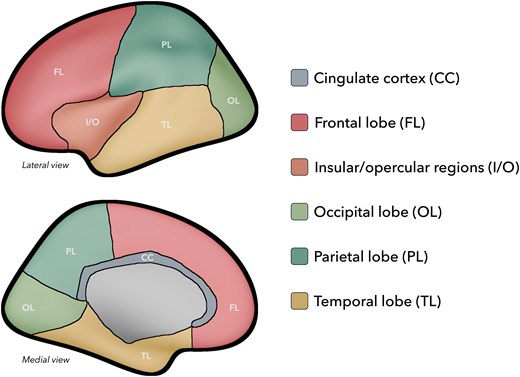
Summary of the spatial distribution of cortical hypertrophy in temporal lobe epilepsy. Cortical regions displaying hypertrophy are colour-coded with respect to the associated brain lobe, cortex or regions. Occipital lobe (OL): cuneus.9 Insular/opercular regions (I/O): rolandic operculum;51 insula.51,53 Parietal lobe (PL): superior parietal lobule;9 angular gyrus (AG).9 Cingulate cortex (CC): rostral anterior cingulate cortex;50 posterior cingulate cortex;42 cingulate cortex;53 cingulum.51 Frontal lobe (FL): inferior frontal gyrus;48,51 supplementary motor area;51 medial prefrontal cortex;53 superior frontal gyrus;51 orbitofrontal cortex.49 Temporal lobe (TL): temporal pole/temporopolar cortex;42,51,52 parahippocampal gyrus;53,55 middle temporal gyrus;9,51 inferior temporal gyrus;51,53 hippocampus;46,55 superior temporal gyrus;53 fusiform gyrus;51,53 medial temporal lobe;9 frontal piriform cortex;9 entorhinal cortex;52 perirhinal cortex.52
Biological basis of cortical hypertrophy
At the tissue level, increased cortical thickness may result from neuronal hypertrophy, abnormal glia, cellular swelling, altered vasculature, or changes in extracellular space and neuropil composition.42 Rather than representing static residual damage, such thickening may reflect an ongoing pathological process,9 with localized gliotic or inflammatory activity promoting tissue swelling, synaptic reorganization and heightened excitability.40,57 Alternatively, although hypertrophy strictly refers to enlargement of existing cells, increased cortical thickness may also reflect hyperplasia, defined as an increase in cell number due to proliferation.58 Evidence for this comes from both animal models59 and human epilepsy studies.60 Increased hippocampal neurogenesis in particular has been reported in TLE, and at times, accompanied by astrocytic hyperplasia (astrocytosis).61 In addition, focal proliferative abnormalities such as multifocal clusters of oligodendroglial hyperplasia (MOGHE) have been identified near the grey-white matter junction in post-resection specimens of patients with frontal62 and temporal63 lobe epilepsy. These findings suggest that, in some regions, increased cortical thickness may reflect ongoing proliferative processes rather than purely reactive or degenerative changes.
Pathophysiological mechanisms of cortical hypertrophy
Whereas the previous section focused on cellular processes that may increase cortical thickness, this section considers broader disease-related and network-level factors that may drive or shape these changes in TLE. These mechanisms are not mutually exclusive and likely reflect interactions between seizure activity, network organization, adaptive plasticity and methodological influences.
One potential contributor is seizure-related activity.42 Recurrent seizures in kindled animal models have been associated with hypertrophy of the neuropil within the molecular layer and hilus. This may result from expansion of dendritic arbors, reactive gliosis with or without accompanying proliferation or hypertrophy of glial processes, increased axonal terminal branching or a combination of these mechanisms.64,65 Similar neuropil changes observed across hippocampal subfields, including CA1 and CA3,66 suggest that such structural remodelling may represent a common consequence of repeated seizure activity.
At a larger scale, the spatial distribution of hypertrophy may depend on the lateralization of the epileptogenic zone. Patients with left hippocampal atrophy tend to show a bilateral pattern of hypertrophy, whereas those with right hippocampal atrophy tend to show ipsilateral changes.53 Similarly, another study reported more widespread hypertrophic regions in patients with left-sided TLE than in those with right-sided TLE. This difference may reflect the involvement of left-hemisphere language networks, although the larger sample size in the left TLE group may also have contributed to these findings.9 Beyond the temporal lobe, hypertrophy in the orbitofrontal cortex may be explained by its extensive connectivity with both deep and cortical temporal lobe structures, thus potentially reflecting abnormal cellular changes.49
A second potential contributor of cortical hypertrophy is compensation. Focal cortical hypertrophy has been reported after both resective surgery45,67,68 and hemispherotomy,69 particularly in contralateral or non-epileptogenic regions. Several interacting mechanisms have been proposed, such as restoration of functional or metabolic disturbances that promote secondary synaptogenesis.67,70 Consistent with this view, diffusion and functional MRI studies in postsurgical patients demonstrate neuroplastic reorganization, with the remaining cortex assuming functions previously supported by resected areas.71-75 For example, in Sturge–Weber syndrome (SWS), postoperative contralateral hypertrophy has been observed and attributed to restoration of cerebral perfusion following interruption of a vascular steal phenomenon. Although this mechanism is specific to SWS and does not directly apply to TLE, an analogous principle may operate, in that removal or disconnection of epileptogenic tissue may relieve remote network dysfunction (e.g. diaschisis or metabolic depression). This would allow functional reorganization, synaptic remodelling and activity-dependent structural growth in the remaining cortex, that may be interpreted as hypertrophy. Although much of this evidence comes from postoperative studies, these mechanisms may reflect fundamental properties of the brain as a dynamic, adaptive system. Accordingly, similar compensatory processes may occur preoperatively in TLE, where network dysfunction, diaschisis or metabolic depression could be partially mitigated through spontaneous or seizure-driven reorganization.76
Third, methodological factors should also be considered. In studies using voxel-based morphometry, one commonly proposed explanation for hypertrophy is reduced grey-white matter contrast, as several hypertrophic regions overlap with areas showing decreased or abnormal white matter.51-55 Additionally, partial volume effects from adjacent structures may contribute to apparent hypertrophy, especially in the cingulate cortex, potentially reflecting grey matter displacement secondary to cerebrospinal fluid expansion.53 Voxel-based morphometry can also be sensitive to inaccuracies in spatial normalization, such that apparent group differences may partly reflect shape or registration mismatches rather than true differences in grey matter.77 Surface-based measures of cortical thickness are also sensitive to grey-white matter contrast and segmentation accuracy. For instance, systematic surface/segmentation extraction errors (often driven by motion, intensity inhomogeneity and local contrast variation) can propagate into cortical thickness outputs and produce spurious group effects. This underscores the need for rigorous quality control procedures, visual inspection and—when appropriate—manual correction.78,79
Finally, additional mechanisms not directly addressed in the studies reviewed may also contribute to cortical hypertrophy in TLE. Genetic factors and channelopathies have been implicated in epilepsy-related alterations of neuronal excitability, synaptic development and cortical organization, and may influence structural brain phenotypes, including regional increases in cortical thickness.80-82 Evidence from these developmental and genetic epilepsy syndromes suggests that such molecular abnormalities can affect neuronal growth, glial responses and network maturation, potentially predisposing certain cortical regions to hypertrophic changes.83,84 While direct links between these mechanisms and cortical hypertrophy in TLE remain limited, they represent plausible contributors that warrant further investigation using multimodal imaging and genetic approaches.85
Clinical relevance of cortical hypertrophy in TLE
Because cortical hypertrophy has been reported sporadically in the TLE literature, its clinical significance remains poorly understood. Beyond being a simple epiphenomenon of recurrent epileptic activity, cortical hypertrophy could represent a marker of distinct disease trajectories in a subset of patients. One hypothesis is that hypertrophy reflects adaptive or compensatory mechanisms within the epileptic network, potentially associated with better preservation of verbal or cognitive functions, independently of seizure burden.86 Conversely, it may correspond to atypical or developmentally altered tissue that follows a different course of progression compared with more classical patterns of cortical atrophy.
Previous studies have linked cortical hypertrophy to several clinical contexts, including structural compensation in the contralateral hemisphere,50 successful epilepsy resective surgery that may reflect cortical remodelling,50 associations with affective symptoms such as depression,49 and specific pathological markers.46 Together, these findings suggest that hypertrophy may reflect a combination of compensatory plasticity, network reorganization and region-specific vulnerability rather than uniform disease severity.
From a clinical perspective, regions of increased cortical thickness could therefore serve as a biomarker to identify patients with different developmental trajectories and clinical outcomes. In addition, if cortical hypertrophy delineates functionally or biologically distinct tissue, it may have implications for presurgical evaluation by helping to refine the boundaries between epileptogenic and potentially compensatory cortex. However, these hypotheses remain speculative and warrant further investigation.
Transdiagnostic perspective on cortical hypertrophy
Cortical hypertrophy observed in TLE is unlikely to represent an isolated phenomenon, as comparable structural increases have been reported across other epilepsy syndromes, neurological disorders and neurodevelopmental conditions. Similar patterns of cortical hypertrophy have been reported in extratemporal focal epilepsies, particularly in frontal lobe epilepsy associated with type II focal cortical dysplasia.87 Proposed mechanisms underlying this hypertrophy include aberrant synaptogenesis within the lesion and its interconnected cortical networks, leading to delayed synaptic pruning and an excess of grey matter.88 Increased cortical neuronal density has also been observed in hemimegalencephaly, where disordered neurogenesis during the late stages of cortical development, together with failure of reorganization mechanisms, may contribute to pathogenesis.89 Outside the field of epilepsy, cortical hypertrophy has additionally been reported in neurodevelopmental disorders.90,91 In autism spectrum disorder, cortical development appears to follow an atypical trajectory, with initially reduced cortical thickness variability followed by increased variability later in development, suggesting altered grey matter maturation, brain overgrowth or reduced age-related synaptic pruning during adolescence.92
Gaps, controversies and future directions
The predominant focus on cortical atrophy in TLE may have led researchers to overlook hypertrophy findings, thereby introducing a potential reporting bias within the literature. However, cortical hypertrophy was not reported in large, multi-site studies from the Enhancing NeuroImaging Genetics through Meta-Analysis (ENIGMA)-Epilepsy consortium.24,25,28,93 This may reflect limited spatial resolution, as ENIGMA-Epilepsy analyses were restricted to 68 cortical grey matter regions rather than vertex-wise measures. Re-analysing such datasets using finer-grained cortical representations may therefore reveal more subtle or localized hypertrophic effects.
Heterogeneity in imaging methodologies and their inherent limitations may also complicate the identification of true cortical hypertrophy in TLE. Many existing studies rely heavily on voxel-based morphometry, underscoring the need for future work incorporating complementary surface- and volume-based measures of cortical thickness. A key unresolved controversy is whether reported hypertrophy reflects a genuine biological phenomenon or, in part, methodological or segmentation-related artefacts.
To minimize methodological challenges that can affect measurements and interpretation of cortical hypertrophy, future studies could benefit from the following considerations. First, as recommended by the Neuroimaging Task Force of the International League Against Epilepsy (ILAE), structural MRI should be acquired using isotropic, millimetric 3D T1-weighted acquisitions.94 Second, following the use of surface-based, post-processing methods, thorough visual inspection of the pial and white matter surfaces is vital.95 This is especially important in epilepsy, where white matter hyperintensities, blurring, motion and lesions can adversely affect automated segmentations.96-98 Although these issues may necessitate manual editing of grey and white matter boundaries, such corrections do not guarantee improved detection of group-level differences in cortical measurements.79 Third, surface-wise grey-white matter contrast or image-quality metrics can be included as covariates in statistical analyses to mitigate potential bias in cortical thickness estimates. Fourth, integrative approaches combining complementary imaging sequences [e.g. T2-weighted fluid-attenuated inversion recovery (T2-FLAIR), quantitative T1 mapping, diffusion MRI], postoperative histopathology and network-based modelling, may help improve the biological interpretation of cortical hypertrophy. Finally, longitudinal designs will be essential to clarify the dynamic evolution and clinical relevance of cortical hypertrophy in TLE.
Conclusions
While cortical atrophy has dominated research in TLE, cortical hypertrophy has received comparatively little attention and merits further study. Despite the limited number of available studies and variability in measurement approaches, hypertrophy has been reported beyond the epileptogenic zone, involving both ipsilateral and contralateral hemispheres; its biological and clinical significance remains unclear. However, as epilepsy is increasingly recognized as a network disorder, these findings offer important insights into how epileptic networks may be modulated. The presence of cortical hypertrophy may reflect compensatory processes, seizure-related remodelling, or other structural alterations, with potential implications for network organization and function. A balanced consideration of both cortical atrophy and hypertrophy will be essential for refining our understanding of TLE as a disorder characterized by complex and dynamic brain remodelling.
Funding
F.F. is funded by the Savoy Foundation for Epilepsy. B.T.N. is supported by the Centre de recherche du CHUS (CRCHUS). B.C.B. acknowledges research support from the National Sciences and Engineering Research Council of Canada (NSERC Discovery-1304413), Canadian Institutes of Health Research (FDN-154298, PJT-174995), Sick Kids Foundation (NI17-039), Helmholtz International BigBrain Analytics and Learning Laboratory (HIBALL), Healthy Brains, Healthy Lives (HBHL), Brain Canada Foundation, Fonds de Recherche du Québec—Santé (FRQS), the Tier-2 Canada Research Chairs Program and the Centre for Excellence in Epilepsy at the Neuro (CEEN). F.L.W.V.J.S. is supported by the National Institutes of Health (R01NS127892) and American Epilepsy Society (846534). S.L. is supported by the CRCHUS, Université de Sherbrooke, the Pediatric Research Foundation, NSERC (Discovery, RGPIN-2025-06138), Fonds de Recherche du Québec—Nature et Technologie (FRQNT, 378036) and the Tier-2 Canada Research Chairs Program.
Competing interests
The authors report no competing interests.
Supplementary material
Supplementary material is available at Brain online.


