


Sir,
We read with great interest the recent article by Zarb et al. (2019) entitled ‘Ossified blood vessels in primary familial brain calcification elicit a neurotoxic astrocyte response’, which describes the cellular environment surrounding brain calcification in a mouse model of primary familial brain calcification (PFBC). PFBC is characterized by mostly symmetrical bilateral calcium phosphate aggregates found in the basal ganglia and/or other brain regions that may be associated with neuropsychiatric, motor, and non-specific symptoms. Pathogenic changes in at least five different genes, SLC20A2, XPR1, PDGFB, PDGFRB, and MYORG cause PFBC (Alvarez-Fischer and Westenberger, 2019; Westenberger et al., 2019). The SLC20A2 and XPR1 genes encode inorganic phosphate (Pi) transporters (PiT2 and XPR1, respectively), which play an important role in Pi, and consequently, in calcium phosphate homeostasis. The protein products of PDGFB and PDGFRB (PDGF-B and PDGFRβ) participate in a signalling pathway important for angiogenesis and bone remodelling; and the MYORG protein is relevant for proper function of brain astrocytes. Notably, in patients and in all three PFBC mouse models (Slc20a2 and Myorg knockdown and Pdgfb hypomorphic mice), calcium phosphate accumulation begins in or around blood vessels. However, the molecular mechanisms eliciting calcification currently remain largely unknown. At least three lines of evidence suggest osteoclasts, bone-resorbing macrophages essential for calcium phosphate homeostasis, have a role in brain calcification and PFBC: (i) zebrafish carrying a mutation in xpr1b, an orthologue of human XPR1, lack microglia and have an abnormal bone structure characteristic of defects in osteoclast function (Meireles et al., 2014); (ii) PiT2 is amply expressed throughout osteoclast differentiation, indicating that it may be responsible for phosphate uptake in osteoclasts (Albano et al., 2015); and (iii) PDGF-B-dependent signalling regulates osteoclast formation (Iqbal and Zaidi, 2007; Vandyke et al., 2010). Thus, brain calcification in PFBC may result from the disturbed clearance of tissue calcification by osteoclasts whose maturation from monocytes/macrophages was impaired.
Accordingly, Zarb et al. (2019) report that brain calcification nodules are surrounded by cells that express a subset of osteoclast markers. To identify these cells, the authors analysed brain slices from a PFBC mouse model (Pdgfb hypomorph, Pdgfbret/ret) (Keller et al., 2013; Zarb et al., 2019). They conclude that altered PDGF-B:PDGFRβ signalling attracts bone-formatting cells (i.e. cells expressing osteoclast, osteoblast, and osteocyte markers) and the deposition of structural bone proteins (e.g. collagen I, osteopontin, osteocalcin). This subsequently leads to an ossification of the neurovascular unit (NVU) and activation of microglia and astroglia (Zarb et al., 2019).
We here investigated whether the maturation of osteoclasts is indeed disturbed in the Pdgfbret/ret mouse model. Furthermore, we explored whether these findings could be recapitulated in PFBC patients carrying a heterozygous deletion of exons 1–3 in PDGFB [c.(?_+1)_(250+1_251-1)del] most likely resulting in no protein translated from this allele (Balck et al., submitted for publication). Finally, we aimed to verify whether the reported effects of PDGF-B on the mitochondrial transmembrane potential (ΔΨm) in cultured smooth muscle cells (SMCs) can be extrapolated to monocytes, as osteoclasts develop from this cell lineage. Namely, it was previously shown that PDGF-B induces ΔΨm hyperpolarization in SMCs subsequently rendering these cells more resistant to apoptosis (Deuse et al., 2014). We wondered whether pathogenic changes in PDGF-B would prevent ΔΨm hyperpolarization and thereby increase susceptibility to cell death.
In line with the findings by Zarb et al., we identified cells from the monocyte lineage, i.e. osteoclasts and macrophages (CD68-positive cells) surrounding calcification nodules in the brain of Pdgfbret/ret mice (Fig. 1A). Furthermore, our analysis of the capacity of monocytes derived from 4-month-old Pdgfbret/ret mice and wild-type littermates to develop into osteoclasts revealed that this differentiation ability is significantly reduced in Pdgfbret/ret mice with respect to both number and size of osteoclasts (n = 8–10 mice per group) (Fig. 1B and C). Moreover, we found that the capacity of monocytes to convert into osteoclasts was significantly decreased in 12-month-old mice in comparison to 4-month-old animals of both genotypes (two-way ANOVA, post hoc Holm-Sidak test). Again, this seemed to be more pronounced in aged Pdgfbret/ret mice in comparison to age-matched wild-type littermates, although the difference failed to be significant. As the function of osteoclasts to remove calcification appears to be more relevant than their number and size, we analysed the capacity of osteoclasts derived from the same animals to remove calcifications using an Osteo-Surface Assay (Fig. 1D, Supplementary Material). For both genotypes, capacity to remove calcifications was decreased in aged compared to young animals as expected. Surprisingly, the capacity to remove calcifications was higher in the younger Pdgfbret/ret mice in comparison to their wild-type littermates, while this finding was the reverse in the aged animals (Fig. 1E and F).
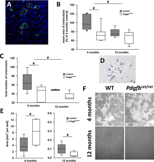
Osteoclast formation is decreased in young Pdgfbret/ret mice. (A) A striatal section of a 1-year-old Pdgfbret/ret mouse stained with DAPI (blue) and anti-CD68 antibody (green). The dotted circle indicates the area of calcification, white arrows point to CD68-positive cells. Note that the CD68 staining is near the calcification sides. (B and C) The capacity of monocytes to develop into osteoclasts in young (4 months) and aged (12 months) Pdgfbret/ret mice (white) and aged-matched wild-type littermates (dark grey), both regarding cell number (B) and cell size (C). (D) The same batch of monocytes plated on a calcified surface and developed into osteoclasts (Osteo Assay Surface Multiple Well Plate from Corning). (E) Representative photographs of the calcified surface (osteomatrix grey) and cleaned spots (white) and (F) the quantification of resorbed calcium deposits. Box plots indicate 25th to 75th percentile, black lines represent median and whisker indicate 5th and 95th percentile. Statistical analysis was performed using one-way ANOVA and post hoc Holm-Sidak test (n = 5–10 mice per group, #P < 0.05).
We next analysed white blood cells from individuals carrying a partial PDGFB deletion [male 41 (middle-aged), female 71 (aged)] (Balck et al., submitted for publication) and age-matched controls [male 46, male 44 (middle-aged), male 63, male 74 (aged)]. Likely because of the small sample number, results lack significance, albeit following the same pattern as that found in mice (Fig. 2A–D). Our analysis of ΔΨm as an indicator of cell death in human monocytes (Supplementary Material) revealed that ΔΨm was significantly decreased in cells from deletion carriers in comparison to control cells, rendering them more susceptible to apoptosis than cells with wild-type PDGFB (two-way ANOVA, post hoc Holm-Sidak test, n = 2–4 per group) (Fig. 2E). In line with this result, we found a significant increase in oxygen consumption after carbonyl cyanide-4-(trifluoromethoxy)phenylhydrazone (FCCP) treatment in the cells carrying a partial PDGFB deletion when compared to controls (Fig. 2F).
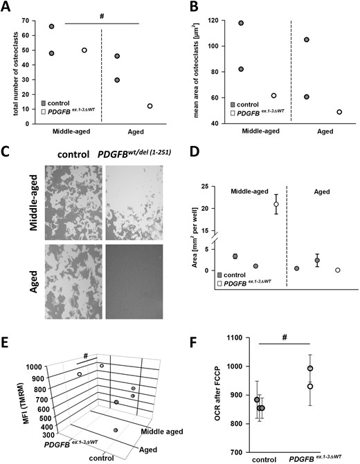
Analysis of monocytes from human PDGFB exons 1–3 deletion carriers. (A and B) The capacity of monocytes to develop into osteoclasts in a 41-year-old PDGFB mutation carrier (white) and age-matched controls (dark grey), (mean: 45, n = 2), and a 71-year-old mutation carrier (n = 1) and controls (mean: 68.5, n = 2), respectively. (C) Representative photographs of the calcified surface and (D) the quantification of resorbed calcium deposits of osteoclasts derived from the same subjects. (E) The mitochondrial transmembrane potential is significantly increased in exon 1–3 deletion carriers (two-way ANOVA, post hoc Holm-Sidak test). (F) The oxygen consumption rate (OCR) of cultured leucocytes is significantly increased in cells derived from mutation carriers compared to age-matched controls. Statistical analysis was performed using two-way (A–E) and one-way (F) ANOVA and post hoc Holm-Sidak test (n = 3 subjects per group, #P < 0.05). MFI = mean fluorescence intensity; TMRM = tetramethylrhodamine methyl ester perchlorate.
Thus, our work indicates that monocytes obtained from a PFBC mouse model are less capable of developing into osteoclasts. Moreover, function of the osteoclasts of aged Pdgfbret/ret mice is diminished in comparison to those generated from monocytes of wild-type littermates. In accordance with these findings, we were able to detect CD68-positive cells surrounding calcium deposits in the brains of Pdgfbret/ret mice. Such cells were not identified in the brains of aged control animals that lack calcium deposits. Therefore, our findings confirm, complement, and expand upon the observations by Zarb et al. (2019). Importantly, monocytes derived from PFBC patients carrying a heterozygous deletion of PDGFB exons 1–3 also exhibited a decreased capacity to mature into functional osteoclasts, although these results lack significance because of small sample size. Interestingly, ΔΨm is significantly hypopolarized in these patient-derived cells with likely reduced PDGF-B levels in comparison to controls. Mitochondrial dysfunction is involved in the induction of apoptosis by releasing apoptogenic factors (Ly et al., 2003; Deuse et al., 2014). In this respect, our findings are in line with previous reports in SMCs showing that treatment with the factor PDGF-B induces ΔΨm hyperpolarization, thereby rendering cultured cells more resistant to apoptosis (Deuse et al., 2014). Although we only show a coincidence of the two phenomena, it is tempting to speculate that ΔΨm hypopolarization and consequent apoptosis at least partially explain the decrease in osteoclast function. Supporting this view, we found that the maximum capacity of oxygen consumption after FCCP treatment is significantly increased in monocytes obtained from PDGFB partial deletion carriers compared to healthy controls. Processes increasing mitochondrial respiratory complexes and the cellular oxygen consumption rate are followed by cellular apoptosis (Wang et al., 2018). Further studies are warranted to unravel the relationship between calcium deposits and clinical manifestation.
Data availability
The data that support the findings of this study are available from the corresponding author, upon reasonable request.
Acknowledgement
Pdgfbret/ret founder pairs were kindly provided by Anika Keller, University of Zürich.
Funding
The study was supported by the Deutsche Forschungsgemeinschaft (FOR2488; to A.W., J.E., and C.K.), the Fritz-Thyssen Foundation (D.A-F.), by intramural funds from the University of Lübeck (C.K., C.S.) and by a career development award from the Hermann and Lilly Schilling Foundation (to C.K.).
Competing interests
The authors report no competing interests.
References
Author notes
Christian Schiemenz and Ana Westenberger contributed equally to this work.


