


Abstract
We present a 15-patient series of awake ‘off-pump’ [without cardiopulmonary bypass (CPB)] coronary artery bypass graft surgery, facilitated by thoracic epidural analgesia (TEA) and femoral nerve block.
Surgery was performed with a conventional median sternotomy. Analgesia was provided with TEA at T1–2 or 2–3 interspace, using bupivacaine 0.5% and sufentanil 1.66 µg ml−1, initially at 20 ml litre−1 until T1–10 dermatomal block was achieved, then maintained at 2–14 ml litre−1 throughout surgery. Femoral nerve block was performed before operation with neuro-stimulation at the saphenous vein harvest site with 10 ml each of bupivacaine 0.25% and lidocaine 2%. Successful awake surgery, avoiding general anaesthesia (GA) with adequate surgical conditions, without CPB was the primary end point.
Fifteen men, mean (sd) age of 63 (9) yr (range 49–81 yr), weight 78 (10) kg, underwent surgery. Three patients (20%) needed conversion to GA: one patient due to insufficient thoracic analgesia, another required initiation of CPB, and the third needed stabilization of the heart for graft suturing due to profound respiratory movements. All three were successfully extubated immediately after surgery. Awake surgery was successful and uneventful in 80% of cases.
Combined TEA and femoral block is a novel anaesthetic technique, and is feasible, for cardiac surgery. However, certain technical limitations need to be overcome to evaluate the full potential of ‘awake’ cardiac surgery.
General anaesthesia (GA) remains the preferred anaesthetic technique for coronary artery bypass grafting (CABG). Procedures to reduce anaesthetic or surgical mortality and morbidity during open heart surgery have continually evolved and new strategies in cardiac anaesthesia now enable immediate extubation (ultra-fast-track anaesthesia). The perioperative use of high thoracic epidural anaesthesia (TEA) as an adjunct to GA has been shown to be beneficial in patients with coronary artery disease.1 Superior pain control with TEA2,3 improves pulmonary function and reduces postoperative pulmonary dysfunction,2,4–7 allowing earlier extubation with less respiratory complications. Further advantages of TEA include T1–T5 sympathicolysis with subsequent improvement of coronary and internal thoracic arterial perfusion,8 decreased heart rate,9 and potential reduction of arrhythmias. Intraoperative haemodynamic stability may be improved through stress response modulation and reduced risk of myocardial ischaemia.2,9–12 Benefits of TEA can persist into the postoperative period, improving recovery, and early mobilization. The off-pump coronary artery grafting (OPCAB) technique allows reduced heparin doses, possibly also reducing perioperative epidural haematoma risk.
Awake cardiac surgery was first described in 2000 by Karagoz and colleagues,13 in which CABG was performed in awake patients without GA using only TEA. Various operative techniques for ‘awake’ cardiac surgery have been described.1,14–24 The early cases were mostly associated with minimally invasive surgical techniques,13,25 or with a limited number of grafts. Some authors reported the use of local anaesthetic infiltration18 or a lumbar subarachnoid block26 to provide sufficient analgesia for saphenous vein harvesting, as TEA clearly cannot. Formally ‘awake’ CABG techniques were only considered applicable to a very limited number of patients, and inappropriate for patients with multiple co-morbidities, requiring multiple grafting for complete revascularization. We follow our recent publication20 of the first experience of combining TEA and femoral nerve block for two cases of awake cardiac surgery; with the first case series of 15 awake OPCAB using this anaesthetic technique.
Methods
After approval by the Institutional Review Board, and with informed written consent, 15 male adult patients with symptomatic coronary artery disease were prospectively enrolled in our study and underwent OPCAB without GA. Patients were considered eligible if they had no contraindications to TEA, and if coronary artery anatomy was favourable for OPCAB, thus were at a reduced risk of conversion to cardiopulmonary bypass (CPB). Exclusion criteria were severely depressed left ventricular ejection function (LVEF<30%), congestive heart failure, preoperative haemodynamic instability or intra-aortic counter pulsation, emergency surgery, acute myocardial infarction (MI), associated valvular disease, moderate to severe pulmonary hypertension (systolic >50 mm Hg), previous cardiac surgery, or patient refusal. The presence of obesity, diabetes, advanced age, chronic renal failure, or any other co-morbidity did not affect the patient selection except for significant impaired lung function (forced expiratory volume in 1 s <50%).17 All routine cardiac medications were continued on the morning of surgery. The heparin infusion was stopped at least 4 h before surgery and clopidogrel more than 7 days before surgery.
The contraindications for epidural catheterization were patient refusal, pre-existing coagulopathy, prothrombin time >40 s, a low platelet count (<100 000), the use of anti-platelet drugs within 5 days before surgery (excluding aspirin), sepsis, or the presence of a skin infection at the site of the catheter.
All catheters were inserted immediately before surgery in the post-anaesthesia care unit (PACU). A peripheral venous line was placed and the patients received light sedation (midazolam 1–2 mg). A TEA was placed at T1–2 or T2–3 interspace, by the midline approach in the sitting position and the catheter secured. After an initial 20 ml bolus, more than 1 h, of bupivacaine 0.5% and sufentanil 1.66 µg ml−1, a T1–10 dermatomal block was achieved. Thereafter, the epidural was continued at 2–14 ml h−1 (after the initial 12 patients, no sufentanil was added for maintenance). Immediately after surgery, bupivacaine 0.06% and fentanyl 3 µg ml−1 would be infused at 4–14 ml h−1 until the second postoperative day.
A single shot femoral block was performed, taking care to elicit a contraction of the internal portion of the quadriceps with a neuro-stimulation threshold of 0.2 mA. A single injection of lidocaine 2%, 10 ml and bupivacaine 0.25%, 10 ml was given. A femoral arterial catheter was placed on the opposite side under local anaesthesia and a central venous catheter by right subclavian approach.
After confirmation of the TEA (T1–10 dermatomes) and femoral block, the patient was transferred to the operating room (OR). Routine standard monitoring included a five-channel ECG with continuous ST-segment analysis (leads II and V5), direct arterial and central venous pressure, pulse oximetry, urinary catheter including temperature monitoring, and end-tidal carbon dioxide via a face mask. The patients were draped in a manner to give unrestricted airway access in the case of an urgent tracheal intubation. Bilateral chest tubes were inserted in the fifth intercostal spaces before sternotomy using 24F drains. Complete midline sternotomy was then performed in a standard fashion, during the inspiratory phase of respiration. Then, the pericardium was opened to inspect the heart and coronary target vessels. All grafts to be used were harvested simultaneously by two surgeons: saphenous vein grafting of the left thigh and left or bilateral internal mammary arteries. At any time during the procedure, inadvertent pleural opening was immediately repaired to avoid a pneumothorax and subsequent pulmonary dysfunction. Anticoagulation was performed at least 2 h after the epidural puncture, using non-fractionated heparin 150 IU kg−1 (activated coagulation time >300 s) appropriate for OPCAB.
To stabilize the heart for OPCAB, the Cor-Vasc System (CoroNéo, Montréal, Canada) was used; in addition, sponges were placed under the left side of the heart to expose the anterior and lateral wall. Occasionally, left epicardial sutures were used to lift the heart and get better access to the lateral wall. Target vessels were temporarily occluded with atraumatic vessel loops for proximal and distal flow occlusion. During CABG, hypotension was defined as a systolic arterial pressure below 70 mm Hg and was treated using manual titration of phenylephrine via microdrip from a 40 µg ml−1 solution—as it is standard for OPCAB in our institution. Depending on the surgeon's choice or the target vessels, revascularization began with the internal mammary artery, usually on the left anterior descending (LAD) artery. For each distal anastomosis, an end-to-side technique was used with a running 7-0 polypropylene suture. After completion of the anastomosis, when mammary grafts were used, the diastolic flow was qualitatively assessed by vascular Doppler. Flow in the venous grafts was assessed using the Cliniflow II electromagnetic blood flow meter (Carolina Medial Electronic Inc., NC, USA). After assessing good graft perfusion, intra-pericardial and mediastinal drainage tubes were inserted and heparin reversed with protamine. Temporary pacing wires were inserted in the right atrial appendage and ventricle, complete haemostasis achieved, then the sternum and skin closed in the standard fashion.
After surgery, all patients were monitored in the PACU for at least 4 h, until the following criteria were met, when they were transferred directly to the cardiac ward: patient alert, awake, haemodynamically stable with no active bleeding, not requiring inotropes (systolic arterial pressure >90 mm Hg), self-ventilating on maximum 50% oxygen (Pao2>10.5 kPa, Paco2<6 kPa) and cooperative with pain scores <4/10. Preoperative, intraoperative, and postoperative data collection were done by the perioperative cardiac anaesthesia research group (PeriCARG). All data are presented as mean (sd) (range).
Results
Fifteen men with symptomatic coronary artery disease and preserved LVEF underwent OPCAB. Patient characteristics are presented in Table 1. Mean age was 63 (9) (49–81) yr, weight 78 (10) kg, and five were obese (BMI>28). Preoperative New York Heart Association functional class (NYHA) was all above 3 (mean 3.3), LVEF was 61% (12%) (30–81%). Four patients were actively smoking and seven quit for at least 3 months before surgery, four patients were diabetic, two had chronic renal failure, eight patients had treated high arterial pressure, and 11 had dyslipidaemia. Midline sternotomy and harvesting of internal mammary arteries were successful in all patients, without respiratory compromise, as all breaches in the pleura were successfully repaired, and no visible pneumothoraces were encountered. Mean operative time was 125 (32) min, number of bypasses was 2.5 (2–4) grafts, and ischaemic time during distal anastomosis was 18.8 (6.5) min (Table 2). Three out of 15 patients needed phenylephrine to maintain systolic arterial pressure above 70 mm Hg throughout CABG with no signs of ischaemia on ECG tracing. There was good haemodynamic stability throughout surgery (Fig. 1). On the basis of the preoperative coronary angiogram, complete revascularization was achieved in all patients.
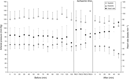
Mean arterial pressure and heart rate throughout surgery, values displayed every 10 min and the lowest of each variable during each ischaemic time during OPCAB.
Patient characteristics. Data are mean (sd) (range)
| Patients | n=15 |
|---|---|
| Age (yr) | 63.4 (8.9) (49–81) |
| Weight (kg) | 78.1 (10.3) (62.4–90.0) |
| BMI | 26.6 (2.2) (22.7–30.7) |
| Preoperative NYHA class | 3.3 (0.5) (3–4) |
| LVEF (%) | 61.1 (11.8) (30–81) |
| Tobacco use: active/quit >3 months (n) | 4/7 |
| High arterial pressure (n) | 8 |
| Dyslipidaemia (n) | 11 |
| Diabetes (n) | 4 |
| Chronic renal failure (n) | 2 |
| COPD (n) | 0 |
Surgical operation and conditions. LIMA, left internal mammary artery; RITA, right IMA; SVG, saphenous vein graft; CPB, cardiopulmonary bypass; AF, atrial fibrillation; BP, blood pressure; ICU, return to intensive care unit. *Patient 3: positioning of the heart for OPCAB led to sudden drop of BP necessitating conversion to on-pump CABG. †Patient 8: TEA provided incomplete thoracic analgesia, elective conversion to GA was performed before sternotomy. Postoperative pain control using TEA was excellent. ‡Patient 12: exaggerated respiratory exertion, stabilization of the heart for OPCAB was impossible, conversion to GA was undertaken
| Patient | Age (yr) | LVEF (%) | Grafts (number) | Ischaemic time (min) | Postoperative complication |
|---|---|---|---|---|---|
| 1 | 65.5 | 60 | LIMA-SVG1 (2) | 12 | |
| 2 | 64.4 | 55 | LIMA-SVG1 (2) | 9 | |
| 3* | 62.8 | 30 | LIMA-SVG1 (2) | CPB 34, aortic cross clamp time 13 | AF |
| 4 | 60.8 | 81 | SVG2 (2) | 14 | AF, 1 pack red blood cells |
| 5 | 81.3 | 60 | LIMA-SVG1 (2) | 10 | ↓BP day1, ICU |
| 6 | 78.7 | 60 | LIMA-SVG2 (3) | 17 | |
| 7 | 72.5 | 70 | LIMA-SVG1 (2) | 14 | |
| 8† | 51.5 | 60 | SVG2 (2) | 26 | |
| 9 | 60.2 | 70 | LIMA-SVG2 (3) | 18 | |
| 10 | 49.9 | 72 | LIMA-RITA-SVG1 (3) | 24 | |
| 11 | 52.6 | 48 | LIMA-SVG1 (2) | 20 | |
| 12‡ | 59.9 | 70 | LIMA-SVG2 (3) | 23 | AF |
| 13 | 61.0 | 60 | LIMA-SVG3 (4) | 30 | |
| 14 | 63.9 | 60 | LIMA-SVG2 (3) | 26 | AF |
| 15 | 65.5 | 60 | LIMA-SVG2 (3) | 20 | AF |
Three patients required GA. One TEA failed to produce sufficient somatosensory block; the patient was electively intubated before sternotomy and extubated immediately after surgery, although TEA still achieved excellent pain control. In the second patient, with an LVEF of only 30%, sternotomy and internal mammary artery harvesting were uneventful, but severe haemodynamic instability during the first distal anastomosis of the LAD required conversion to CPB. Endotracheal intubation was performed, the patient underwent coronary bypass grafting uneventfully and was extubated immediately after surgery. In the third patient with TEA using bupivacaine 0.5% and sufentanil 2 µg kg−1, a ventilatory frequency of 12–14 min−1 during surgery made it impossible to create the necessary stabilization required for OPCAB. GA was achieved; immediate extubation after surgery was successful and the further postoperative course uneventful.
Postoperative analgesia was achieved using TEA with bupivacaine 0.06% and fentanyl 3 µg ml−1 at 4–12 ml h−1. All patients were transferred to the PACU and then to the cardiac ward. The TEA was removed on postoperative day 3. Atrial fibrillation was observed after operation in five patients; only one patient received 1 unit of red blood cells. In one patient, low arterial pressure on postoperative day 1 required the transfer to the ICU for monitoring and treatment. This patient was hypovolaemic and normal arterial pressure returned with crystalloid volume repletion. The mean hospital length of stay was 5.2 (1.2) (4–8) days. There was no mortality or major morbidity, and no complication related to TEA.
All patients were asked whether they were satisfied with having stayed awake during surgery and whether they would chose this technique again. All patients were highly satisfied and would choose this technique again. Interestingly, the patients who were converted to GA said they would try ‘awake’ cardiac surgery again as a first approach.
Discussion
We present the first case series of ‘awake’ cardiac surgery using a median sternotomy approach with a combined TEA and femoral block. This series demonstrates the feasibility of the technique and certain limitations which might be addressed in order to offer this choice of anaesthesia to more patients.
It was surprising that after recruiting the first few patients, they talked about their ‘positive’ experience with this new technique to other patients. It appeared that patients who have already undergone surgery under regional anaesthetic techniques, such as endoscopic transurethral prostrate surgery, were quite willing to undergo cardiac surgery under TEA. In addition, as known to many anaesthesiologists, the fear of ‘not waking up’ after surgery is a main concern and the possibility of staying awake during surgery seemed very attractive. Careful psychological assessment of the patient's suitability for ‘awake’ cardiac surgery is important as it is for other types of surgeries and not everyone can be suited for this technique. There were no definite criteria other than the pre-requisites outlined previously. However, these pre-requisites are similar to other types of surgery where the anaesthesiologist can make a conscious decision to approach or not a certain patient for a regional technique. Unusually nervous patients and patients ‘who absolutely want to sleep’ are never good candidates for regional techniques. It was decided early on that the insertion of all catheters and the testing of TEA and regional block efficiency were to be made before the patients entered the OR; this was performed in the postoperative care unit since there are no induction room facilities in our hospital setting. This had the dual advantage of allowing early familiarization with the PACU nursing team and reassurance that effective TEA would be achieved before transfer to the OR. Nursing staff in the OR were essential in providing a reassuring atmosphere throughout surgery.
The initial choice of TEA mixture of LA and opioid was according to the description in the literature. After 12 patients, however, it was noted that the continuous use of sufentanil in the epidural mixture resulted in sedation and a typical ‘opioid’ respiration pattern (large tidal volume and low frequency); this compromised the stabilization of the heart in one patient who required GA. Therefore in the rest of the patients, after first bolus of mixture of LA and sufentanil, bupivacaine 0.5% alone was used for subsequent analgesia. Preoperative TEA testing with ice or pin-prick can be misleading; a ‘patchy’ block remaining undiscovered, as in patient number 8. However, careful reassessment by the surgeons for early recognition of patchy block was carried out before skin incision, sternotomy, and after each step of surgery; despite this, a single episode of insufficient anaesthesia occurred which required GA.
In the ‘limited’ awake cardiac surgery literature, several approaches to deal with pneumothorax are described. One is to leave the pleural space widely open whenever a hole in its integrity is detected;13 another technique implies closure of the pleural space with or without insertion of thoracic drains whenever this occurs.19,27 Preparation of the left internal mammary artery is considered more demanding without opening the pleural space. However, our cardiac surgeons noticed that the lung is more retracted into the pleural space during spontaneous respiration, than during intermittent positive pressure ventilation (IPPV). This actually made mammary artery preparation easier, which has been noted previously.19 In ‘Conventional’ cardiac surgery and in more than 20% of awake OPCAB cases presented so far,19 the pleural space may be opened at any stage of surgery, making it necessary to insert thoracic drains at the end of surgery.
We offered the patients the choice between supplemental sedation or music via headphones—as we would in other types of surgery performed under regional anaesthesia. Sedation already accompanied TEA using bupivacaine and sufentanil, but if required, very low propofol infusion rates easily sedated patients.
OPCAB is prone to cause significant arterial pressure drops during the positioning of the heart. In conventional surgery, the anaesthesiologist can treat these changes by careful titration of phenylephrine. Our extensive experience with more than 2000 OPCAB cases and manual titration of phenylephrine via microdrip from a 40 µg ml−1 solution achieves a better fine-tuning of the arterial pressure than with conventional syringe drivers. We used the same strategy for awake OPCAB. Whereas, arterial pressure fluctuations of 40–50 mm Hg in conventional OPCAB are tolerated, this causes severe problems in spontaneously breathing patients, resulting in restlessness18,19,28 later progressing to respiratory distress.
We made sure that the manoeuvring of the heart and evaluation of the distal grafting sites were performed very carefully in order to avoid arterial pressure or heart rate drops, necessitating close cooperation between anaesthesiologists and cardiac during awake cardiac surgery. It is therefore imperative that the anaesthetic-cardiac surgery team have extensive combined experience and understanding with this technique. Awake cardiac surgery might be less suitable for those patients with a reduced ejection fraction, as the lower ejection fraction increases the risk of severe haemodynamic changes during OPCAG heart positioning and grafting.
It is imperative that all drugs and devices necessary for rapid intubation are readily available during awake cardiac surgery. All cases were performed with the same anaesthesiologist (T.H.) and two anaesthetic technicians with whom all aspects and particularities of awake OPCAB were discussed before the study was undertaken. Conversion of awake into conventional OPCAB does not preclude immediate extubation after surgery.
Maintenance of body temperature is a key aspect to facilitate immediate extubation. This was performed in the routine way: elevated OR temperature and heating blankets for lower body. However, it was noted that most of our awake patients maintained body temperature easily and some preferred to have the lower heating blankets turned off during surgery. Body temperature was maintained above 36° during surgery in all patients.
It is interesting to note that all patients were highly satisfied and would want to undergo the same surgery again if necessary. In general, patients who had undergone previous procedures under local or regional anaesthesia were most likely to ask for a similar approach in cardiac surgery.
Awake cardiac surgery is clearly in its infancy. To determine the safety of this technique in comparison with OPCAB under general anaesthesia, many more patients need to be studied. In addition, future studies should focus on the impact of awake cardiac surgery on cognitive assessment and recovery, its relation to neuro-hormonal responses during surgery. Owing to unresolved problems with on-pump awake cardiac surgery, especially temperature management and apnoea at the beginning of extracorporeal circulation,29,30 OPCAB seems to be the main domain for this technique.
Acknowledgement
The authors are thankful for the enormous support of all the nursing staff of Hôtel-Dieu, especially the PACU nurses and the operating room staff, during the course of this proect.


