


We are inspired by the article entitled “The Anatomy and Clinical Application of Preorbital Septum Fiber,” by Zhao et al.1 This article is the first to describe the preorbital septum fiber as a distinct layer. We have occasionally found this preorbital septum fiber in our practice as well. Surgeons debate about whether to excise this fiber during blepharoplasty or not. However, some facts about the preorbital septum fiber should be fully understood before making the final judgment on what to do with it. Based on the preorbital septum fiber’s description by Zhao et al,1 we went to the cadaveric laboratory to study the fiber further. This study was conducted in accordance with the Declaration of Helsinki. We would like to share what we found. A summary of further findings of preorbital septum fiber is demonstrated in the accompanying Video (available online at www.aestheticsurgeryjournal.com).
First, the preorbital septum fiber is not found in every cadaver, and it can be found in only one eye or in both eyes. Figure 1 illustrates a 60-year-old female fresh cadaver with preorbital septum fiber on her left upper eyelid. There is no preorbital septum fiber on her right upper eyelid.
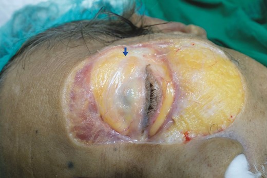
An illustration of a 60-year-old female fresh cadaver with preorbital septum fiber on her left upper eyelid. The preorbital septum fiber is marked with a blue arrow.
Second, the boundary of the preorbital septum fiber is ambiguous, lacking significant origin and insertion. The preorbital septum fiber is prominent laterally and fades away medially. The preorbital septum fiber is adhered to the orbital septum separating from the orbital fat.
Third, the preorbital septum fiber was sent for histologic study which found that the preorbital septum fiber was located anteriorly to the orbital septum and was a different layer to the orbital septum. This finding is supported by previous studies2,3 describing the existence of a fibroadipose layer located anteriorly to the orbital septum and posteriorly to the orbicularis muscle.
Fourth, the preorbital septum fiber was found to be present not only on the upper lid but also on the lower eyelid, as shown in the Video. The preorbital septum fiber at the right lower eyelid was demonstrated in a 65-year-old male embalmed cadaver. The preorbital septum fiber was not found to be present on the right upper lid or on the left side.
Regarding the further study of preorbital septum fiber on cadavers, it seems that this fiber plays no significant role in eyelid function. The dissection of this fiber during blepharoplasty would not result in substantial adverse outcomes and it can be excised. We hope that this further study on cadavers can provide additional essential information on the preorbital septum fiber and that these findings will augment the innovative work of Zhao et al.1
Disclosures
The authors declared no potential conflicts of interest with respect to the research, authorship, and publication of this article.
Funding
The authors received no financial support for the research, authorship, and publication of this article.
References
Author notes
Dr N. Wongkietkachorn is a Lecturer of Plastic Surgery in private practice in Bangkok, Thailand

