


 https://orcid.org/0000-0003-1386-6180

Abstract
Background
Ventilator-associated pneumonia (VAP) is a frequent complication of invasive mechanical ventilation (IMV), yet its determinants in patients ventilated for non-pulmonary indications remain poorly characterized.
Methods
Data were derived from the PULMIVAP multicenter prospective cohort (ClinicalTrial: NCT04849039), conducted in 8 Northern Italian intensive care units (January 2021–April 2024). All adults receiving IMV for non-pulmonary indications were enrolled if expected to require IMV >48 hours and antibiotic-free for ≥3 days preceding intubation. Clinical and therapeutic data were collected for up to 15 days of IMV; microbiological sampling was performed according to clinical indication. The primary outcome was time to first VAP within 15 days of IMV (VAP15). Associations between baseline characteristics, time-varying exposures, and VAP15 were evaluated. Thirty-day mortality from IMV initiation was a secondary outcome.
Results
Among 406 patients, 148 (36%) developed VAP15, at a median of 6 days (IQR 4.0–7.5) with an incidence rate of 43.2 per 1000 ventilator-days at risk (95% confidence intervals [95% CI], 36.4–49.9). Non-neurologic diagnosis (adjusted hazard ratio [aHR] 0.51; 95% CI, 0.31–0.86), female sex (aHR, 0.52; 95% CI, 0.36–0.75), and early antibiotic initiation (days 1–2 of IMV, aHR, 0.65; 95% CI, 0.45–0.93) were independently associated with lower risk of VAP15. Predominant pathogens were Staphylococcus aureus, Klebsiella spp., and Haemophilus influenzae; multidrug-resistant organisms were uncommon. VAP15 showed no competing relationship or significant association with mortality (overall, 20.4%).
Conclusions
In patients receiving IMV for non-pulmonary indications, non-neurologic diagnosis, female sex, and early antibiotic exposure were associated with lower observed VAP incidence, though causality cannot be inferred. Risk-stratified prevention strategies warrant further investigation into this population.
BACKGROUND
Ventilator-associated pneumonia (VAP) is a frequent and serious healthcare-associated infection in intensive care units (ICUs), often leading to prolonged invasive mechanical ventilation (IMV) and extended hospital stay [1, 2]. Although its independent contribution to mortality remains debated, VAP imposes a substantial burden through increased morbidity and healthcare costs [3].
A better understanding of how VAP affects specific patient populations is essential for improving prevention, management strategies, and outcomes. Most available evidence derives from studies conducted in general ICU populations, where patients ventilated for non-pulmonary conditions, particularly those with neurological injuries, are underrepresented [4, 5]. However, VAP incidence appears particularly high in this subgroup, reaching up to 71% among patients with severe traumatic brain injury and 76% in those with stroke [6, 7]. Several factors may contribute to this increased susceptibility, including impaired airway protection, aspiration risk prior to intubation, and brain injury-associated immune dysfunction [8].
Despite the high burden of VAP in this population, clinical risk factors, microbiological characteristics, and potential preventive strategies remain incompletely characterized. Addressing these gaps may help refine risk stratification and inform targeted prevention strategies.
Using data from the PULMIVAP multicenter prospective cohort, which enrolled patients receiving IMV for non-pulmonary indications with systematic daily collection of clinical and therapeutic data, we aimed to (1) describe the incidence of first VAP occurred within 15 days of IMV, (2) identify clinical and pharmacological factors associated with its occurrence over time, (3) characterize the microbiological patterns identified during the first 15 days of IMV, and (4) analyze the 30-day mortality from IMV initiation.
METHODS
Study Design and Setting
Data were derived from a multicenter, prospective, observational cohort study (PULMIVAP; ClinicalTrials.gov identifier NCT04849039) approved by the Italian Milan Area 2 Ethics Committee (#533_2019, approval date: 5 June 2019).
PULMIVAP was designed primarily to characterize pulmonary microbiota and its longitudinal dynamics in patients intubated for non-pulmonary conditions; for that aim, patients who developed a first VAP within 15 days of IMV (VAP15) were compared with those who did not, matched 1:1 within centers by sex, age (±10 years), and reason for intubation. The microbiota analyses are reported separately [9].
A prespecified secondary objective of PULMIVAP, and the focus of the present analysis, was the identification of clinical and pharmacological factors associated with VAP15. For this analysis the matched design was not retained: the full enrolled cohort of patients intubated for non-pulmonary conditions was used. A study-specific operational definition of VAP, agreed by all participating centers before enrollment, was applied; the development and comparison of candidate definitions has been described previously [10].
Because enrollment was governed by the microbiota objective, which required continuation until 73 matched VAP15/no-VAP15 pairs were accrued, no a priori sample size or power calculation was performed for the risk-factor objective reported here.
PULMIVAP Cohort
All adult patients consecutively admitted to 8 ICUs (all but one were general ICUs) in Northern Italy between January 2021 and April 2024 were eligible if they were expected to require IMV for >48 hours and had not received antibiotics for at least 3 days preceding intubation. This last criterion was adopted because the primary aim of the parent study was microbiota analysis.
Patients with respiratory distress or pneumonia at ICU admission were excluded. Notably, the study period overlapped with the COVID-19 pandemic, which affected the enrollment of patients intubated for non-pulmonary conditions.
ICU admission diagnosis was recorded and classified as neurologic (including structural and hypoxic brain injury) or non-neurologic, based on the initial clinical assessment (Supplementary Table 1).
Finally, for the present analysis, patients receiving <3 days of IMV were excluded, as VAP cannot be diagnosed before day 3 of ventilation.
VAP prevention measures followed local standard-of-care protocols across participating centers.
Clinical Variables Data Collection
Baseline data were collected on the day of IMV initiation (enrollment) and included demographic characteristics, IMV initiation setting, ICU admission diagnosis, clinical parameters (septic shock diagnosis, Glasgow Coma Scale score, and APACHE II score), and medical history focusing on major comorbidities (diabetes mellitus, liver failure, chronic inflammatory or autoimmune diseases, respiratory diseases, cardiovascular diseases, chronic renal failure [including dialysis], and immunocompromised status).
Selected clinical data were recorded, in accordance with routine clinical practice, for up to the first 15 days of IMV, which were defined a priori as the study follow-up window; therefore, only variables routinely assessed and documented as part of standard care were recorded. Missing daily values indicate that the corresponding measurement was not performed on that day and that clinicians relied on the most recent available measurement.
Recorded variables included those used for daily VAP assessment: the worst body temperature, white blood cell count, ventilatory parameters (positive end-expiratory pressure, fraction of inspired oxygen [FiO2], and PaO2/FiO2 ratio), presence of new or worsening respiratory secretions, and radiological findings suggestive of pulmonary deterioration. Tracheostomy, main surgical procedures, and selected pharmacologic therapies (antibiotics, gastroprotective agents, corticosteroids, antiepileptics, antihypertensives, anti-inflammatory drugs, and sedatives) were also recorded using the same approach. Respiratory and blood cultures were obtained at the discretion of the treating team according to clinical indication; no protocol-mandated surveillance sampling was performed.
Antibiotic use was recorded daily throughout follow-up; however, the indication for administration was not documented.
Outcomes
For the present analysis, the outcome of interest was time to first VAP15. Daily data were collected during the first 15 days of IMV, and time to first VAP was ascertained over the risk period from day 3, the earliest day on which VAP can be diagnosed, to day 15 of IMV, censoring at extubation, ICU discharge, or death, whichever occurred first.
The 15-day window of the parent microbiota study was chosen because first VAP episodes occur predominantly within the first 2 weeks of IMV [11] and because patients with neurologic conditions typically require a mean IMV duration of approximately 14 days [12, 13].
The exposures of interest comprised baseline characteristics and time-varying clinical and pharmacological factors; the microbiological characteristics of VAP15 episodes were also described.
Thirty-day mortality from IMV initiation was assessed as a secondary outcome. Relevant outcome data were extracted from the end-of-study form, which captured events occurring after the 15-day follow-up window, from day 16 of IMV until ICU discharge. The recorded variables included the occurrence of at least one VAP episode during this period, date of IMV discontinuation, date of ICU discharge, and date of death. These variables are summarized in Supplementary Results.
VAP and Multidrug Resistance Definitions
Attending physician's clinical diagnosis of VAP was recorded daily by the local investigator; given the observational nature of the study, this assessment governed clinical management, and it was the primary outcome for all analyses reported. Notably, before study initiation, investigators from all participating centers agreed on a study operational definition derived from international recommendations; the development and comparison of candidate definitions has been reported previously [10] and the criterion is reproduced in the Supplementary Methods. A single quantitative threshold for microbiological confirmation, bacterial growth ≥104 CFU/mL in endotracheal aspirate, was agreed by all participating centers and applied across sites. This threshold lies at the more sensitive end of the recommended range and was chosen to maximize detection of early episodes in a population sampled under variable antibiotic exposure.
A microorganism was considered multidrug-resistant (MDR) if non-susceptible to at least one agent in 3 or more antimicrobial categories for Enterobacterales or if methicillin resistance was present for Staphylococcus aureus [14].
Statistical Analysis
Demographic and clinical characteristics assessed at intubation or within the first 2 days of IMV, a period during which VAP cannot be diagnosed by definition and hereafter referred to as baseline covariates, were summarized as counts and percentages for categorical variables and as medians with first and third quartiles (Q1–Q3) for continuous variables.
Continuous variables were modeled as continuous covariates in regression analyses; for descriptive purposes, their distribution according to selected cutoffs was also reported and hazard ratios were estimated.
For baseline variables, the proportion of missing values for each variable is reported in Tables. As missing data were infrequent, no specific statistical method was applied to handle them; instead, missing values were retained as a separate dummy category to avoid excluding patients from the multivariable analyses. This category was not assigned any clinical interpretation. For time-varying variables, the most recently available value was carried forward until a new measurement became available, reflecting the information available to the treating clinician at each time point. Drug exposure, tracheostomy, and surgical procedures were recorded as discrete events and were considered complete by design; absence of a recorded event was interpreted as absence of exposure. For the time-to-VAP15 analysis, risk time accrued from day 3 of IMV (delayed entry), consistent with the definition that VAP cannot be diagnosed before day 3.
Follow-up ended at the earliest of VAP diagnosis, extubation, death, or day 15 of IMV.
Daily clinical variables (tracheostomy, surgical procedures, and selected treatments) were modeled using 2 exposure variables: an early exposure indicator (presence on days 1 and/or 2 of IMV) and a time-varying covariate from day 3 onward capturing exposure during follow-up. From day 3 to day 15 of IMV, exposure status was updated daily according to each patient's exposure history up to that time. For these time-varying exposures, patients were considered exposed (covariate = 1) from exposure initiation until interruption or discontinuation. Exposure status immediately reverted to unexposed (covariate = 0) following interruption and returned to exposed upon re-initiation. This process was repeated for any subsequent interruptions and re-initiations, allowing the same patient to contribute person-time to both the exposed and unexposed categories at different stages of follow-up. Thus, the time-varying covariate reflected current exposure status throughout the risk period. Working examples of time-varying covariate coding for drugs are shown in Supplementary Figure 1 (Supplementary Methods).
To preserve causal inference, tracheostomy, antibiotic, and nonsteroidal anti-inflammatory drug use on the day of VAP diagnosis were handled to minimize reverse causation (see Supplementary Methods, Management of antibiotic and NSAID for details).
The cumulative incidence of VAP15 was calculated for the overall cohort and by center. Incidence rates were also estimated as the number of VAP15 events per 1000 ventilator-days at risk; both measures are presented with 95% confidence intervals (CIs).
Regarding time to VAP15 analysis, the proportional hazards assumption was assessed for each baseline covariate (see Supplementary Methods for details). Given the heterogeneity in VAP15 incidence across centers, reflecting differences in patient case mix, management strategies, and the impact of the COVID-19 pandemic, Cox proportional hazards models including a center-level frailty term (random intercept) were used.
Baseline characteristics were first evaluated in univariable models, and multivariable models were subsequently constructed to identify factors independently associated with VAP15. Variables were initially screened based on clinical relevance and prior knowledge; among these, variables showing a univariable association with the outcome (HR, P < .20) were considered. Final model selection was guided by the corrected Akaike information criterion, favoring clinically plausible and parsimonious models while maintaining an adequate events-per-variable ratio. The only exceptions were age and APACHE II score (as an indicator of disease severity), which were included in the final model based on their clinical relevance, irrespective of statistical significance. See Supplementary Methods for details.
For drug exposures, early and current variables were simultaneously included in frailty Cox models when estimated (≥1 VAP15 event occurred in the exposure category); each drug was analyzed separately. For drugs significantly associated with VAP15, stratified analyses assessed whether early initiation modified the effect of subsequent exposure (see Supplementary Methods for details). Confounders included in the multivariable models are reported in the table footnotes.
For the secondary outcome, 30-IMV-day mortality, a 3-state illness-death (multistate) model was applied to estimate transitions from ICU to VAP, ICU to death, and VAP to death, allowing estimation of transition-specific hazards and assessing the relationship between VAP15 and 30-IMV-day mortality (see Supplementary Methods for details).
A multivariable frailty Cox model including VAP15 as a time-varying covariate was also fitted to account for potential confounding. The covariates selection process followed the same strategy used for the VAP15 analysis.
Follow-up was defined from initiation of IMV until death, ICU discharge, or 30 days after IMV initiation, whichever occurred first.
Analyses were performed with SAS version 9.4. A 2-sided P value < .05 was considered significant.
The study is reported in accordance with the Strengthening the Reporting of Observational Studies in Epidemiology (STROBE) statement (Supplementary material).
Patient Consent Statement
The study was conducted in accordance with the Declaration of Helsinki and was approved by the Milan Area 2 Ethics Committee (533_2019, 5 June 2019). Because the study population consisted of patients undergoing IMV who were by definition unable to provide informed consent at the time of enrollment, and because the study was observational, involved no intervention, and no additional risk, the Ethics Committee approved a deferred-consent procedure in accordance with Italian legislation on non-interventional research in emergency and critical care settings. Written informed consent to the continued use of data and samples was sought from each patient upon recovery of decision-making capacity. Data from patients who died or did not regain capacity before ICU discharge were retained under this approved procedure. One patient withdrew consent following recovery of decision-making capacity and was excluded from the analysis; no patient was excluded on grounds of inability to consent.
RESULTS
Study Population
A total of 472 patients were enrolled across 8 ICUs, of whom 406 were included in the final analysis (Figure 1). Enrollment over the 40-month study period is detailed in Supplementary Figure 2.
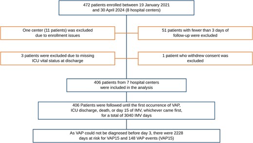
Study flowchart.
Baseline characteristics of the 406 patients included in the analysis are summarized in Table 1. In approximately 72% of cases IMV was initiated in-hospital (emergency department or ICU).
Baseline Characteristics of the Study Cohort
| All Patients (n = 406) | |
|---|---|
| Center, n (%) | |
| A | 87 (21%) |
| B | 75 (18%) |
| C | 72 (18%) |
| D | 57 (14%) |
| E | 55 (14%) |
| F | 36 (9%) |
| G | 24 (6%) |
| Year of enrollment, n (%) | |
| 2021–2022 | 270 (67%) |
| 2023–2024 (Jan–Apr) | 136 (33%) |
| IMV start location, n (%) | |
| Community | 113 (28%) |
| Emergency department | 161 (40%) |
| ICU | 132 (32%) |
| ICU admission diagnosis, n (%) | |
| Non-neurologic | 100 (25%) |
| Neurologic | 306 (75%) |
| Structural brain injury | 268 (66%) |
| Hypoxic brain injury | 38 (9%) |
| Sex at birth, n (%) | |
| Male | 240 (59%) |
| Female | 166 (41%) |
| Agea | |
| Median (Q1–Q3), y | 62.5 (51.0–72.0) |
| 18–55 y, n (%) | 132 (33%) |
| 56–68 y, n (%) | 135 (33%) |
| 69–87 y, n (%) | 139 (34%) |
| BMIa | |
| Median (Q1–Q3) | 25.0 (23.0–28.0) |
| 18–24.9, n (%) | 181 (45%) |
| 25–29.9, n (%) | 171 (42%) |
| 30–50, n (%) | 54 (13%) |
| Cigarette smoking, n (%) | |
| No | 300 (74%) |
| Yes | 106 (26%) |
| At least one comorbidity, n (%) | |
| No | 177 (44%) |
| Yes | 229 (56%) |
| Diabetes mellitus, n (%) | |
| No | 348 (86%) |
| Yes | 58 (14%) |
| Liver failure, n (%) | |
| No | 391 (96%) |
| Yes | 15 (4%) |
| Chronic inflammatory/autoimmune diseases, n (%) | |
| No | 368 (91%) |
| Yes | 38 (9%) |
| Respiratory diseases, n (%) | |
| No | 372 (92%) |
| Yes | 34 (8%) |
| Cardiovascular diseases, n (%) | |
| No | 222 (55%) |
| Yes | 184 (45%) |
| Chronic renal failure (including dialysis), n (%) | |
| No | 381 (94%) |
| Yes | 25 (6%) |
| Immunocompromised status, n (%) | |
| No | 394 (97%) |
| Yes | 12 (3%) |
| Septic shock diagnosis, n (%) | |
| No | 391 (96%) |
| Yes | 15 (4%) |
| GCS Scorea | |
| Median (Q1–Q3) | 7.0 (4.0–12.0) |
| 3–8, n (%) | 230 (57%) |
| 9–13, n (%) | 91 (22%) |
| 14–15, n (%) | 85 (21%) |
| APACHE II Scorea | |
| Median (Q1–Q3) | 16.5 (11.0–21.0) |
| 0–10, n (%) | 70 (17%) |
| 11–20, n (%) | 187 (46%) |
| 21–40, n (%) | 137 (34%) |
| Missing | 12 (3%) |
| PEEPa | |
| Median (Q1–Q3) | 5.0 (5.0–7.0) |
| 3–5, n (%) | 239 (59%) |
| 6–8, n (%) | 135 (33%) |
| 9–18, n (%) | 32 (8%) |
| FiO2a | |
| Median (Q1–Q3) | 40 (30.0–45.0) |
| 21–40, n (%) | 273 (67%) |
| 41–50, n (%) | 96 (24%) |
| 51–100, n (%) | 37 (9%) |
| PaO2/FiO2a, n (%) | |
| Median (Q1–Q3) | 310.0 (239.3–390.0) |
| 88–200, n (%) | 54 (13%) |
| 201–300, n (%) | 135 (33%) |
| 301–800, n (%) | 216 (53%) |
| Missing | 1 |
| Deep sedationb (RASS-5), n (%) | |
| No | 145 (36%) |
| Yes | 261 (64%) |
| Any antibiotic, n (%) | |
| No | 199 (49%) |
| Yes | 207 (51%) |
| Standard beta-lactams | 95 (23%) |
| Antipseudomonal beta-lactams | 49 (12%) |
| Carbapenems | 10 (2%) |
| Anti-MRSA agents | 7 (2%) |
| Combination therapy | 61 (15%) |
| Antiepileptics, n (%) | |
| No | 257 (63%) |
| Yes | 149 (37%) |
| Antihypertensives, n (%) | |
| No | 286 (70%) |
| Yes | 120 (30%) |
| NSAID, n (%) | |
| No | 328 (81%) |
| Yes | 78 (19%) |
| Steroids, n (%) | |
| No | 325 (80%) |
| Yes | 81 (20%) |
| Tracheostomyb, n (%) | |
| No | 402 (99%) |
| Yes | 4 (1%) |
| ICP and/or EVDb, n (%) | |
| No | 277 (68%) |
| Yes | 129 (32%) |
| Other neurosurgical or maxillofacial surgeryb, n (%) | |
| No | 335 (83%) |
| Yes | 71 (17%) |
| Orthopedic surgeryb, n (%) | |
| No | 391 (96%) |
| Yes | 15 (4%) |
| Open thoracoabdominal/vascular surgery with drain placementb, n (%) | |
| No | 370 (91%) |
| Yes | 36 (9%) |
| Interventional and endovascular radiologyb, n (%) | |
| No | 350 (86%) |
| Yes | 56 (14%) |
| Other surgeryb,c, n (%) | |
| No | 394 (97%) |
| Yes | 12 (3%) |
| Number of surgeriesb, n (%) | |
| No surgery | 180 (44%) |
| 1 surgery | 142 (35%) |
| 2 surgeriesc | 75 (19%) |
| 3 surgeriesd | 9 (2%) |
| Positive blood culture | |
| No/not collected | 394 (97%) |
| Yes | 12 (3%) |
Baseline variables were assessed at intubation or within the first 2 d of mechanical ventilation, when VAP cannot be diagnosed by definition.
Antibiotic subcategories are not mutually exclusive; patients receiving combination therapy are also counted under each individual class. Therefore, subcategory counts sum to more than the number of patients receiving any antibiotic.
APACHE II Score, Acute Physiology and Chronic Health Evaluation II score; IMV, invasive mechanical ventilation; BMI, body mass index (kg/m2); GCS, Glasgow coma score; EVD, external ventricular drain; For PEEP (Positive End-Expiratory Pressure, cm H2O), FiO2 (Fraction of Inspired Oxygen), and PaO2/FiO2 (the ratio of arterial oxygen partial pressure—PaO2, in mmHg—to the fractional inspired oxygen—FiO2, expressed as a fraction); ICP, intracranial pressure monitoring; ICU, intensive care unit; NE: not estimable (no events); NSAID, nonsteroidal anti-inflammatory drug.
aCutoffs were derived from the distribution of the study population or based on clinically meaningful thresholds.
bCharacteristics presented at intubation or within day 2 of mechanical ventilation (period during which VAP cannot be diagnosed by definition).
cTwo surgeries: 32 patients underwent ICP and/or EVD + Interventional and endovascular radiology; 31 patients underwent ICP and/or EVD + Other neurosurgical or maxillofacial surgery; 7 patients underwent Open thoracoabdominal/vascular surgery with drain placement + orthopedic surgery; 3 patients underwent Other neurosurgical or maxillofacial surgery + Interventional and endovascular radiology; 1 patient underwent Other neurosurgical or maxillofacial surgery + orthopedic surgery; 1 patient underwent Open thoracoabdominal/vascular surgery with drain placement + liver transplantation.
dThree surgeries: 8 patients underwent ICP and/or EVD + Other neurosurgical or maxillofacial surgery + Interventional and endovascular radiology; 1 patient underwent ICP and/or EVD + Open thoracoabdominal/vascular surgery with drain placement + orthopedic surgery.
The cohort comprised 306 patients (75%) admitted with a neurologic diagnosis, 268 (66%) with structural brain injury (cerebrovascular and traumatic brain injury) and 38 (9%) with hypoxic brain injury following cardiac arrest, while 100 patients (25%) had non-neurologic diagnoses, chiefly polytrauma and isolated trauma, septic shock without a pulmonary source, gastrointestinal or abdominal conditions, and postsurgical care. In every case the indication for intubation was depressed consciousness, airway protection, hemodynamic instability, or planned postoperative ventilation; patients with respiratory distress or pneumonia at ICU admission were excluded by protocol. Further details are provided in Supplementary Table 1.
The median age was 62.5 years (Q1–Q3: 51–72), 59% of patients were male, about 55% had a body mass index ≥25, and 26% were active smokers. Overall, 56% of patients had at least one preexisting comorbidity.
Regarding baseline therapies and procedures (days 1 and/or 2 of IMV), 64% of patients were in deep sedation and 51% (207/406) received antibiotics, regardless of subsequent continuation. Overall, 56% underwent surgery during the first 2 days of IMV, including intracranial pressure monitoring and/or external ventricular drain placement in 32% of cases.
Among the 406 patients analyzed, 148 (36.5%) VAP15 were diagnosed.
The median time to onset was day 6 (Q1–Q3, 4.0–7.5).
The overall incidence rate of VAP15 was 43.2 per 1000 ventilator-days at risk (95% CI, 36.4–49.9) and varied across centers (Supplementary Figure 3).
Notably, the majority of first VAP observed in the cohort (148/157, 94%) occurred within the first 15 days of IMV.
Baseline Characteristics and VAP15 Occurrence
Associations between baseline characteristics and the risk of VAP15 are presented in Table 2.
First Ventilator-associated Pneumonia Within 15 Days of Mechanical Ventilation, According to Baseline Characteristics
| VAP15 | Total Ventilator-Days At Risk | HRa (95% CI) | aHRb (95% CI) | |
|---|---|---|---|---|
| Year of enrollment | ||||
| 2021–2022 | 105 | 1552 | 1 (ref) | 1 (ref) |
| 2023–2024 (Jan–Apr) | 43 | 676 | 0.86 (0.59–1.26) | 0.86 (0.59–1.26) |
| IMV start location | ||||
| ICU | 42 | 661 | 1 (ref) | 1 (ref) |
| Emergency department | 57 | 844 | 1.14 (0.76–1.73) | 1.13 (0.74–1.72) |
| Community | 49 | 723 | 1.26 (0.82–1.92) | 1.12 (0.72–1.75) |
| ICU admission diagnosis | ||||
| Neurologic | 127 | 1640 | 1 (ref) | 1 (ref) |
| Non-neurologic | 21 | 588 | 0.54 (0.33–0.88) | 0.51 (0.31–0.86) |
| Sex at birth | ||||
| Male | 102 | 1287 | 1 (ref) | 1 (ref) |
| Female | 46 | 941 | 0.57 (0.40–0.81) | 0.52 (0.36–0.75) |
| Age, y | ||||
| 18–55 | 47 | 729 | 1 (ref) | 1 (ref) |
| 56–68 | 57 | 706 | 1.26 (0.85–1.87) | 1.29 (0.86–1.94) |
| 69–87 | 44 | 793 | 0.87 (0.58–1.33) | 0.94 (0.60–1.46) |
| BMI group | ||||
| 18–24.9 | 61 | 937 | 1 (ref) | 1 (ref) |
| 25–29.9 | 69 | 946 | 1.11 (0.79–1.57) | 1.12 (0.78–1.61) |
| 30–50 | 18 | 345 | 0.94 (0.55–1.59) | 1.19 (0.69–2.06) |
| Smoking | ||||
| No | 116 | 1722 | 1 (ref) | 1 (ref) |
| Yes | 32 | 506 | 0.99 (0.67–1.48) | 0.88 (0.59–1.32) |
| Comorbiditiesc | ||||
| No comorbidity | 67 | 940 | 1 (ref) | 1 (ref) |
| At least one comorbidity | 81 | 1288 | 0.97 (0.69–1.34) | 1.08 (0.74–1.58) |
| Diabetes mellitus | 19 | 316 | 0.97 (0.58–1.63) | 1.06 (0.60–1.89) |
| Liver failure | 4 | 75 | 0.77 (0.28–2.14) | 0.92 (0.32–2.61) |
| Chronic inflammatory | 13 | 207 | 0.92 (0.50–1.68) | 1.21 (0.62–2.34) |
| Respiratory diseases | 8 | 164 | 0.70 (0.34–1.47) | 0.82 (0.38–1.76) |
| Cardiovascular diseases | 67 | 1051 | 1.01 (0.71–1.42) | 1.10 (0.74–1.64) |
| Chronic renal failure | 4 | 177 | 0.38 (0.14–1.04) | 0.42 (0.15–1.19) |
| Immunocompromised | 2 | 87 | 0.36 (0.09–1.48) | 0.41 (0.10–1.72) |
| Septic shock diagnosis | ||||
| No | 147 | 2130 | 1 (ref) | 1 (ref) |
| Yes | 1 | 98 | 0.13 (0.02–0.93) | 0.21 (0.03–1.62) |
| GCS score groupd | ||||
| 3–8 | 93 | 1277 | 1 (ref) | 1 (ref) |
| 9–13 | 36 | 472 | 1.03 (0.69–1.52) | 1.06 (0.71–1.57) |
| 14–15 | 19 | 479 | 0.64 (0.38–1.06) | 0.78 (0.45–1.37) |
| APACHE II score group | ||||
| 0–10 | 27 | 376 | 1 (ref) | 1 (ref) |
| 11–20 | 69 | 1075 | 0.78 (0.49–1.24) | 0.79 (0.49–1.26) |
| 21–40 | 49 | 708 | 0.84 (0.51–1.38) | 0.92 (0.54–1.56) |
| Missing | 3 | - | ||
| PEEPe | ||||
| 3–5 | 97 | 1288 | 1 (ref) | 1 (ref) |
| 6–8 | 40 | 754 | 0.83 (0.56–1.17) | 0.87 (0.58–1.30) |
| 9–18 | 11 | 186 | 0.84 (0.44–1.61) | 0.88 (0.46–1.70) |
| FiO2e | ||||
| 21–40 | 102 | 1468 | 1 (ref) | 1 (ref) |
| 41–50 | 35 | 549 | 1.00 (0.68–1.48) | 1.10 (0.73–1.64) |
| 51–100 | 11 | 211 | 0.82 (0.44–1.54) | 0.91 (0.47–1.76) |
| PaO2/FiO2e | ||||
| 301–800 | 74 | 1150 | 1 (ref) | 1 (ref) |
| 201–300 | 56 | 756 | 1.14 (0.80–1.61) | 1.03 (0.71–1.49) |
| 88–200 | 18 | 310 | 0.99 (0.59–1.68) | 1.24 (0.72–2.14) |
| Deep sedation (RASS-5) | ||||
| No | 51 | 728 | 1 (ref) | 1 (ref) |
| Yes | 97 | 1500 | 0.93 (0.57–1.52) | 0.99 (0.62–1.59) |
| Any antibiotic | ||||
| No | 81 | 1000 | 1 (ref) | 1 (ref) |
| Yes | 67 | 1228 | 0.71 (0.50–0.99) | 0.65 (0.45–0.93) |
| Standard beta-lactams | 41 | 543 | 0.98 (0.66–1.46) | 0.88 (0.59–1.31) |
| Antipseudomonas beta-lactams | 11 | 293 | 0.49 (0.26–0.94) | 0.45 (0.23–0.88) |
| Carbapenems | 0 | 53 | NE | NE |
| Anti-MRSA agents | 2 | 39 | 0.54 (0.13–2.25) | 0.55 (0.13–2.31) |
| Combination therapy | 12 | 432 | 0.40 (0.22–0.75) | 0.35 (0.18–0.67) |
| Antiepileptics | ||||
| No | 87 | 1392 | 1 (ref) | 1 (ref) |
| Yes | 61 | 836 | 0.95 (0.66–1.39) | 0.85 (0.58–1.25) |
| Antihypertensives | ||||
| No | 105 | 1597 | 1 (ref) | 1 (ref) |
| Yes | 43 | 631 | 1.11 (0.75–1.65) | 1.41 (0.88–2.27) |
| NSAID | ||||
| No | 116 | 1794 | 1 (ref) | 1 (ref) |
| Yes | 32 | 434 | 1.12 (0.76–1.67) | 0.94 (0.63–1.41) |
| Steroids | ||||
| No | 128 | 1730 | 1 (ref) | 1 (ref) |
| Yes | 20 | 498 | 0.56 (0.34–0.90) | 0.66 (0.40–1.09) |
| Tracheostomyf | ||||
| No | 148 | 2205 | 1 (ref) | 1 (ref) |
| Yes | 0 | 23 | NE | NE |
| Surgeries | ||||
| ICP and/or EVDf | ||||
| No | 84 | 1512 | 1 (ref) | 1 (ref) |
| Yes | 64 | 716 | 1.45 (1.02–2.05) | 1.34 (0.93–1.91) |
| Other neurosurgical or maxillofacial surgeryf | ||||
| No | 117 | 1798 | 1 (ref) | 1 (ref) |
| Yes | 31 | 430 | 0.96 (0.64–1.45) | 0.99 (0.65–1.49) |
| Orthopedic surgeryf | ||||
| No | 144 | 2130 | 1 (ref) | 1 (ref) |
| Yes | 4 | 98 | 0.84 (0.31–2.32) | 1.42 (0.46–4.34) |
| Open thoracoabdominal/vascular surgery with drain placementf | ||||
| No | 143 | 1968 | 1 (ref) | 1 (ref) |
| Yes | 5 | 260 | 0.34 (0.14–0.85) | 0.49 (0.18–1.32) |
| Interventional and endovascular radiologyf | ||||
| No | 122 | 1942 | 1 (ref) | 1 (ref) |
| Yes | 26 | 286 | 1.33 (0.85–2.07) | 1.15 (0.72–1.83) |
| Other surgeryf | ||||
| No | 148 | 2140 | 1 (ref) | 1 (ref) |
| Yes | 0 | 88 | NE | NE |
| Number of surgeriesf | ||||
| No surgery | 58 | 920 | 1 (ref) | 1 (ref) |
| 1 surgery | 53 | 810 | 1.04 (0.71–1.51) | 1.25 (0.84–1.85) |
| 2–3 surgeries | 37 | 498 | 1.11 (0.72–1.72) | 1.12 (0.71–1.76) |
| Blood positive culture | ||||
| No/not collected | 147 | 2167 | 1 (ref) | 1 (ref) |
| Yes | 1 | 61 | 0.23 (0.03–1.67) | 0.28 (0.04–2.03) |
Ventilator-days at risk (from day ≥3 of IMV), crude (HRs) and adjusted hazard ratios (aHRs), and 95% confidence intervals (CIs).
Antibiotic subcategories are not mutually exclusive; patients receiving combination therapy are also counted under each individual class. Therefore, subcategory counts sum to more than the number of patients receiving any antibiotic.
Abbreviations: aHR, adjusted hazard ratio; BMI, body mass index; CI, confidence interval; EVD, external ventricular drain; GCS, Glasgow coma score; HR, hazard ratio; ICP, intracranial pressure monitoring; ICU, intensive care unit; IMV, invasive mechanical ventilation; NE, not estimable (no events); NSAID, nonsteroidal anti-inflammatory drug; VAP15, ventilator-associated pneumonia within day 15 of IMV.
aHRs estimate obtained applying multivariable Cox model considering center as a hierarchical factor accounted for as random intercepts (frailty model) and single risk factor at a time.
bHRs estimate obtained applying frailty Cox model considering as covariates age, sex at birth, APACHE II score, ICU admission diagnosis, Early Start antibiotic and antihypertensives during the follow-up period.
cHRs were calculated using Cox proportional hazards models, considering 3 categories for comorbidities: absence of the specific condition and no other comorbidities (reference category), absence of the specific condition but presence of other comorbidities, and presence of the specific condition.
dHRs estimate obtained applying frailty Cox model considering as covariates age, sex at birth, ICU admission diagnosis, Early Start antibiotic and antihypertensives during the follow-up period.
eFor PEEP (Positive End-Expiratory Pressure), FiO2 (Fraction of Inspired Oxygen), and PaO2/FiO2 (the ratio of arterial oxygen partial pressure—PaO2, in mmHg—to the fractional inspired oxygen—FiO2, expressed as a fraction). Cutoffs for PEEP, FiO2, and PaO2/FiO2 were based on tertiles of the distribution among patients free of VAP through day 15 of mechanical ventilation.
fCharacteristics presented at intubation or within day 2 of mechanical ventilation (period during which VAP cannot be diagnosed by definition).
Patients admitted with a non-neurologic diagnosis had a significantly lower risk of VAP15 than those admitted for neurologic conditions (aHR, 0.51; 95% CI, 0.31–0.86; 21 events per 588 IMV-days at risk vs 127 per 1640 IMV-days at risk). Female sex was also associated with a lower risk of VAP15 compared with male sex (aHR, 0.52; 95% CI, 0.36–0.75; 46 events per 941 vs 102 per 1287 IMV-days at risk).
Patients who received antibiotics on day 1 and/or day 2 of intubation (irrespective of subsequent continuation) developed VAP15 less frequently than those who did not (aHR, 0.65; 95% CI, 0.45–0.93; 67 events per 1228 vs 81 per 1000 IMV-days at risk).
Among the 207 patients receiving early antibiotics, 31 (15.0%) discontinued therapy within the first 2 days. Of these, 17 (54.8%) received no further antibiotics during follow-up, and 10 of those 17 (59%) subsequently developed VAP. Given the small size of this subgroup, no further analyses were performed (data not shown).
No other baseline characteristic was significantly associated with VAP15 after adjustment for potential confounders and severity at enrollment (APACHE II).
Time-Varying Exposures and VAP15 Occurrence
Associations between time-varying exposures during follow-up (day ≥3 of IMV) and the risk of VAP15 occurrence are presented in Table 3, whereas, for descriptive purposes, the distribution of VAP15 according to exposure status is shown in Supplementary Table 2.
Time-Varying Exposures During Follow-up (Day ≥3 of Mechanical Ventilation) and VAP15: Crude and Adjusted Hazard Ratios With 95% Confidence Intervals
| HRa (95% CI) | aHRb (95% CI) | |
|---|---|---|
| Deep sedation (RASS-5) | ||
| No | 1 (ref) | 1 (ref) |
| Yes | 1.41 (0.96–2.08) | 1.19 (0.80–1.78) |
| Any antibiotic | ||
| No | 1 (ref) | 1 (ref) |
| Yes | 0.79 (0.54–1.14) | 0.77 (0.53–1.11) |
| Antiepileptics | ||
| No | 1 (ref) | 1 (ref) |
| Yes | 1.28 (0.77–2.13) | 1.28 (0.77–2.13) |
| Antihypertensives | ||
| No | 1 | 1 |
| Yes | 0.60 (0.38–0.93) | 0.55 (0.35–0.87) |
| NSAID | ||
| No | 1 | 1 |
| Yes | 1.31 (0.90–1.91) | 1.23 (0.84–1.81) |
| Steroids | ||
| No | 1 | 1 |
| Yes | 1.07 (0.61–1.89) | 1.09 (0.60–1.98) |
| Tracheostomy | ||
| No | 1 (ref) | 1 (ref) |
| Yes | 0.89 (0.47–1.68) | 0.88 (0.47–1.68) |
| Surgeries | ||
| ICP and/or EVDd | ||
| No | 1 (ref) | 1 (ref) |
| Yes | 1.45 (0.35–5.93) | 1.77 (0.41–7.53) |
| Other neurosurgical or maxillofacial surgery | ||
| No | 1 (ref) | 1 (ref) |
| Yes | NE | NE |
| Orthopedic surgery | ||
| No | 1 (ref) | 1 (ref) |
| Yes | NE | NE |
| Open thoracoabdominal/vascular surgery with drain placement | ||
| No | 1 (ref) | 1 (ref) |
| Yes | NE | NE |
| Interventional and endovascular radiology | ||
| No | 1 (ref) | 1 (ref) |
| Yes | 0.82 (0.26–2.60) | 0.84 (0.27–2.68) |
| Other surgery | ||
| No | 1 (ref) | 1 (ref) |
| Yes | NE | NE |
| Number of surgeries | ||
| No surgery | 1 (ref) | 1 (ref) |
| 1 surgery | 1.45 (0.35–5.93) | 1.79 (0.42–7.61) |
| 2–3 surgeries | NE | NE |
| Blood positive culturec | ||
| No/not collected | 1 (ref) | 1 (ref) |
| Yes | 0.82 (0.44–1.50) | 0.94 (0.51–1.76) |
aHR, adjusted hazard ratio; BMI, body mass index; CI, confidence interval; GCS, Glasgow coma score; HR, hazard ratio; ICU, intensive care unit; IMV, mechanical ventilation; NE, not estimable (no events); VAP15, ventilator-associated pneumonia within day 15 of IMV.
aHRs estimate obtained applying multivariable Cox model considering center as a hierarchical factor accounted for as random intercepts (frailty model), and for each single risk factor at a time its own early exposure.
bHRs estimate obtained applying frailty Cox model considering as covariates age, sex at birth, APACHE II score, ICU admission diagnosis, Early start antibiotics, antihypertensives during the follow-up period, and for each single risk factor at a time its own early exposure.
cOnly the first positive blood culture from day ≥3 of IMV onward was considered.
dICP: intracranial pressure monitoring; EVD: external ventricular drain.
Overall, 67% of patients (271/406) received antibiotics at least once from day ≥3 of IMV until the end of follow-up. The indication for each prescription was not captured by the case report form.
After excluding prescriptions administered exclusively on the day of VAP diagnosis to minimize reverse causation, and after adjustment for confounders, antibiotic exposure showed no significant association with VAP15 occurrence (aHR, 0.77, 95% CI, 0.53–1.11).
Exposure to antihypertensive therapy from day 3 onward was associated with a reduced risk of VAP15 (aHR, 0.55; 95% CI, 0.35–0.87), with VAP15 occurring in 54 of 181 exposed patients compared with 94 of 225 patients who were never exposed during at-risk follow-up (Supplementary Table 2).
No other exposure was significantly associated with VAP15 after adjustment for potential confounders.
Microbiology
During the first 15 days of IMV, 172 of 406 patients (42%) had at least one endotracheal aspirate culture with growth ≥104 CFU/mL, and 68 had a positive blood culture. The distribution of bacterial genera across the whole cohort, by specimen type, is reported in Supplementary Table 3. Respiratory sampling was available in 64% of VAP15 (95/148), with microbiological confirmation in 82% of these (78/95), yielding 107 isolates (Figure 2). The most frequent genera were S. aureus (23%), Klebsiella spp. (18%), Haemophilus influenzae (14%), Pseudomonas aeruginosa (9%), and Escherichia coli (7%), together accounting for nearly three-quarters of the 107 isolates. Only 4 patients with VAP15 harbored MDR organisms: methicillin-resistant S. aureus, ESBL-producing Serratia spp. and Enterobacter spp., and a carbapenem-resistant E. coli.
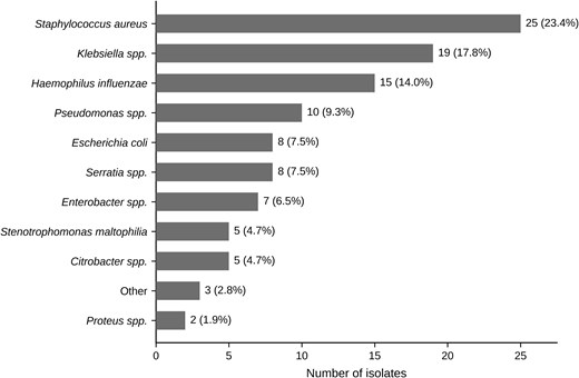
Bacterial genera identified at VAP15 diagnosis: n = 107 isolates (≥104 CFU/mL) from endotracheal aspirates collected within the preceding 2 days of diagnosis in 78/148 VAP15 patients (52.7%). The number of isolates and their corresponding proportion (out of the 107 total isolates) are reported for each bacterial genus.
Thirty-Day Mortality
Overall, the 30-day mortality from IMV initiation was 20.4% (83/406). In the multistate illness-death model, no significant competing relationship between VAP15 and death was observed. VAP15 was not significantly associated with subsequent mortality (transition VAP to death: HR = 1.20; 95% CI, 0.77–1.86), indicating no evidence that VAP15 substantially modified the hazard of death within 30 days from IMV start (Supplementary Table 4).
Similar results were observed when VAP15 was entered as a time-varying covariate in the frailty multivariable Cox model (aHR for VAP15 was 0.83; 95% CI, 0.52–1.33).
DISCUSSION
In this multicenter prospective cohort of patients receiving IMV for non-pulmonary indications, 148 of 406 patients (36.5%) developed a first episode of VAP during the 15-day observation period, an incidence rate of 43.2 per 1000 ventilator-days at risk. Non-neurologic admission diagnosis and female sex were independently associated with a lower risk of VAP15, as was early antibiotic exposure. In the time-varying exposures analysis, antihypertensive therapy was associated with a reduced risk of VAP15. Finally, VAP15 occurrence was not associated with mortality, noting that 94% of VAP observed occurred within the first 15 days of IMV.
The overall high incidence of first VAP in our cohort is best interpreted against studies of comparable ICU populations. It exceeds the rate reported in large Italian ICU cohorts that included all patients admitted to general ICUs [15], but those cohorts differ substantially from ours: three-quarters of our patients were intubated for a neurologic indication, with a median Glasgow Coma Score of 7, and were exposed to impaired airway protection, aspiration, prolonged intubation, and brain injury-induced immune dysfunction. These factors are known drivers of increased VAP risk [4, 5]. Of note, our results are consistent with studies analyzing similar ICU populations. In a randomized trial of ceftriaxone prophylaxis conducted in comatose brain-injured patients, early VAP occurred in 32% of the placebo group within the first week of ventilation [7], against 36.5% over a 15-day window here.
We also observed a significant sex-related difference, with male patients experiencing VAP more frequently than female, consistent with previous reports [15–17]. Biological mechanisms may partly explain this finding, as estrogens have been shown to enhance innate and adaptive immune responses, whereas androgens may exert immunosuppressive effects [18, 19]. In addition, behavioral and lifestyle factors, including differences in smoking prevalence and comorbidities, may also contribute to the observed sex differences [20].
Although our study did not investigate mechanistic pathways, these findings support consideration of sex in VAP risk stratification.
The role of antibiotic exposure in VAP prevention remains debated. In our cohort, we observed that early antibiotic exposure (within the first 2 days of IMV initiation) was associated with a lower incidence of VAP, whereas exposure modeled as a time-varying covariate during follow-up showed a weaker, nonsignificant association. These findings are directionally consistent with prior data: prophylactic antibiotics have been shown to reduce VAP incidence without a clear effect on mortality [21], with most evidence derived from patients with neurological injury [22–25] and recent trials in cardiac arrest and acute brain injury reporting lower early VAP incidence when antibiotics were given at or shortly after intubation [7, 26].
However, this association must be interpreted with caution, for two reasons. First, the indication for antibiotic administration was not recorded, so confounding by indication cannot be excluded. Second, antibiotic exposure was not independent of VAP ascertainment. Since the diagnosis ultimately relied on the physician's clinical judgment, early initiation or continuation of antibiotic therapy may have attenuated the fever, leukocytosis, and secretion changes on which that diagnosis depends, reducing the likelihood of VAP detection rather than its actual incidence. Our results nonetheless indicate that targeted and timely antibiotic strategies in selected high-risk patients warrant evaluation in trials designed for that purpose.
An association between antihypertensive exposure during follow-up and a reduced risk of VAP15 was also observed. This result should be considered exploratory and hypothesis-generating. Antihypertensive agents were captured as a single, undifferentiated category, so the signal cannot be attributed to any specific drug class; in particular, it cannot be linked to angiotensin-converting enzyme inhibitors, for which a protective effect against pneumonia has been proposed through enhancement of swallowing and cough reflexes [27]. Moreover, although adjustment for baseline severity and the competing-risk analyses did not eliminate the association, residual time-dependent confounding remains plausible because treatment initiation may itself reflect clinical stabilization. No causal or drug-class-specific interpretation can therefore be made from these data.
The microbiological profile observed during the first 15 days of IMV warrants attention. Although the predominance of S. aureus and Klebsiella spp. was broadly consistent with previous Italian ICU data [15], the relatively high proportion of H. influenzae, the lower prevalence of P. aeruginosa, and the very limited detection of MDR organisms together describe a microbiological profile characterized by a predominance of pathogens commonly reported in early-onset VAP and a low prevalence of multidrug resistance. This pattern is most plausibly explained by the cohort's defining features: patients had no respiratory disease at admission and, by design, had received no antibiotics in the 3 days before intubation, and follow-up was restricted to early-onset episodes within 15 days of IMV. Such conditions select against prior antimicrobial pressure and against MDR colonization. Similar profiles have been reported in early-onset VAP among patients with cardiac arrest and brain injury [7, 26]. If confirmed in larger and less selected populations, this lower-resistance microbiological profile may be relevant to the design of empirical treatment strategies for early VAP, although our findings should not be extrapolated to patients with substantial prior antibiotic exposure or later-onset disease.
Finally, VAP was not associated with increased 30-day mortality from IMV initiation in our cohort. This is in line with previous studies suggesting that, in heterogeneous ICU populations, mortality is primarily driven by underlying disease severity rather than VAP itself [2, 15]. However, the prognostic impact of VAP may vary across specific patient subgroups, and disease-specific effects cannot be excluded [28].
This analysis has several limitations. First, PULMIVAP was designed primarily for microbiota analysis and was not powered for a risk-factor model, with consequently limited subgroup analyses, particularly of specific antibiotic classes. Second, VAP was diagnosed clinically, and microbiological confirmation was not available for all episodes, reflecting real-world practice. Third, the indication for antibiotic administration was not recorded, with possible confounding by indication. Fourth, the exclusion of patients with respiratory disease at admission and of those treated with antibiotics within 3 days before intubation, along with the follow-up limited to 15 days, defines a selected population, limiting generalizability. Moreover, the 15-day data collection window precluded analysis of further outcomes that could add information on the burden of VAP. Fifth, VAP-prevention measures were not systematically recorded. Finally, decisions to withhold or withdraw life-sustaining treatment, and do-not-resuscitate orders, were not recorded. In such population, these decisions are frequent and are driven by neurological prognosis, potentially constituting a competing pathway to death independent of infection. This should be considered when interpreting our finding for 30-day mortality, and future studies in this population should record treatment-limitation decisions prospectively. Notably, our study enrolled only patients with an expected duration of IMV>48 hours, which may have limited this condition.
Strengths include the prospective multicenter design, the systematic daily collection of clinical and therapeutic data, the focus on a relatively homogeneous population of patients ventilated for non-pulmonary conditions, and the use of time-dependent modeling to account for dynamic exposures.
CONCLUSIONS
In patients receiving IMV for non-pulmonary indications, non-neurological diagnosis, female sex, and early antibiotic exposure were associated with a lower observed incidence of first VAP. These are associations observed in a cohort assembled for a different primary purpose and not powered for risk-factor estimation. They identify candidate populations and a candidate intervention, timed early antibiotic administration in patients ventilated for non-pulmonary, predominantly neurological, indications, for evaluation in appropriately designed prospective studies.
Supplementary Data
Supplementary materials are available at Open Forum Infectious Diseases online. Consisting of data provided by the authors to benefit the reader, the posted materials are not copyedited and are the sole responsibility of the authors, so questions or comments should be addressed to the corresponding author.
Notes
Acknowledgments. The authors wish to thank the The members of the PULMIVAP Study Group: Infectious Diseases Unit, Foundation IRCCS Ca' Granda Ospedale Maggiore Policlinico, Milano, Italy: Federica Sole Golfetto, Anna Tonizzo, Davide Mangioni. Monza Scientific Direction, Fondazione IRCCS San Gerardo dei Tintori: Francesca Elli. Neurointensive Care Unit, Department of Neuroscience, Fondazione IRCCS San Gerardo dei Tintori: Silvia Del Bianco. Department of Medicine and Surgery, University of Milano-Bicocca: Alessia Vargiolu. Laboratory of Probiogenomics, Department of Chemistry, Life Sciences and Environmental Sustainability, University of Parma, Parma, Italy: Christian Milani, Giulia Alessandri, Gabriele Andrea Lugli. Department of Anesthesia, Intensive Care and Emergency, ASST Spedali Civili University Hospital, Brescia: Lorenzo Pilati. Department of Medical and Surgical Specialties, Radiological Sciences and Public Health, University of Brescia, Antonino Panacea, Nicola Latronico, Gloria Erbici. Department of Anesthesia and Intensive Care, A. Manzoni Hospital, ASST Lecco: Alberto Steffanoni, Martina Viganò, Cristina Panzeri, Daniele Cerani. Neuroscience Intensive Care Unit and Department of Anesthesia, Critical Care and Emergency, Foundation IRCCS Ca' Granda Ospedale Maggiore Policlinico, Milano: Fabrizio Ortolano, Giacomo Grasselli, Sara Mazzarello, Chiara Fiorentini. Department of Anesthesia and Intensive Care Medicine, Grande Ospedale Metropolitano Niguarda, Milano: Gabriele Bassi, Riccardo Giudici, Elisabetta Zago. Department of Anesthesia and Intensive Care, Azienda Ospedaliero Universitaria Maggiore della Carità, Novara: Francesca Grossi, Martina Fracazzini, Nello De Vita, Stefania Guido, Eduardo Rocca. Anesthesia and Intensive Care Unit, University Hospital of Padua, Padua: Federico Geraldini, Marzia Grandis, Sabrina Calandra. Department of Medicine (DIMED), University of Padua, Padua, Italy: Alessandro De Cassai. Department of Anesthesia and Intensive Care, Parma University Hospital, Parma: Marco di Lecce, Bianca Maria Mainini, Emanuele Sani. Tettamanti Center, Fondazione IRCCS San Gerardo dei Tintori, Monza: Alessio Stevano. Federica Sole Golfetto, Anna Tonizzo, Davide Mangioni (Infectious Diseases Unit, Foundation IRCCS Ca’ Granda Ospedale Maggiore Policlinico, Milano, Italy); Francesca Elli, Silvia Del Bianco, Alessia Vargiolu (Monza Scientific Direction and Neurointensive Care Unit, Fondazione IRCCS San Gerardo dei Tintori and Department of Medicine and Surgery, University of Milano-Bicocca), Christian Milani (Laboratory of Probiogenomics, Department of Chemistry, Life Sciences and Environmental Sustainability, University of Parma, Parma, Italy); Antonino Panacea, Nicola Latronico, Lorenzo Pilati, Gloria Erbici (Department of Anesthesia, Intensive Care and Emergency, ASST Spedali Civili University Hospital, Brescia), Alberto Steffanoni, Martina Viganò, Cristina Panzeri, Daniele Cerani (Department of Anesthesia and Intensive Care, A. Manzoni Hospital, ASST Lecco), Fabrizio Ortolano, Giacomo Grasselli, Sara Mazzarello, Chiara Fiorentini (Neuroscience Intensive Care Unit and Department of Anesthesia, Critical Care and Emergency, Foundation IRCCS Ca’ Granda Ospedale Maggiore Policlinico, Milano); Gabriele Bassi, Riccardo Giudici, Elisabetta Zago (Department of Anesthesia and Intensive Care Medicine, Grande Ospedale Metropolitano Niguarda, Milano), Francesca Grossi, Nello De Vita, Stefania Guido, Eduardo Rocca (Department of Anesthesia and Intensive Care, Azienda Ospedaliero Universitaria Maggiore della Carità, Novara), Federico Geraldini, Alessandro De Cassai, Marzia Grandis, Sabrina Calandra (Anesthesia and Intensive Care Unit, University Hospital of Padova, Padova), Marco di Lecce, Bianca Maria Mainini, Emanuele Sani (Department of Anesthesia and Intensive Care, Parma University Hospital, Parma).
Author contributions. Conceptualization: L. A., L. C., E. Pa., and A. B.; Methodology: L. A., L. C., E. Pa., and A. B.; Formal analysis: L. C. and I. O.; Software: L. C. and I. O.; Writing—original draft: L. A., L. C., E. Pa., and I. O.; Writing—review & editing: F. M., L. M., F. T., C. A., C. Bur., C. Bug., M. F., A. F., G. G., R. F., M. M., A. M., E. Pi., L. T., R. V., T. Z., and M. V.; Supervision: G. C., A. G., and A. B.; Funding acquisition: L. A. All authors reviewed and approved the final manuscript.
Data availability statement. The data underlying this article cannot be shared publicly to protect the privacy of individuals who participated in the study. The data will be shared on reasonable request to the corresponding author.
The members of the PULMIVAP Study Group: Infectious Diseases Unit, Foundation IRCCS Ca’ Granda Ospedale Maggiore Policlinico, Milano, Italy: Federica Sole Golfetto, Anna Tonizzo, Davide Mangioni. Monza Scientific Direction, Fondazione IRCCS San Gerardo dei Tintori: Francesca Elli. Neurointensive Care Unit, Department of Neuroscience, Fondazione IRCCS San Gerardo dei Tintori: Silvia Del Bianco. Department of Medicine and Surgery, University of Milano-Bicocca: Alessia Vargiolu. Laboratory of Probiogenomics, Department of Chemistry, Life Sciences and Environmental Sustainability, University of Parma, Parma, Italy: Christian Milani, Giulia Alessandri, Gabriele Andrea Lugli. Department of Anesthesia, Intensive Care and Emergency, ASST Spedali Civili University Hospital, Brescia: Lorenzo Pilati. Department of Medical and Surgical Specialties, Radiological Sciences and Public Health, University of Brescia, Antonino Panacea, Nicola Latronico, Gloria Erbici. Department of Anesthesia and Intensive Care, A. Manzoni Hospital, ASST Lecco: Alberto Steffanoni, Martina Viganò, Cristina Panzeri, Daniele Cerani. Neuroscience Intensive Care Unit and Department of Anesthesia, Critical Care and Emergency, Foundation IRCCS Ca’ Granda Ospedale Maggiore Policlinico, Milano: Fabrizio Ortolano, Giacomo Grasselli, Sara Mazzarello, Chiara Fiorentini. Department of Anesthesia and Intensive Care Medicine, Grande Ospedale Metropolitano Niguarda, Milano: Gabriele Bassi, Riccardo Giudici, Elisabetta Zago. Department of Anesthesia and Intensive Care, Azienda Ospedaliero Universitaria Maggiore della Carità, Novara: Francesca Grossi, Martina Fracazzini, Nello De Vita, Stefania Guido, Eduardo Rocca. Anesthesia and Intensive Care Unit, University Hospital of Padua, Padua: Federico Geraldini, Marzia Grandis, Sabrina Calandra. Department of Medicine (DIMED), University of Padua, Padua, Italy: Alessandro De Cassai. Department of Anesthesia and Intensive Care, Parma University Hospital, Parma: Marco di Lecce, Bianca Maria Mainini, Emanuele Sani. Tettamanti Center, Fondazione IRCCS San Gerardo dei Tintori, Monza: Alessio Stevano.
Financial support. This work was supported by the Italian Ministry of Health [Ricerca Finalizzata, Giovani Ricercatori, grant number GR-2018–12365988 to L. A.].
References
Author notes
Potential conflicts of interest. G. C. has received research grants and consulting fees from Integra LifeSciences and NeurOptics. All other authors: No reported conflicts of interest.


Comments