-
PDF
- Split View
-
Views
-
Cite
Cite
I-Ming Chen, Chun-Che Shih, Extending hybrid approach to residual Stanford type A dissecting aortic aneurysm, Interactive CardioVascular and Thoracic Surgery, Volume 7, Issue 5, October 2008, Pages 794–796, https://doi.org/10.1510/icvts.2008.176842
Close - Share Icon Share
Abstract
Residual Stanford type A dissecting aortic aneurysm was frequently encountered several years after emergent repair. Surgical approach remained challenging and hazardous, not only due to the extensive involvement of the dilated false lumen but also the high comorbility of redo sternotomy and extensive thoraco-abdominal procedure. We present a modified hybrid technique incorporating arch replacement with bifurcated graft to relocate supra-aortic branches followed by anastomosis with reverse sleeve graft of elephant trunk over distal arch. After stent graft insertion over proper landing zone, all the communicating holes could be sealed and the compressed true lumen of descending aorta would be fully dilated. This technique not only simplified some laborious situations but also simultaneously resolved the entire thoracic dissection segment with an acceptable and optimal midterm result.
1. Introduction
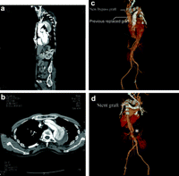
Life-threatening acute Stanford type A aortic dissection can be successfully repaired by emergent ascending aortic replacement. However, residual type A aortic dissection arising from the proximal aortic arch to the descending and abdominal aorta was frequently noted and gradually became enlarged during the follow-up period (Fig. 1a,b ). These remaining dissections may cause some lethal complications such as rupture or malperfusion of vital organs due to compression of the true lumen by the enlarged false lumen. Traditional operative methods caused surgeons to encounter very difficult situations, including redo-sternotomy with concomitant thoracotomy or the clamshell approach in order to repair these large-scale dissecting aortic aneurysms [1]. Furthermore, the redo operation worsened the bleeding complication and prolonged operative time, leading to increased surgical mortality and morbidity. We introduce a new modified hybrid technique to deal with these residual dissections by supra-aortic relocation with bifurcated graft, followed by arch replacement using the elephant trunk procedure with reversed sleeve graft, then conclude with the incorporation of stent graft implantation.

Preoperative CT: (a) Sagittal view: the dissection arising from the distal end of the previous replaced graft. (b) Axial view: dissection flap involving the entire arch with the largest diameter exceeding 6 cm. Post-operative 3-D CT reconstruction: (c) anterior view of CT with 3-D reconstruction seven days after repair (d) post view. *Communicating hole just below the celiac artery level.
2. Materials and methods
2.1. Surgical technique
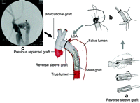
Cardiopulmonary bypass was set up by double arterial cannulations of femoral and right axillary artery as well as femoral venous cannulation. Limb ischemia was also prevented by insertion of another perfusion catheter from the arterial cannulas toward the distal arteries. Redo median sternotomy was executed by an oscillating saw. The adhesion was divided to make well exposures from ascending aorta to distal arch. The innominate artery and left common carotid artery were occluded with Fogarty balloon and transected consecutively. The intimal flaps were tacked down by 5-0 polypropylene running suture with ‘sandwich’ Teflon pledgets. The bilateral limbs of 24–12 Ultramax Y graft (Atrium Medical, Hudson, NH) was trimmed to relocate these arch branches in the end-to-end fashion during the cooling period of cardiopulmonary extracorporeal support (Fig. 2 ).

Depiction of hybrid surgical procedure: after reverse sleeve graft was prepared as in Fig. a, the entire arch is replaced with distal elephant trunk technique Fig. b, and complete angiogram after stent graft deployment is shown in Fig. c. ▾, anastomosis site of reverse sleeve graft; LSA, left subclavian artery.
Cardiac arrest was achieved under the protection of antegrade cardioplegia infusion. After halting the femoral arterial inflow, systemic circulation was ceased when rectal temperature cooled down to 25 °C. Both the cerebrum hemispheres were protected with continuous 25 °C cold blood flow (1000 ml/min) through the right axillary arterial perfusion after clamping the main trunk of bifurcational graft. Arch aortotomy was explored from the ascending aorta just distal to the previous replaced graft to the distal aortic arch between the left common carotid artery and left subclavian artery. Another straight Ultramax graft of the proper size (usually 26 or 28 mm) was prepared for arch reconstruction. The graft was rolled up as a sleeve graft in the inside-out manner, the modified elephant trunk technique for clearer surgical field recommended by Hsieh et al. [2], then inserted into the true lumen of distal aorta (Fig. 2a). After completion of the anastomosis with 3-0 polypropylene sutures, the free end of the reversed sleeve graft was withdrawn (Fig. 2b). The femoral arterial inflow was restarted again after clamping the reversed sleeve graft. During re-warming, the main trunk of bifurcated graft was anastomosed with the previous replaced graft and the reversed sleeve graft in a T-shape manner (Fig. 2c).
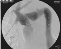
After successful weaning off cardiopulmonary bypass, the femoral arterial cannula was removed and the delivery system for the proximal components of the thoracic stent graft (Zenith® TX2™, thoracic endoprostheses) introduced through the arteriotomy. Under fluoroscopic guidance, the delivery system was advanced into the reversed sleeve graft and securely incorporated after stent graft deployment (Video 1 ).

2.2. Surgical results
This modified hybrid technique required on average 1.58 endograft implantations and has been applied successfully in 15 consecutive patients from January 2006 to December 2007 with a mean age of 67.1±10.2 (42–77) years and a mean interval to previous operations of 4.5 years. An Institutional Review Board – approved retrospective chart was also conducted. These residual dissections all arise from the distal anastomosis of previous replaced ascending aortic graft with patent and progressive enlarged false lumens. Numerous communicating holes between intimal flaps could be identified not only over the thoracic but also the abdominal aorta and even iliac artery. The average maximal size of these dissecting aneurysms was 6.2 cm in diameter, which were involved mostly in the aortic arch and descending aorta. Six of them had a history of Marfan syndrome. None of them suffered with preoperative malperfusion of vital organs. The mean cardiopulmonary bypass time, aortic clamp time and circulatory arrest time was 283, 87 and 25 min, respectively. The mean operative time was 399 min with mean contrast injection of 121 ml. The mean intra-operative blood loss was 856 ml. The mean durations of intensive care unit and hospital stay were 8.2 and 15.7 days, respectively, without any surgical mortality. Neither severe stroke nor paraplegia was found postoperatively. Three of our patients needed temporary hemodialysis due to oligouria and acute renal insufficiency. Superficial wound dehiscence occurred in two of our patients. All of our patients received regular follow-up at our outpatient department after discharge and progressively thrombosed false aneurysm over the entire thoracic aortic segment was shown in the following chest computed tomography scan.
3. Discussion
Residual Stanford type A dissecting aortic aneurysm is a troublesome surgical condition in patients who had previously undergone emergent ascending aortic repair. Although acceptable results have been proposed in proximal reoperation after repaired acute Stanford type A dissection [1], repair of residual Stanford type A with involvement of aortic arch and descending aorta, in which redo sternotomy and concomitant thoracotomy were inevitable, continues to be a formidable challenge. In our new hybrid technique, compromise of the cerebral blood supply was minimized under the biarterial inflow of extracorporeal assist, particularly during the reconstruction of the left common carotid artery with a bifurcated graft [2,3]. The reversed sleeve graft technique also provided a good surgical visual field when carrying out distal aortic anastomosis and ultimately resulted in better hemostasis despite requiring one additional anastomosis from graft to graft [2,3]. The reversed sleeve graft created a longer and safer proximal landing zone (mean length of 8.5 cm) and avoided potential intima injury during stent graft deployment. Furthermore, after secure incorporation with the proximal component of the Zeniths TX2 stent graft, it facilitated correction of folded, kinked and squeezed Dacron graft segments in the compressed true lumen. Since all of the communicating holes between the true and false lumen of entire thoracic aorta that would have been completely occluded by the stent graft and thrombosis of false lumen proceeded more quickly (Fig. 1c,d). This rationale also showed great potential in halting dilatation progress and may even induce aneurysm shrinkage after reducing the endotension of false aneurysm.
Although the residual communicating holes of abdominal involved segment still exist, the dilatation progress of false aneurysm seemed to slow down during the follow-up period and even regressed with partial thrombosis. This may also be the theoretical hypothesis of the very low incidence of paraplegia after hybrid endovascular repair. Therefore, this modified hybrid technique not only resolved the dissection involvement of the entire thoracic aorta without requiring a laborious thoracotomy but also yielded an acceptable and optimal midterm result.
This work was supported by grants from the National Science Council, Taiwan SC-95-2341-B-010-028-MY3 and 94-2314-B-010-057; Taipei Veterans General Hospital, Taiwan V95C1-049, V95E1-010, V95ED1-008, V95-S22-005 Taipei, Taiwan.


