-
PDF
- Split View
-
Views
-
Cite
Cite
Alex Frydrychowicz, Ernst Weigang, Mathias Langer, Michael Markl, Flow-sensitive 3D magnetic resonance imaging reveals complex blood flow alterations in aortic Dacron graft repair, Interactive CardioVascular and Thoracic Surgery, Volume 5, Issue 4, August 2006, Pages 340–342, https://doi.org/10.1510/icvts.2006.129577
Close - Share Icon Share
Abstract
A thorough knowledge of both vascular anatomy and hemodynamics is of high interest for the understanding of the severity of the underlying disease, the therapeutic decision-making and follow-up. In this context, flow-sensitive 3D magnetic resonance imaging (MRI) offers the possibility to simultaneously acquire detailed information about vascular hemodynamics and morphology. Recent methodological progress, extensive validation of the technique and combination with advanced 3D blood flow visualization enables nowadays for a detailed depiction of normal and altered flow characteristics in large arteries such as the thoracic and abdominal aorta. We report the comprehensive MR analysis of hemodynamic alterations in an otherwise healthy patient who underwent a Dacron graft repair after traumatic rupture of the proximal descending aorta. Flow-sensitive time-resolved 3D MRI was employed to analyze the effects of the implantation of a Dacron prosthesis on local vascular hemodynamics. Despite the unsuspicious appearance on angiographic images, 3D blood flow visualization revealed the development of complex and substantially altered systolic blood flow within the graft. These initial findings might in future enrich vascular diagnostics, therapeutic decision making, graft design, or serve as a comprehensive research tool.
1. Introduction
For the evaluation of alterations or pathological changes in vascular geometry, the knowledge of local vascular hemodynamics within the aorta is of high interest. Specifically, for subsequently performed surgical interventions, the assessment of blood flow characteristics may offer new insights into differences between surgical techniques, long term durability of procedures, or serve as a predictive tool in assessment of diseases and therapeutic follow-up.
The intrinsic sensitivity of magnetic resonance imaging (MRI) offers the possibility of direct quantitative assessment of three-directional blood flow velocities simultaneous with the underlying morphological information. After its initiation, the qualitative and quantitative assessment of blood flow using magnetic resonance imaging (MRI) was consecutively investigated throughout the years. The underlying blood flow encoding technique dates back about 20 years to the beginning of MRI [1] and has been extensively validated [2,3]. Recent advances in flow sensitive MRI provide the opportunity to analyze the dynamics and spatial distribution of local and global blood flow within a three-dimensional (3D) volume covering the entire vascular structure of interest [4]. If coupled with interactive advanced 3D visualization, ECG and respiration synchronized time-resolved 3D MR imaging permits the detailed evaluation of vascular hemodynamics in pathologically altered or surgically repaired vascular regions [5–7].
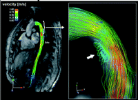
We report time-resolved flow sensitive 3D MR imaging and advanced 3D blood flow visualization results for a 23-year-old patient with Dacron prosthesis in the proximal descending aorta (placed 2 years before after traumatic aortic rupture). For comparison, altered flow features can be compared to findings in an age-matched volunteer. Fig. 1 depicts time-resolved 3D particle traces originating from a user-selected plane in the ascending aorta. Normal, laminar-like flow without retrograde or vortical movements can be seen. In the patient, the delineation of the graft can clearly be appreciated in morphological MR images (Fig. 2A , yellow arrows). Despite the unsuspicious appearance of extent and location of the graft, 3D blood flow visualization revealed complex blood flow alterations such as the development of accelerated and complex helical blood flow and vortex formation during systole.

Time-resolved 3D particle-trace visualization of blood flow at three time points in the systole of a healthy individual without alterations of the aortic geometry. Laminar-like flow distribution over the aortic arch, in the supraaortic branches and the descending aorta can be appreciated. No evolvement of vortices, roll-like flow formations or retrograde flow are visible.

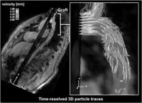
A. Contrast-enhanced MR-angiography covering the ascending (AAo) and descending (DAo) thoracic aorta in a patient with implanted prosthesis for repair of the proximal descending aorta after traumatic rupture with clear delineation of the implanted prosthesis (yellow arrow). The white rectangle indicates the magnified area in B-D for which time-resolved 3D particle trace visualization was performed. B-C. Systolic blood flow patterns visualized as time-resolved particle-traces in three successive time frames. Complex blood flow and a marked helix formation (white arrows) within the graft are clearly visible. Color coding corresponds to the measured local velocities. The 3-dimensional temporal evolution of blood flow can be most clearly appreciated in the supplementary movie file.
Figs. 2B-D and 3 illustrate extensive changes in flow patterns in the descending aorta associated with the graft repair. Blood flow acceleration (change in color coding of 3D stream-lines and particle traces) within the graft as expected from the lack of compliance can clearly be appreciated for all visualization modes.

Systolic blood flow visualized as stream-lines provide an overview over blood flow velocities and hemodynamic alterations including the vortex formation (white arrow) at the small curvature in the proximal descending aorta. Color coding corresponds to the local velocity magnitude.
However, additional substantially altered flow characteristics could be detected, including the formation of a roll-like vortex at the proximal ostium of the prosthesis which is situated close to the small curvature of the aortic arch (Fig. 3, white arrow). Further, the investigation of the temporal evolution of blood flow in 3D (3D particle traces, Fig. 2B-D) additionally illustrates highly complex flow through and distal to the graft. Locally altered vascular hemodynamics led to a considerable helical flow pattern as indicated by the white arrows (Video 1 ).

3D particle trace visualization of the hemodynamic alterations in a patient with a Dacron graft repair after traumatic rupture of the proximal descending aorta. On the left an overview is given. On the right, massive turbulences and flow acceleration can be appreciated. Measured flow velocities are color-coded. A roll-like tubular turbulence at the inner curvature can be seen as shown in Fig. 3.
Whether the flow disturbances associated with a surgical intervention such as prosthesis implantation are due to the graft, its material and surface, and altered vessel compliance or include additional effects related, e.g. to the technique of the suture performed for the graft repair, remains speculative. Also, the influence of dynamic cardiovascular parameters such as stroke volume, heart rate, cardiac output and blood pressure may not be underestimated and could influence the extent of the reported changes. Nevertheless, the results indicate the substantial influence of prosthesis repair on local hemodynamics. Even though graft location and size seem inconspicuous in angiography images, quite remarkable changes in local blood flow patterns were detected in this patient. These changes in blood flow and their correlation with graft material, length, location and surgical technique may therefore have the potential to further understand the influence of alterations in vascular geometry and aid surgical planning, optimal graft placement, graft design, or serve as a research tool.
In conclusion, these preliminary pictorial results demonstrate and illustrate the advantage of the complete spatial and temporal coverage of flow sensitive MRI. This technique permits the analysis of complex flow features in 3D, which may otherwise remain undetected or only be partially and incompletely analyzed. The proposed technique has the potential to greatly enhance the understanding of blood flow characteristics associated with a particular pathology and associated surgical treatment [8,9]. Thereby, it may provide better insights into genesis, progression and prognosis of vascular pathologies or treatment after evaluation in comparative studies with larger cohorts of patients with similar pathologies or therapies.
2. Additional information on flow sensitive MRI and blood flow visualization
The patient was referred to MRI for a routine follow-up evaluation of a Dacron prosthesis repair in the proximal descending aorta after traumatic rupture. A healthy volunteer was included for comparative purposes. From both individuals written informed consent was obtained prior to the MRI investigation. The study has been approved by the local ethics review committee.
Flow sensitive MRI examinations were performed on a 3T MR system (Magnetom TRIO, Siemens, Germany) using an rf-spoiled gradient echo sequence with interleaved 3-directional velocity encoding, which has been validated previously [4]. Data were acquired during free breathing and were prospectively gated to the ECG cycle. Respiration control was achieved using navigator gating. Further parameters were: flip angle α=15°, venc=150 cm/s, spatial resolution (3.2×2.1×5.0) mm, TE=3.5 ms, TR=5.6 ms, temporal resolution=45 ms. Blood flow visualization was performed using a commercially available software package (EnSight, CEI, Apex NC, USA). Various modes of depiction of measured flow characteristics included time-resolved 2D vector graphs and 3D particle traces as well as 3D stream-lines [10]. 3D stream-lines resemble a path through the vessel at a certain point in time. This path is a tangent to the respecting acquired velocity vectors. The concept of time-resolved 3D particle traces is based on imaginary mass-less particles originating from a user-chosen area of interest. The set of depictable time-frames is predefined by the temporal resolution during data acquisition. 2D-vector graphs are used for high detail analysis of local hemodynamic features. Each vector resembles the temporal evolution of 3-directional blood flow velocity over the RR-cycle.
Dr. Markl received funding by the ‘Deutsche Forschungsgemeinschaft’ Grant MA 2383/3-1


