-
PDF
- Split View
-
Views
-
Cite
Cite
Pascal Defaye, Adama Kane, Peggy Jacon, Jean François Obadia, Inadvertent implantable cardioverter defibrillator lead placement in the left ventricle: long-term follow-up at nine years and management by minimally-invasive surgery, Interactive CardioVascular and Thoracic Surgery, Volume 12, Issue 3, March 2011, Pages 492–493, https://doi.org/10.1510/icvts.2010.250209
Close - Share Icon Share
Abstract
We describe the case history of a 43-year-old male with type 1 Brugada syndrome. He was fitted with an implantable cardioverter defibrillator (ICD) for primary prevention nine years ago. After admission for inappropriate shocks, an abnormal position of the lead was discovered. Further investigations (chest X-ray and transesophageal echocardiography) showed that the ICD lead was in fact in the left ventricle. The ICD lead was removed successfully using video-assisted thoracoscopic surgery.
1. Case report
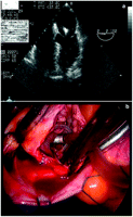
A 43-year-old male implanted with a single-chamber implantable cardioverter defibrillator (ICD) was hospitalised after two inappropriate shocks. The device had been placed nine years prior for primary prevention of type 1 Brugada syndrome. A transthoracic echocardiography (TTE) suggested an unusual position of the defibrillation lead not previously suspected by the antero-posterior (AP) chest X-ray. A subsequent lateral chest X-ray showed that the lead was probably pointing posterior towards the left ventricular (LV) cavity (Fig. 1b : arrow). This abnormal position was later confirmed by the TEE: the defibrillation lead was seen crossing the patent foramen ovale (PFO), the mitral valve with a final insertion on the lateral wall of the left ventricle (LV) (Fig. 2b ). Please note that the patient was entirely asymptomatic and there was no thromboembolic event in the nine years of follow-up.

Apical (a) and lateral (b) chest X-ray showing ICD lead pointing posterior towards the left ventricular cavity (arrow). ICD, implantable cardio- verter defibrillator.

TEE: lead in left ventricle through the patent foramen ovale and the mitral valve (a), video-endoscopic: lead through the patent foramen ovale (b). TEE, transesophageal echocardiography.
What decision should have been made for this inadvertently positioned lead that had been implanted in the LV for nine years?
It could be left in situ assuming that it would not pose a risk.
Long-term anticoagulant therapy could have been initiated because of a hypothetical risk of embolism, without recommending removal of the lead.
The lead could be removed. Since it was implanted in the LV nine years previously, the option of endovascular removal more or less assisted by a laser extraction sheath posed a potential embolic risk. Therefore, removal of the lead would have to be done surgically [1].
The inadvertent placement of an ICD lead in the LV via a PFO is well known, but the long-term effects are rarely described [2]. It creates diagnostic and management issues, as in the current case. While the follow-up consultation of the ICD is of little assistance in detecting an abnormal position of the lead in the left cavities, the diagnosis is most frequently suggested by chest X-ray and confirmed by a TTE.
Removal is controversial since some medical reports have advised long-term follow-up without complications [3]. However, there is evidence that the leads positioned in the LV can be associated with short- or long-term thrombo- embolic complications [3].
Percutaneous removal of the LV lead exposes the patient to the risk of embolism as well as the risk of injury in the region of the PFO, the mitral valve and the LV. Conversely, if chosen, anticoagulant therapy is likely to cause long-term bleeding complications. Therefore, we decided to use minimally-invasive surgical approach since it carried the lowest risk. The lead was removed successfully using video-assisted thoracoscopic surgery with extra-corporeal circulation and aortic clamping (Fig. 2b) and the PFO was sutured at the same time. The patient recovered uneventfully postprocedure.


