


Abstract
Lipothymoma is a very rare thymic benign tumor. A 55-year-old male, who underwent thymectomy four years previously, presented with cavitating pulmonary tuberculosis.
1. Introduction
Lipothymoma is a rare anterior mediastinal mass which usually presents as very large tumors [1]. It is characterized by almost symmetrical bilobar growth extending to both pleural cavities. Its morphological features and the fat consistency help in preoperative radiological diagnosis. Prognosis is very good. In this paper, we report a rare long-term complication of pulmonary tuberculosis post-thymectomy.
2. Case presentation
A 55-year-old male presented with hemoptysis, fever and weight loss. He had undergone thymectomy for a huge lipothymoma four years previously through a median sternotomy. Recent chest X-ray (CXR) and computed tomography (CT)-scan (Fig. 1a,b ) showed aggressive cavitating tuberculous infection affecting both lungs. His old chest X-ray post-tumor excion was normal (Fig. 1c). Revision of the old chest X-ray (Fig. 2a ) revealed a huge mediastinal tumor resembling cardiomegaly. The old CT-scan (Fig. 2b) delineated a well-defined fatty tumor surrounding a normal sized heart. Histopathology showed lipothymoma and gross appearance (Fig. 2c), weighing 5.5 kg. The postoperative course was smooth and he was followed uneventfully for more than three years. He was not exposed to any tuberculous patients. He noticed recent loss of weight and intermittent episodes of pyrexia. He presented to the emergency room with hemoptysis. Sputum cultures were positive for Mycobacterium tuberculosis. He responded well to anti-tuberculous, anti-tussive, and tranexamic acid treatment and did not require embolization or surgical intervention.
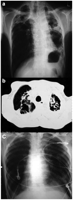
CXR (a) and CT-scan (b) showing multiple cavities destroying both lungs, more evident on the left lung, (c) showing CXR after excision of the tumor.
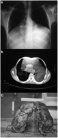
CXR (a) and CT-scan (b) showing the huge fatty tumor surrounding a normal sized heart. The thymoma was compressing the heart and filling both lower pleural cavities. (c) The tumor after excision.
3. Conclusion
Tuberculous infection complicating late follow-up of a thymectomy has not been reported before in the literature. This may be a coincidental finding or may have been attributed to decreased cellular immunity. Thymolipoma is an uncommon benign neoplasm that accounts for 2.9% of thymic tumors [2]. Thymolipomas usually grow slowly and can attain large sizes before the diagnosis is made. Compression of adjacent structures, the heart, great vessels, lungs and tracheobronchial tree, are their main presenting symptoms. Images on X-rays can resemble cardiomegaly as in our case [3]. This benign neoplasm can be associated with some autoimmune disorders, such as myasthenia gravis, systemic lupus erythematosus, hypogammaglobulinemia, Graves' disease and red cell aplasia [4, 5]. The first reported case of a lipoma of the thymus was published by Lange in 1916 but the term lipothymoma was first introduced by Hall in 1948 [6]. Biopsy is not indicated and the only curative treatment is complete excision via sternotomy, anterolateral thoracotomy or Clamshell incision.
