-
PDF
- Split View
-
Views
-
Cite
Cite
Ingo Breitenbach, Aschraf El-Essawi, Marcel Anssar, Wolfgang Harringer, Handling of extensive aneurysm of the aorta with bronchomalacia in a Marfan patient, Interactive CardioVascular and Thoracic Surgery, Volume 11, Issue 5, November 2010, Pages 706–707, https://doi.org/10.1510/icvts.2010.245449
Close - Share Icon Share
Abstract
Surgical management of extensive aneurysms of the aorta still remains demanding. Herein, we describe the successful treatment of a 59-year-old Marfan patient with extensive aneurysm of the aorta complicated by bronchomalacia.
1. Introduction
Bronchomalacia caused by external compression of an aortic aneurysm is a rare disease requiring individual treatment. We describe a patient in whom this complication was successfully handled by implantation of an endobronchial stent and advanced second stage operation.
2. Case report
A 59-year-old female with an unrecognised Marfan's syndrome and an uneventful past history was referred to our hospital with the vague symptoms of fatigue, pyrexia, tachycardia and thoracic pain.
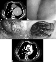
Chest radiography showed a large homogenic shadow projecting onto the left upper lobe of the lung. Computer tomography identified the mass as a large aneurysm of the ascending aorta and aortic arch with a maximal diameter of 10.5 cm. Furthermore, an old type B-dissection with an extend-I thoracoabdominal aneurysm (Crawford classification) was identified (Fig. 1a ). The respiratory function test revealed moderate obstructive pulmonary disease [forced expiratory volume in 1 s (FEV1) 1.37 l, 45% of expected and vital capacity (VC) 1.76 l, 49% of expected]. Echocardiography revealed Barlow's disease with prolapse of both mitral valve leaflets resulting in severe mitral regurgitation as well as moderate aortic regurgitation secondary to root dilatation.

(a) Preoperative chest CT-scan. Left main bronchus (1) is displaced and compressed by massive aortic aneurysm of the thoracic aorta (3). Right main bronchus (2). (b) Bronchoscopy showing collapsed left main bronchus. (c) Nitinol stent implantation into the left main bronchus with residual collapse due to compression by the aneurysm. (d) Complete competent left main bronchus after early second step operation. (e) CT-scan at one-year follow-up with stented left main bronchus (1) and replacement of the aorta with a dacron prostheses (3). Right main bronchus (2).
Because of the extent of the disease a two-stage operation incorporating an elephant trunk in the first stage with subsequent replacement of the descending aorta in a second stage was chosen. Following median sternotomy cardiopulmonary bypass was established via the right subclavian artery and both the superior and inferior vena cava. The mitral valve was reconstructed by a quadrangular resection and sliding plasty of the posterior leaflet and the implantation of artificial chordae onto the prolapsing segment of the anterior leaflet. In addition, the annulus was stabilized by a 36-mm Carpentier–Edwards Physio Annuloplasty Ring (Edwards Lifesciences, Irvine, CA, USA). In moderate hypothermic circulatory arrest at 28 °C with selective antegrade cerebral perfusion a total arch replacement including an elephant trunk was accomplished followed by a valve sparing aortic root replacement using the reimplantation technique. Intraoperative echocardiography revealed no residual aortic or mitral regurgitation. Following an initially uneventful postoperative course the patient had to be reintubated on the second postoperative day due to respiratory insufficiency. Fiber-optic bronchoscopy identified a collapsed left main bronchus as the cause for the respiratory insufficiency (Fig. 1b). A Nitinol Ultraflex tracheobronchial stent (Boston Scientific, Ballybrit, Ireland) was implanted, however, this did not resolve the collapse completely (Fig. 1c), due to compression by the aneurysm of the descending aorta. Decision for an early second stage procedure was taken. The descending aorta was replaced down to the celiac trunk at the 20th postoperative day, while the patient was still on mechanical respiratory support. The operative procedure was uneventful and postoperative bronchoscopy showed a completely patent left main bronchus (Fig. 1d). Following a prolonged weaning the patient was able to leave the intensive care unit on the 50th postoperative day. At one-year follow-up following the initial procedure the patient was in good health (NYHA I) showing regular results in the computed tomography (CT)-scan (Fig. 1e). Because the patient denied additional intervention the stent was still left in place.
3. Discussion
The treatment of patients with extensive aneurysm of the aorta remains a strategic surgical challenge [1]. To facilitate staged surgery for the aortic arch and the descending aorta. Borst et al. introduced the elephant trunk technique in 1983 [2]. In the first stage, the ascending aorta and arch are replaced via a median sternotomy and a free-floating extension of the arch prostheses is left behind in the proximal descending aorta. Three months later, the descending aorta is replaced via a left lateral thoracotomy.
To complete the surgical treatment in a single stage the frozen elephant trunk technique was introduced in 2003 by Karck and colleagues [3]. In this case, our choice was a staged operative procedure over a three-month interval facilitated by the elephant trunk technique because of the extent of the aneurysm that reached down to the celiac trunk. A single stage procedure using frozen elephant trunk technique could not be used because of the need of a landing zone for the stent graft at the distal end.
Bronchomalacia resulting from external compression by an aortic aneurysm has been published in several cases [4–6]. Nevertheless, there is no standardized approach for the treatment of this complication.
Comer and colleagues showed that left main bronchus compression due to an extend-I thoracoabdominal aneurysm could be resolved by implantation of an uncovered expandable metallic stent. Nevertheless, a weaning of the patient from mechanically ventilation was unsuccessful and the patient died due to pneumonia in the following days [7]. Slonim et al. reported a successful stent implantation in a patient with tracheo-bronchial stenosis. Weaning after implantation of the patient from mechanically ventilation was successful but the patient died 14 days after later due to rupture of the aneurysm [8].
Ewert and colleagues reported a 75-year-old patient who was successfully treated with stent implantation into the trachea but after extubation the stent was not strong enough to resist the external pressure of the aneurysm as in our case. The patient died due to bronchopneumonia [9].
In our case, the implantation of a stent improved the patency of the left main bronchus but did not resolve it completely, presumably, due to persistent external compression by the descending aortic aneurysm as Ewert et al. [9] reported. Only the advanced second stage procedure resolved the residual collapse. Primarily diagnosing this problem with the option of concomitant temporary endo-bronchial stenting and if necessary – hence residual clinically significant collapse persists – advancing the second stage procedure to the same hospital stay appear to be adequate strategic options when facing such complex surgical challenge. Stent implantation without an additional surgical approach remains a palliative and symptomatic option for patients who denied surgery.


