-
PDF
- Split View
-
Views
-
Cite
Cite
Marc Hartert, Manfred Dahm, Achim Neufang, Christian-Friedrich Vahl, Minimum cause – maximum effect: the travelogue of a bullet, Interactive CardioVascular and Thoracic Surgery, Volume 11, Issue 5, November 2010, Pages 698–700, https://doi.org/10.1510/icvts.2010.245100
Close - Share Icon Share
Abstract
This case report involves a 57-year-old male, accidentally shot in the chest with a small bore firearm. The bullet entered the left hemithorax, disrupting the left internal mammarian artery. It then penetrated the anterior wall of the right ventricle causing a pericardial tamponade. After leaving the base of the right heart it perforated the diaphragm, the liver, the spleen and the pancreas. Finally, it penetrated the abdominal aorta 3 cm proximally to the coeliac trunk and reached its final position paravertebrally. This case report illustrates that the management of even minimum gunshot wounds requires a maximum variety of surgical skills.
1. Introduction
As violence is not a part of everyday life in Europe, thoracic and/or abdominal gunshot wounds are rather exceptional. The extent of this problem is difficult to estimate as gunshot injuries are often linked with traumata from rifles or other large firearms. Due to their extreme variability, such wounds are counted among the most challenging surgical interventions related to penetrating traumata [1–3]. They are associated with a wide range of injuries varying from minor soft-tissue damages to deep wounds affecting multiple organ systems. This case report involves a 57-year-old male, accidentally shot in the chest with a small bore firearm. Despite the objectively small size of the bullet, the projectile left an impressing thoracoabdominal battlefield.
2. Case
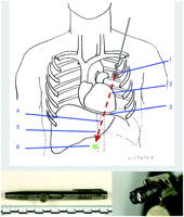
A 57-year-old male was presented to the emergency department via paramedics after having sustained a self-inflicted gunshot wound. Twenty years ago, the patient, a car dealer, had found a shooting pen (calibre .22 lr) in a car he had been preparing for sale (Fig. 1 , lower panel). On March 3rd 2009, he coincidentally had recovered the weapon and began to experiment with it. By mistake, a shot went off. Upon examination, a single entrance wound at the left mid-paramanubrial area without any detectable exit wound was observed; the bullet remained stuck in the body. The shooting pen was presumably held to the upper left chest with the barrel pointing downwards. Echocardiography performed in the ER revealed a haemodynamically significant pericardial effusion. In the abdomen, no sonographically relevant liquid was detectable. Due to cardiovascular instability further diagnostics were deferred. Clinical findings as well as signs of shock made an emergency operation obligatory.

Upper panel: Trajectory of the bullet. The bullet perforated the left anterior chest and entered the left hemithorax transmediastinal, disrupting the left internal mammarian artery (1). It then penetrated the anterior wall of the right ventricle causing a pericardial tamponade (2). After leaving the heart dorsal through the right ventricle it emigrated via the diaphragm (3) into the liver, the spleen and the pancreas (4). Finally, it punched through the abdominal aorta 3 cm proximally to the coeliac trunk (5) and ended paravertebrally (6). Lower panel: shooting pen (on the left); projectile calibre .22 lr (on the right).
The passage of the projectile resembled an odyssey through the human body (Fig. 1, upper panel). First, it hit the left internal mammarian artery (IMA), and then traversed the right ventricle by entry via the anterior wall and dorsobasal exit. After an emergency median sternotomy, the ventricular laceration was sewed with patch-armed sutures, followed by mending of the left IMA. Anticipating merely a thoracic wound path, an incessant bleeding required further intensive inspection. On its way through the thorax, the bullet caused a transit wound in the diaphragm. Following laparotomy, a large amount of bloody fluid was obtained from the abdominal cavity. Reaching the abdomen, the projectile hit the left lobe of the liver, the spleen and the pancreas. As source of continuous arterial bleedings a shot right through the aorta about 3 cm proximal to the coeliac trunk was detected. The bullet reached its final destination paravertebrally without causing any further damage. First, the aortic lesions (front and back wall) were repaired with Teflon®-coated single button sutures. Therefore, the diaphragm was dissected and the aorta was cross-clamped twice (x-clamp times: 30 min/12 min). That followed a spleenectomy as well as surgical treatment of the liver and pancreas bleedings. An additional exploration of the colon, the small bowel and the stomach revealed no additional injuries. The projectile, palpable near the vertebral column, was removed and transferred to the forensic department.
Due to an increasing circulatory disorder and continuous bleeding via the abdominal drainage, re-laparotomy was inevitable on the first day postoperatively. At this, a bleeding of the tail of pancreas was detected and overstitched. An extended stay on the intensive care unit was necessary due to respiratory insufficiency based on enterococcic pneumonia, ultimately causing indispensable tracheotomy. A progressive respiratory improvement allowed de-cannulation on the 21st postoperative day. Two days later, the patient was transferred to the general ward. A CT-scan confirmed a good operative result without indications of any relevant pathology. No indication of cardiac insufficiency or any other functional deficits led to the patient's discharge on the 28th postoperative day. Follow-up after six and 12 months showed normal cardiac function at echocardiography and no surgically related pathology at CT-scan.
3. Discussion
This case report exemplifies the uniqueness of bullet wounds: minimum injuries that – at least at first sight – appear to be straightforwardly manageable may culminate in a maximum venture for all surgical disciplines. Particularly, thoracoabdominal gunshot injuries represent some of the most demanding injuries surgeons have to face due to the impending polytraumatic effect of the projectile [2]. The prognostic outcome is related to early diagnosis and operative repair.
Surgical care of gunshot wounds of the heart and the descending thoracic or abdominal aorta focuses on substantial and continuous blood loss [4]. Resuscitation management of these penetrating injuries involves massive volume replacement of colloid and crystalloid solutions as well as of blood [5]. Associated injuries to liver, stomach and lungs may increase difficulties concerning resuscitation, the risk of infection and pulmonary complications. The aortic destruction path associated with gunshot wounds frequently induces significant damage to the vessel wall, necessitating temporary aortic occlusion and prosthetic graft repair [3]. Angiography, frequently necessary in blunt aortic lacerations, is not always possible due to the patient's haemodynamic instability. Considering the progress of ultrasonography, it is strongly recommend as initial evaluation tool [6]. Early surgical intervention may be the only diagnostic procedure at hand, as rapid operative control of the haemorrhagic site is the most effective resuscitation manoeuvre [7]. Intraoperatively, the surgeon must be ready for every eventuality. The patient needs to be prepared from neck to mid-thigh in case another body cavity has to be accessed. The two most critical decisions to make during the management of thoracoabdominal gunshot wounds are timing and correct order of access to the body cavity affected. Overlooked injuries are generally the result of (1) the failure of an initially correct exploration of the proper body cavity due to unclear indications or (2) a subordination of an extensive examination of such patients as their critical condition requires the vital need for damage control [5]. Misdiagnosis of transdiaphragmatic injuries often occur, as these injuries vex even the most experienced trauma surgeon [3]. The major causes of inappropriate sequencing are: (1) persistently unexplained hypotension disregarded by the surgical findings in the initial cavity accessed, and (2) misleading chest tube output, both of which were considered to be indications of an incorrect access order to the respective body cavity [8]. The surgeon must attempt to follow injury trajectories and examine the diaphragm and pericardium for bulging or penetration. Chest tube output as well as peak airway pressures must be tracked intraoperatively. Transabdominal pericardial window, diagnostic peritoneal lavage, re-insertion of a new chest tube, and even intraoperative use of image converters are valuable tools for diagnosing an injury in an adjacent body cavity [9]. Intelligent use or a combination of these procedures may avoid an opening of another body cavity as the physiological implications can be devastating and may promote hypothermia and its sequelae (acidosis and coagulopathy).
Presented at the 39th Annual Meeting of the German Society for Thoracic and Cardiovascular Surgery, Stuttgart, Germany, 14th–17th February 2010.
We are very grateful to Katrin Pitzer-Hartert for writing assistance and schematic drawing. We thank the Forensic Department of the Police Headquarters Mainz and the State Office of Criminal Investigation of Rhineland-Palatinate for appropriation of forensic details.


