


Abstract
Transapical aortic valve implantation is an evolving technology for treating high-risk patients with symptomatic aortic stenosis. The transition to a catheter based implantation technique inherits one fundamental change: the native valve stays in place and is no longer removed. The selection of the correct plane of the aortic annulus, therefore, is mandatory. In addition, exact alignment of the sheath and catheters according to axis of the ascending aorta is imperative for correct implantation. That is why any additional movements have to be avoided. To aid in better workflow, we developed an easy-to-use cheap holder for the introduction sheath. By using a rigid table mount instrument holder the sheath can easily be fixed in the desired orientation, abolishing any movement and reducing the X-ray load to the implanting surgeon.
1. Introduction
Transapical transcatheter aortic valve implantation (TA-TAVI) with the Sapien transcatheter heart valve (Edwards Lifesciences, Irvine, CA, USA) has been increasingly performed in the last years. Its concept, safety and effectiveness have been proven by multicenter trials [1] and in clinical reality [2–4].
One of several important issues is the fixation of the introduction device (sheath) with respect to the apex of the left ventricle. During the implantation process, the sheath has to be kept stable without any further movement, which would negatively influence the correct position of the valve. The sheath is kept in place directly at the thoracic wall by the implanting surgeon or the surgical assistant. The proximity of the apex, hence the thoracic wall, to the target area (aortic valve) and thus to the X-ray beam, leads to an extra load of X-ray to the hand holding the sheath.
In this report, we describe a simple and cheap, but effective method for having a third ‘hand’ during deployment of a transcatheter aortic valve, thus abolishing any unnecessary movement of the device, and reducing the X-ray load of the implanting surgeon and potentially making it a real single surgeon procedure.
2. Materials and methods
For a TA-TAVI procedure, a minithoracotomy right above the left ventricular apex is performed under general anesthesia. The implantation technique of the Sapien valve following the usual steps has been described elsewhere [1]. By means of an aortic root angiography an angle of fluoroscopy is selected, in which the plane of the native valve shows as a line and all three sinus are depicted at equal height.
Over the extra-stiff guide wire the 26 Fr Ascendra Introducer sheath (Edwards Lifesciences, Irvine, CA, USA) is inserted. Under fluoroscopy, the sheath is positioned correctly aligning the left ventricular–sheath axis to the axis of the ascending aorta as straight as possible. This can be achieved be lifting or moving the sheath as necessary.
As soon as the correct position of the sheath has been found, the sheath is fixed to a Rigid Table Mount Instrument Holder (iron assistant, Estech, Lage Zwaluwe, The Netherlands) mounted on the rail of the operating table (Figs. 1 and 2 ). Thus, a fixed connection between the sheath and the patients heart is accomplished. If needed, the sheath can be repositioned easily, by loosening the screws within seconds. From this step onward, no need for further stabilisation by hand is required.
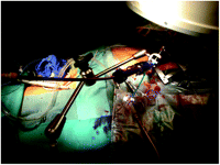
Intraoperative view of the introduction sheath fixed to the third hand.
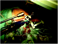
Intraoperative close view of the introduction sheath fixed to the third hand.
The catheter with the crimped valve is then inserted and positioned under fluoroscopic guidance, being double verified with transesophageal echo. When starting rapid pacing, a root injection is performed, to verify the correct positioning or to adjust the valve finally by slightly moving the catheter, if needed. During the same rapid pacing period the valve is deployed, by inflation of its balloon.
3. Results
This technique was successfully performed in seven consecutive patients (mean age 80.1±4.9 years; four male) undergoing TA-TAVI. Three of them had undergone aortocoronary bypass grafting in the past. One patient had a degenerated aortic valve bioprosthesis, which indication performing a valve-in-valve procedure. Patients presented with a mean logistic EuroSCORE of 44.7±18.8. After insertion of the 26 Fr sheath into the left ventricle, the sheath was fixed to the third hand and the proper axial alignment was chosen. Throughout the whole implantation process, the sheath stayed stable without any movement.
In all patients, the placement of the Sapien valve was positioned and implanted correctly.
4. Discussion
Due to the strict educational and learning process, a TAVI program can be safely implemented in cardiac surgery [4]. However, several differences compared to conventional aortic valve replacement are evident. Surgeons particularly are not used to handling catheters routinely. In the beginning, most attention therefore is paid to all catheter movements and the stable position of the introduction sheaths is sometimes neglected. In addition, surgeons often have to cope with unfavorable positioning of the screens for hemodynamics, echo and X-ray. This leads to a rotated body position of the surgeon; while watching the screen on their left-hand side, the devices have to be moved in the opposite direction. This most likely cause unexpected movements of the sheath.
Furthermore, the proximity of the holding hand to the X-ray beam and the position of the surgeon close to the chest result in an unnecessary X-ray load.
For this reason we developed a method to overcome these shortcomings.
This device enables the introduced sheath to stay stable throughout the whole implantation procedure. It allows positioning the sheath in a correct axial direction as needed, abolishing the risk of movement of the surgical assistant. Thus, the implanting surgeon can freely use both hands for guiding wires as well as catheters and positioning the aortic valve. The catheter on which the crimped valve is mounted can be moved easily throughout the whole procedure. In the case of an unexpected situation, the screws of the instrument holder can be loosened in seconds, detaching the sheath immediately.
Though the TAVI procedure is well established, it has to be noted, that there are several different concepts for the workflow during the procedure: in some centers the sheath is held by the surgical assistant, the surgeon handles the catheter and the guide wire and the cardiologist inflates the balloon. Other surgeons are in favor of holding the sheath by themselves with the right hand, positioning the valve with the left hand. The surgical assistant look after the guide wire and inflates the balloon. We follow the later strategy. Additionally, for both situations, the radiation load to the hands is minimized.
This technique is also helpful for new as well as experienced centers to reduce movement, X-ray load and personnel.
