


Abstract
Aortic cross-clamping during cardiac operations may injure the vessel wall and cause tissue lesions. This experimental study analyses the influence of the intravascular and external pressure and the duration of aortic cross-clamping on endothelial tissue damage. Fresh porcine aortas (n=20) were tested with intravascular pressures from 30 to 80 mmHg. The external clamp pressure, necessary to occlude the aorta, was applied by using the second cog of a commercial aortic clamp and cross-clamping was performed for 1 and 30 min. The observed pressure curves were compared to the histological findings. For occlusion of the aorta, an external pressure of at least 10-fold higher than the intravascular pressure (max. 812 mmHg) had to be applied. After 30 min of clamping, a complete endothelial destruction was observed, irrespective of intra-aortic pressure. The aortic media remained intact. After 1 min clamping, fractions of intact endothelial cells were left, ranging from 40 to 70% at different intra-aortic pressures. These results indicate that endothelial tissue lesions due to aortic cross-clamping are not avoidable, even in moderate clamp application. The duration of aortic cross-clamping but not intravascular pressure is the pivotal factor. The integrity of the aortic media can be preserved if low-force cross-clamping is achieved.
1. Introduction
In the majority of heart operations, it is necessary to clamp the ascending aorta below the aortic arch (‘aortic cross-clamping’) to isolate the heart from circulation. Some studies suggested that aortic cross-clamping causes ‘new’ lesions in the pinched off area [1, 2]. Every time a vessel is manipulated, the possibility of plaque rupture, local intimal damage like intimal tears and flaps, and thrombus formation, related to activation of the coagulation system during and after the clamping has to be considered. Evidence suggests that aortic manipulation, particularly the application or removal of the aortic cross-clamp, may dislodge atheromatous, highly prothrombotic material from the ascending aorta, with the potential hazard of embolization to the brain and other organs [3]. Additionally, preexisting damage of the vessel influences the dimension of the lesion [4]. Further important factors which may influence the dimension of the clamp lesions are the clamp duration [5], the intravascular aortic pressure at the time of cross-clamping and the external pressure applied by the clamp [6, 7]. The aim of this study was to evaluate the influence of the cross-clamping duration, the internal aortic pressure as well as the external pressure applied by the clamp on the aortic tissue especially on the aortic endothelium during heart operations.
2. Methods
Twenty hearts were harvested from freshly sacrificed pigs (80–100 kg, age <1 year) at a local abattoir. The aorta and the aortic valve were mobilized and separated from the rest of the heart. Small vessels as well as the brachiocephalic trunk, the subclavian and carotid arteries were ligated and the descending aorta was shortened to a length of ∼10 cm distal to the left subclavian artery. For experimental testing, a Forgarty clamp (Ulrich Medical, Ulm, Germany) incorporating a Hydrojaw Insert Set Hydra 86 (Edwards Lifesciences, Irvine, CA) was equipped with four strain gauges to measure the forces at the clamp during cross-clamping. By means of a closed fluid-filled rubber dummy of the aorta, these forces were calibrated to values of pressure, for better correlation. The experimental set-up consisted of a water reservoir connected to the distal end of the descending aorta. By adjusting the height of the reservoir the pressure could be varied, achieving intravascular aortic pressures of 30, 40, 50, 60, 70 and 80 mmHg for the experiments. The clamp was set at the ascending aorta between brachiocephalic trunk and coronary arteries. The exact location of the clamp was marked for later histological examination. The aorta was clamped at each pressure for 1 and 30 min, respectively, using the first two cogs (of seven) of the clamp. After the measurements the ascending aorta was opened longitudinally and segments of the marked areas were separated using a two-bladed knife. An unclamped reference sample was taken from each aorta and was treated the same way. After fixation, the tissue samples were incorporated into paraffin and cut into 4 μm slices with a microtome. The slices were stained by Hematoxylin/Eosin, Masson-Goldner and Richardson. The samples were analysed by microscope and thermionic microscope. Photographs were taken using an Axio Cam (Carl Zeiss MicroImaging GmbH, Göttingen, Germany).
Quantitative analysis was done by counting the intact endothelial cells along a marked length of 500 μm cross-sectional endothelial tissue at the clamped samples and the unclamped reference. The relation between the numbers of cells of clamped to unclamped is expressed as percentage of intact endothelium.
3. Results
With the current test setup as well as with the majority of commercially available aortic clamps, a continuous increase of external pressure could not be applied. Occlusion of the clamp and thus the resulting external pressure was only possible at discrete niveaus, depending on the number of engaged cogs.
With clamp occlusion at the first cog, leakage of water occurred at an intravascular pressure of 60 mmHg or more. Occlusion of the clamp at the second cog resulted in water-tight occlusion of the aorta at every intravascular pressure. Fig. 1 shows an example of the external pressure course during aortic clamping. Each cog occlusion of the clamp leads to a pressure peak followed by a constant plateau below the peak value. The mean external pressure at the second cog was 554.8±75.1 mmHg (range: 411–811 mmHg). Variation of the intravascular pressure between 30 and 80 mmHg did not affect the external pressure.
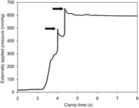
Example plot of the externally applied pressure. The arrows indicate the first and second cog occlusion of the clamp, followed by a plateau below the peak value.
Histologic analysis revealed that aortic clamping for even 1 min resulted in endothelial disruption and intimal detachment (Fig. 2 , left). The media and adventitia showed no abnormalities as compared to the unclamped reference probe. An influence of the intravascular pressure on endothelial integrity was visible when aortic clamping was done for 1 min. At intravascular pressures of 30 and 40 mmHg, >50% of the endothelial cells of the investigated area were left intact, whereas higher intravascular pressures (50–80 mmHg) resulted in an endothelial cell loss of >50% (P=0.002) of the basic value (Fig. 3 ). After application of the aortic clamp for 30 min no intact endothelium was visible, irrespective of the intravascular aortic pressure at cross-clamping (Fig. 2, right). The aortic media further turned out to be intact (Fig. 4 ).
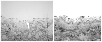
Cross-sectional samples of the aortic wall after clamping (Richardson staining). From left: partial disruption of the endothelial layer (aortic pressure 30 mmHg, 1 min clamping time) and complete destruction of the endothelial layer (aortic pressure 60 mmHg, 30 min clamping time).
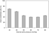
Percentage of intact endothelial cells at different intra-aortic pressures (30–80 mmHg) for a clamping duration of 1 min.
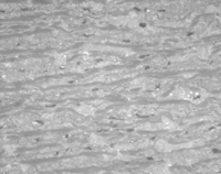
Cross-sectional sample of the aortic media after 30 min clamping (aortic pressure 80 mmHg, Richardson staining) with no visible abnormalities at the clamp area.
4. Discussion
The main finding of this study is that a certain amount of aortic tissue damage is unavoidable when cardiac surgery necessitates the use of aortic cross-clamping. Even in a moderate clamp application (using only the first two cogs – of seven – of the clamp) a complete endothelial disruption at the site of clamp application could be observed after 30 min clamp duration, irrespective of the intravascular pressure during clamping. The aortic media, however, could be preserved.
Several studies analysed the influence of the external clamping pressure on femoral and carotid arteries [6, 7]. The results suggest that there may be a threshold for histologic injury following vascular clamping. Consequently, the idea of a pressure-controlled and ballon-jawed vessel has been proposed to provide atraumatic vessel occlusion [6]. Transferring this idea to aortic cross-clamping, our results indicate that this concept will be of less importance because a continuous increase of external pressure cannot be applied with commercially available aortic clamps. Occlusion of the clamp and the resulting external pressure is possible only at discrete niveaus, depending on the number of engaged cogs. With clamp occlusion at the first cog, leakage of water occurs at an intravascular pressure of 60 mmHg and higher. To keep the aorta occluded an external applied pressure ∼10-fold greater than the intravascular aortic pressure was required. Hence, the external clamp pressure is determined by the aortic pressure and of little variability. While clamping of small muscular vessels with a pressure-controlled vascular clamp may avoid vascular damage, the ascending aorta needs a relatively high occlusion pressure (up to 810 mmHg).
The aortic pressure at the time of clamp application has not been investigated in relation to the cross-clamping time so far. Comparing the endothelial damage at different intravascular pressures for a clamping duration of 1 min, we detected that an amount of up to 70% of endothelial cells remained intact when the pressure in the aorta was below 40 mmHg at clamping. If the aortic pressure was above 50 mmHg, intimal damage was seen in the majority of investigated sections. However, if the same experiment was performed with a clamping period of 30 min, there were no intact endothelial cells detectable at the area of clamping, regardless of the aortic pressure at the application of the clamp. In the event that a cross-clamping period shorter than 30 min will rarely occur during cardiac surgery, the observed differences in endothelial damage in the investigated pressure range is without clinical relevance.
Depending on the aortic pressure at clamping, after 1 min only 70 to 40% of endothelial cells remained intact, hence removal and re-positioning of the clamp as performed in intermittent aortic cross-clamping [8, 9] seems to aggravate damage in comparison to single cross-clamping. Concluding from our results, each repositioning of the clamp is expected to cause new lesions. This is in agreement with an investigation demonstrating histologic changes immediately after clamping [7].
However, aortic tissue damage due to clamping may not result in permanent weakness of the vessel. Dobrin described that the observed lesions in the pinched off area persisted for at least six months but were not associated with chronic mechanical changes [7]. First, endothelial proliferations after traumatic injury occur after two days [10]. Regeneration of the endothelium occurs between 5 and 14 days [10–13], depending on the dimension and extent of the lesion. Several mechanisms for endothelial regeneration are discussed including migration of endothelial cells from the lesion verges and circulating progenitor cells [14, 15]. Also, in our experiments, the aortic media showed no microscopically visible abnormalities, assuming that the structural integrity of the aortic wall could be preserved if a slightly moderate (‘just enough’) cross-clamping is performed.
A limitation of the study should be taken into account. The investigations had been done with aortas from young and healthy animals, which do not represent pathological conditions of aortic tissue. However, in a diseased aorta, the observed tissue lesions will be presumably more pronounced, so much more care should be taken during aortic cross-clamping in such cases.
Summarizing the results, endothelial damage during aortic cross-clamping could not be avoided, mainly due to the high external pressure applied by the clamp even in moderate clamp application, whereas the media remained intact in low-level clamping. Endothelial lesion is dependent to clamp duration. The lesion is complete after 30 min of clamping. The intravascular aortic pressure at cross-clamping is of minor importance, whether a reduction of intra-aortic pressure may be helpful to reduce the risk of embolization remains to be investigated.
