


Background: Leadless pacemaker systems are an important upcoming device in clinical rhythmology. Currently two different products are available with the Micra system (0.8 cubic centimetres, 2.0 g capsule with 25.9 mm in length and an outer diameter of 6.7 mm) being the most used in the clinical setting to this date. The possibility to perform magnetic resonance imaging (MRI) is an important feature of modern pacemaker devices.
Even though the Micra system is suitable for MRI, little is yet know on its impact on artifacts within the images.
Purpose: The aim of our ex vivo study was to perform cardiac MRI to quantify the appearing artifacts in size and to evaluate if artifacts limit or inhibit the assessment of the surrounding myocardium.
Methods: After ex vivo implantation of the leadless pacemaker (LP) in porcine model, hearts were filled with saline solution and fixed on wooden sticks upon a plastic container. The model was examined at 1.5 and at 3 Tesla using common conventional sequences and T2 mapping sequences as well. In addition, conventional X-rays and a CT scans were performed.
Results: Correct implantation using the steerable device was feasible in all porcine hearts used in our ex vivo study. The figure shows MR images in "cine-like" sequences (SSFP-Sequence), here, the Micra pacemaker system produces a "shamrock-shaped" artifact that masks a small focal area without compromising the surrounding myocardium. The occurring artifact was larger at 3 Tesla (17.1% in long axis and 25.2% in short axis, expressed as % of total myocardial area) than compared to images obtained at 1.5 Tesla (14.8% in long axis and 17.8% in short axis).
A similar result was found for the scar-sequence. The late enhancement (scar-) sequences were slightly more prone to artifacts at 3.0 Tesla, leaving the right ventricle and the septum affected by a bright, hyperintense perifocal rim (12.4% and 12.1% at 1.5 Tesla vs. 11.9% and 14.9% at 3.0 Tesla). However, a large proportion of the left ventricular myocardium still remained accessible for image analysis. Similar findings were generated at 1.5 T. Only the perifocal hyperintense rim artifacts in the Turbo-spin-echo (TSE) sequences with selective fat suppression (SPIR) were visually slightly smaller (10.2% vs. 19.9%). No relevant differences were found in perfusion and T2 map sequences.
Conclusions: The use of the Micra system in cardiac MRI appeared to be feasible. We think that performing the most common sequences in Micra patients is possible, even though the right ventricle and the surrounding septal region might be overshadowed by occurring artefacts. In our opinion, the most important clinical MRI evaluations (the detection of major ischemic areas or inflammatory processes) will still be possible in Micra patients. We suggest the use of 1.5 Tesla will be the preferred method in clinical practice.
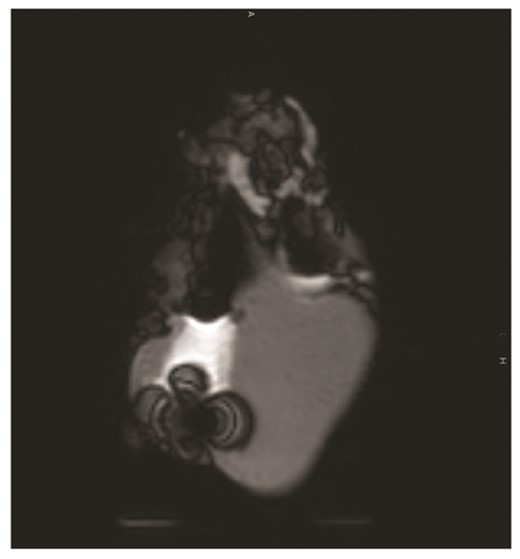
Abstract P437 Figure.
