


43
The features of cardiac electrical and structural remodeling in elite soccer athletes.
Abstract
Background: Regular, intensive exercise like soccer results in physiological biventricular cardiac adaptation. Ethnicity is an established determinant of structural and electrical remodeling of heart. Furthermore, cardiac remodeling has not been characterized in Asian.
Methods: We assessed clinical profile and cardiac dimensions and function in 45 national soccer player (53% male; mean age 25.4 ± 4.1 years) of south Korea, free of cardiovascular disease, participating in endurance disciplines, who experienced particularly intensive and uninterrupted training for 10 to 20 years.
Results: 45 national football team players were evaluated by use of ECG and echocardiography. Results were compared with 180 sedentary control subjects without any medical illness. There were no significant differences in left ventricular parameters between elite athlete and control subjects. Subjects of both group exhibited similar LV dimensions. But, RV dimensions were significantly smaller in controls than in athletes (proximal outflow tract, 27.9±5.5 versus 30.8±5.3 mm, P<0.001; longitudinal dimension, 81.6±9.5 versus 85.8±9.6 mm, P<0.001). RV enlargement compatible with diagnostic criteria for arrhythmogenic RV cardiomyopathy was observed in Elite athlete. Anterior T-wave inversion was present in 14.3% of athletes versus 3.7% of controls (P<0.001). Marked RV enlargement with concomitant anterior T-wave inversion was observed in 5.0% of athletes versus 1.8% of controls (P=0.01).
Conclusions In young elite soccer athletes, endurance training over long periods of time (up to 20 years) was not associated with deterioration in LV function, significant changes in LV morphology. However, RV dimension and electrical remodeling are prominent in soccer player, a little different from previous report containing white or black athlete.
44
Electrocardiographic findings in unselected Chinese adolescents during preparticipation screening
Abstract
Purpose: Resting electrocardiogram (ECG) is used for detecting silent heart disease during preparticipation screening of adolescents.
Methods: Medical history, physical examinations and 12-lead resting ECG were performed in 19,389 adolescents. Those with positive findings were further examined by echocardiography and Holter.
Results: ECG abnormalities were detected in 2,947 (15.3%) participants. Thirty (0.2%) students were diagnosed as WPW syndrome and 1,155 (6.0%) as early repolarization. Eight participants were suspected as long QT syndrome (QTc ≥480 ms) and another 11 had suspected short QT syndrome (QTc ≤330 ms)(see table). There were ectopic cardiac rhythm (n=121, 0.3%), and severe sinus arrhythmia (resting heart rate <50 beats/min) (n=126, 0.6%). T-wave inversion was detected in 24 adolescents in right precordial leads (V1–V3) and 5 in lateral precordial leads (V4–V6).
Conclusion: The prevalence of abnormal ECG patterns is relatively high in the study cohort.
Table Electrocardiogram abnormalities
| 14–16y (n=9705) | 17–20y (n=9684) | P value (age) | |
|---|---|---|---|
| Severe sinus bradycardia (RHR <50 bpm) | M 31 (0.6%) | M 61 (1.2%) | 0.001 |
| F 12 (0.2%) | F 22 (0.5%) | ||
| D 0.004 | D 0.001 | ||
| Prolongated QTc (QTc >480 ms) | M: 2 | M 1 | NS |
| F: 4 | F 1 | ||
| D NS | D NS | ||
| Short QTc (QTc <330 ms) | M 2 (0.04%) | M 1 (0.02%) | NS |
| F 4 (0.08%) | F 4 (0.09%) | ||
| D NS | D NS | ||
| Type A WPW | M 4 (0.1%) | M 5 (0.1%) | NS |
| F 2 (0.0%) | F 5 (0.1%) | ||
| D NS | D NS | ||
| Type B WPW | M 4 (0.1%) | M 4 (0.1%) | NS |
| F 4 (0.1%) | F 2 (0.0%) | ||
| D NS | D NS | ||
| Early repolarization pattern | M 417 (8.5%) | M 602 (11.4%) | 0.001 |
| F 95 (2.0%) | F 41 (0.9%) | ||
| D 0.001 | D 0.001 |
D=Difference in frequency between girls and boys.
45
Prognostic significance of an automated ECG QRS score quantifying myocardial scar burden in patients presenting with symptoms suggestive of acute myocardial infarction
Abstract
Introduction: The Selvester QRS score estimating myocardial scar burden from the 12-lead ECG has been shown to be a predictor of cardiovascular mortality, but is sophisticated and impractical for manual calculation. We hypothesized that an automated version of the QRS score might be used to risk stratify patients presenting with symptoms suggestive of acute myocardial infarction (AMI).
Methods: We prospectively enrolled 1711 consecutive patients with symptoms suggestive of AMI. The QRS score was automatically calculated from a digital 12-lead ECG's recorded at presentation to the ED. The primary endpoint was all-cause mortality during 3 years of follow-up.
Results: AMI was the final diagnosis in 19% of patients, with 3% having STEMI and 16% NSTEMI. Median QRS Score was higher in patients with AMI compared to those without (5 (IQR 2-8) vs. 3 (IQR 2-7), p<0.001). More extensive myocardial scar as indicated by a higher automated QRS score was significantly associated with higher mortality after 3 years (Survival rates 93%, 91%, 86% for patients with a QRS score <5, 5-9 and >=10, p<0.001).

47
Clinical importance of lead aVR in arrhythmogenic cardiomyopathy
Abstract
Arrhythmogenic cardiomyopathy (AC) is a cardiac entity with prominent right ventricular involvement. Lead aVR is a limb lead directed to the right ventricle thus producing suspicious changes. Other ECG criteria such as right precordial T inversions, epsilon waves and localized right precordial QRS prolongation are unspecific, rare findings and only documented in ECG writing techniques of 50mm/s that is unusual in most countries. QRS fragmentation is an ECG criterion recently published in arrhythmogenic cardiomyopathy that is described in a setting of other diseases. The value of QRS fragmentation in arrhythmogenic right ventricular cardiomyopathy is completely unspecific.
Method: In a cohort of 385 patients with typical arrhythmogenic cardiomyopathy (212 males, mean age 46.3 ± 13,1 years) the electrocardiographic morphology in lead aVR was analysed. Localized right precordial QRS prolongation was seen in these patients in 98%, T wave inversions were found in 55%, typical epsilon waves in 24% and QRS fragmentation in 85% of cases. 1496 probands without heart disease (859 males in a age range of 18–82 years) were evaluated as a control group.
Results: In patients with typical ARVC a deep Q wave of 0.3 mV or more, a small R wave 0.2mV or less and inverted T waves in were found in 373 cases (97%). In the control group the same QRS and T wave morphology was present in 284 probands (18.9%). Specificity was low, but negative predictive value was nearly 100%. Conclusion: ECG changes in lead aVR could be identified in 97% of cases presenting with a deep Q wave of 0.3 mV or more, a small R wave 0.3 mV or less and inverted T waves representing electroanatomic scar and myocardial atrophy of the right ventricle. ECG abnormalities in lead aVR represent another significant marker of AC when other ECG markers such as epsilon waves and right precordial T inversions are present.
48
Fragmented QRS complexes in patients with hypertrophic cardiomyopathy: a marker of myocardial fibrosis detected by cardiac magnetic resonance imaging with gadolinium enhancement
Abstract
Purpose: Fragmented QRS complexes (fQRS) have been shown to be a sign of myocardial fibrosis/scarring and subsequent depolarization abnormality in patients with dilated cardiomyopathy, cardIac sarcoidosis and repaired cardiac tetralogy. The aim of this study was to evaluate the association between the fQRS and the late gadolinium enhancement (LGE) on CMR in patients with HCM.
Methods: The 12-lead ECGs of 191 patients with HCM who underwent CMR with gadolinium were analysed for the presence of fQRS. fQRS was defined as the presence of additional deflections on the beginning or top of R wave (R′), or notching/fragmentation in the nadir of the R or S wave in 2 contiguous leads. Patients with typical bundle branch block pattern and/or with QRS ≥ 120 ms (n=31) were excluded from analysis.
Results: Of the remaining 160 patients, 64 (40%) had fQRS on 12-lead ECG and 102 (63.8%) had LGE on CMR. Patients with and without fQRS were of similar gender (69% vs. 73% male respectively, p=0.52) and age (56±16 vs. 57±14 years respectively, p=0.78). LGE was significantly more prevalent in patients with fQRS complexes than patients without fQRS complexes (n=47, 73% vs. n=55, 57%, p=0.037). The positive predictive value of fQRS for LGE on CMR was 73.4%, with a specificity of 70.6%, sensitivity of 46% and negative predictive value of 42.7%. Patients with fQRS complexes had also longer QRS duration (101ms± 16ms vs. 92ms±13ms, p=0.001) supporting depolarization abnormality/delay in these patients.
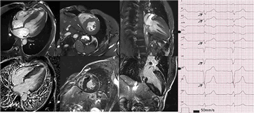
fQRS complexes and LGE on CMRI
49
Electrocardiographic features of disease progression in arrhythmogenic right ventricular cardiomyopathy/dysplasia
Abstract
Introduction: Arrhythmogenic right ventricular dysplasia (ARVC/D) is considered a progressive cardiomyopathy. However, data on the clinical features of disease progression are limited. The aim of this study was to assess 12-lead surface electrocardiographic (ECG) changes during long-term follow-up, and to compare these findings with echocardiographic data in patients with ARVC/D.
Methods: Baseline and follow-up ECGs (25 mm/s, 0.1mV/mm) of ARVC/D patients from three Swiss tertiary care centers were recorded at standard lead positions, analyzed with digital calipers by two blinded observers, and correlated with findings from transthoracic echocardiography.
Results: A baseline ECG was available in 111 patients. The median follow-up was 1476 days (IQR 697-3354). ECG progression was significant for epsilon waves (baseline 14% vs. follow-up 31%, p=0.01) and QRS duration (111ms vs. 114ms, p=0.04; Figure). Six patients with repolarization abnormalities according to the 2010 Task Force Criteria (TFC) at baseline did not display these criteria at follow-up, whereas in all patients with epsilon waves at baseline these depolarization abnormalities also remained at follow-up. TWI in inferior leads were common (36% of patients at baseline), and were significantly associated with major repolarization abnormalities (p=0.02), TWI in lateral precordial leads (p=0.05), and definite ARVC/D (p=0.05).

50
Prevalence and prognosis role of wide QRS and of QRS narrower than normal
Abstract
Introduction: Very narrow QRS has been described whose prevalence and clinical relevance in the normal adult population is unknown.
Methods: 546 healthy men between 50 and 60 yo (group 1) and 373 similar patients with coronary artery disease (368 men, EF < 50% in 40%, group 2) underwent signal averaged ECG allowing precise measurement of QRS duration. Patients and subjects with bundle branch block were excluded. All cause mortality was determined after 17±3 years follow-up.
Results: Mean QRS duration was 97±13 ms for group 1 (67-164) and 103±16 ms (71-171) for group 2. 85/546 group 1 subjects (16%) had QRS < 85 ms and 23/546 (4%) had QRS > 120 ms. Tenth percentile was 84 ms and 90th percentile was 111 ms. 44/373 group 2 patients (12%) had QRS < 85 ms and 44/373 (12%) had QRS > 120 ms. QRS were larger in case of lower EF, lack of previous angioplasty and multivessel disease.
All cause mortality in group 1 was 10,4 % (57/546): 6/85 in case of QRS < 85 ms (7%) and 2/23 (9%) in case of QRS > 120 ms (p=ns compared to normal QRS duration). HR for all-cause mortality in case of QRS < 85 ms was 0,75 (95 % CI 0.32-1.76, p = 0,52) and 0,86 (95% CI 0.21-3.53, p = 0,84) for QRS > 120 ms.
All cause mortality in group 2 was 29 % (109/373): 7/44 in case of QRS < 85 ms (16%) and 22/44 (50%) in case of QRS > 120 ms (p=0.002 when compared to normal QRS duration). Adjusted HR for all-cause mortality in case of QRS < 85 ms was 0,65 (95 % CI 0.29-1.45, p = 0,29) and 1.73 (95% CI 1.02-2.94, p = 0,05) for QRS > 120 ms.
Late potentials (LP) were considered present (2 positive of 3 criteria) in SA-ECG in 116/546 group 1 subjects (21%). LP were present in 100/373 group 2 patients (27%) and were significantly related to multivessel disease, altered EF, lack of revascularization or of angioplasty. LP were more frequently observed in case of QRS > 120 ms in both groups. LP were nor related to all-cause mortality in both groups.
Conclusion: QRS “narrower than normal” (< 85 ms) can be observed in a significant proportion of healthy males between 50 and 60 years old and in similar proportion of patients with ischemic heart disease. In opposition to QRS > 120 msec which are independantly related to a higher all-cause mortality in coronary artery disease patients, QRS < 85 ms were not linked to prognosis in any group.
51
Electrocardiographic features in patients presenting with non-ischaemic cardiomyopathy and ventricular tachycardias
Abstract
Purpose: In patients with non-ischaemic cardiomyopathy (NICM) and ventricular tachycardias (VTs), two different scar patterns, antero-septal and infero-lateral, were identified on the basis of magnetic resonance imaging (MRI) and voltage map studies. Additionally, patients showed different degree of left ventricular (LV) impairment, which were termed early and dilated cardiomyopathy.
We investigated whether widely available 12-lead electrocardiogram (ECG) may help to determine scar location, without the necessity of expensive and invasive tests, such as MRI and electranatomical mapping.
Methods: The baseline 12-lead ECGs of 108 consecutive patients with NICM undergoing catheter ablation for drug-refractory VTs were analyzed. Seventy-two fulfilled diagnostic criteria for dilated cardiomyopathy (left ventricular ejection fraction, LVEF <45%) and 36 for early cardiomyopathy (LVEF >45%). On the basis of low voltage areas (<8mV) on unipolar voltage map, the scar was classified as predominantly antero-septal (n=59) or infero-lateral (n=49).
Results: In case of early cardiomyopathy, a 3-step algorithm including PR interval <170 ms or QRS voltage in all inferior leads <0.6 mV or the presence of q wave in lateral leads identified infero-lateral scar with 92% of sensitivity and 90% of specificity, respectively.
In patients with dilated cardiomyopathy, a 4-step algorithm including paced ventricular rhythm or PR duration >230 ms or QRS duration >170 ms or r wave ≤0.3 mV in lead V3 in case of narrow QRS, identified antero-septal scar with 92% and 81% of sensitivity and specificity, respectively.
The two algorithms were further verified in a validation cohort (constituted by 30 patients), with the 3-step algorithm reaching 80% of sensitivity and 100% of specificity and the 4-step algorithm 100% and 85% of sensitivity and specificity, respectively.
Conclusions: In patients with NICM presenting with VTs, baseline 12-lead ECG analysis allows determining the location of the VT-related scar both in case of preserved and impaired LV function with good accuracy.
52
Electrocardiographic criteria and outcome in patients with arrhythmogenic right ventricular cardiomyopathy
Abstract
Current literature in arrhythmogenic right ventricular cardiomyopathy shows that electrocardiographic markers such as epsilon waves and the amount of T wave inversions of electroanatomic scar size predicts arrhythmic risk. The amount of T wave inversions with increasing electroanatomic scar size were normal T waves, negative T waves in V1 to V3, negative T waves in V1–V3 extending to lateral leads and negative T waves in both precordial and inferior leads. This study was conducted in a large number of patients to correlate ECG findings to the outcome of each patient.
Method: In 321 patients (207 males, mean age 46.7 ± 11.3 years) the amount of T-wave inversions and outcomes of the patients were correlated. T-wave inversions in 4 leads or more (high risk group) were found in 61 patients and T-wave inversions in a maximum of 3 leads (so-called low risk group) could be revealed in 260 patients.
Results: In 38 out of 61 patients with T-wave inversions in 4 or more leads recurrent ventricular tachycardia or ventricular fibrillation occurred. In the low risk group at least 35 out of 260 patients were characterized by ventricular tachycardia or ventricular fibrillation. In the high risk group VT/VF were present in 62%; in the low risk group VT/VF occurred in 15%. These results were highly statistically significant with a p value < 0.000001. Specificity was high with 86%; negative predictive value was higher with 90%. Conclusions: It is known that the amount of T-wave inversions characterizes scar size thus predicting arrhythmic risk. In this analysis it was shown that the amount of T-wave inversions had a strong correlation to outcomes of the patients. T-wave inversion is an excellent parameter to predict ventricular tachycardia or ventricular fibrillation in the course of the patients with arrhythmogenic right ventricular cardiomyopathy.
