


Abstract
Exercise and dobutamine stress may induce acute left atrial volume index (LAVI) dilatation in 1 out 10 patients. The effect of vasodilator stress in LAVI remains unknown.
To assess the feasibility and functional correlates of LAVI change during dipyridamole stress echocardiography (SE).
We studied 149 patients (99 male, age 66±10 years, ejection fraction 59±8%, 64 with previous myocardial infarction), who underwent dipyridamole ABCDE-SE. LAVI was measured with the biplane disk summation method at rest and peak stress: LAVI-dilators were defined as those with stress-rest increase ≥6.8 ml/m2. Criteria for abnormal response of ABCDE-SE were: stress-induced changes in regional wall motion abnormalities (RWMA) for step A; B-lines at peak stress ≥2 for step B (4-site simplified scan, each site scored from 0= A-lines or black lung to 10= white lung for coalescing B-lines); reduced left ventricular contractile reserve (LVCR, peak/ rest based on force) ≤1.1 for step C; abnormal coronary flow velocity reserve (CFVR) ≤2.0, assessed by pulsed wave Doppler sampling in left anterior descending coronary artery for step D; abnormal heart rate reserve (HRR, peak/rest heart rate) ≤1.22 for step E.
LAVI dilation occurred in 13 patients (9%). The positivity was for step A: RWMA in 6 pts (4%); step B: presence of peak B-lines in 12 pts (8%); step C: abnormal LCVR in 14 pts (9%); step D: reduced CFVR in 17 pts (11%) and step E: abnormal HRR in 46 pts (31%). LAVI dilators showed significantly higher incidence of positivity of steps A-B-C-D (see figure) compared to patients without LAVI dilatation.
Evaluation of LAVI change during vasodilator SE is feasible, and LAVI dilatation is more frequently found with ischemic (step A), wet (step B), weak (step C) and cold (step D) heart.
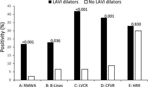
Figure 1
Type of funding source: None

