


Abstract
Fractional flow reserve (FFR) is recommended to guide revascularization of native coronary arteries. Revascularization decision-making for saphenous vein grafts (SVGs), however, is based on angiographic estimation of lesion severity, as studies exploring the value of FFR to detect perfusion imaging defined myocardial ischemia in SVGs are scarce.
We aimed to define an optimal FFR threshold and compare its diagnostic performance with the traditional FFR cutoff and with percentage diameter stenosis (%DS) in patients evaluated for SVG disease using myocardial perfusion imaging (MPI) as the reference standard.
Symptomatic patients with prior coronary artery bypass grafting (CABG) who underwent single-photon emission computed tomography, positron emission tomography or stress perfusion cardiac magnetic resonance imaging, and had FFR measurements of one or more SVGs were included. Angiographic lesion severity of the SVG was measured using 2D quantitative invasive coronary angiography. We allocated and matched the myocardial territory subtended by the SVG to compare the values of FFR and %DS with ischemia determined by MPI.
Included were 77 patients (mean age 72 ± 7 years; 86% male) with 91 SVGs. A total of 38 SVGs (42%) supplied an ischemic coronary territory. FFR and %DS correlated moderately (r= -0.51, P<0.001). The optimal cutoff value of FFR was ≤0.94, resulting in a sensitivity, specificity, positive predictive value (PPV), negative predictive value (NPV) and accuracy of 63%, 75%, 65%, 74% and 70%, respectively (Figure 1). Corresponding values of FFR≤0.80 and DS%≥50 are shown in Figure 1 as well. Sensitivity, NPV and accuracy of FFR ≤0.94 outperformed that of FFR≤0.80, whereas specificity was lower. FFR≤0.94 outperformed %DS≥50 only in terms of sensitivity.
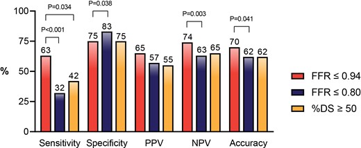
Diagnostic performance of FFR and %DS
Author notes
Funding Acknowledgements: None.

