


Abstract
Assessments of coronary disease activity with 18F-sodium fluoride positron emission tomography (18F-NaF PET) and radiomics-based precision coronary plaque phenotyping derived from contrast-enhanced computed tomography (CT) have both been shown to enhance risk stratification in patients with coronary artery disease (CAD). To date, no study has investigated whether these two promising methods (which can be obtained during a single imaging session on a hybrid PET/CT scanner) are interchangeable or can provide superior predictive performance when used in combination.
We sought to investigate whether the prognostic information provided by latent morphological radiomic coronary plaque features and assessments of disease activity by 18F-NaF PET are complementary in prediction of myocardial infarction.
Patients with known CAD underwent coronary 18F-NaF PET and CT angiography on a hybrid PET/CT scanner. Coronary 18F-NaF uptake was determined by the coronary microcalcification activity (CMA). We performed quantitative plaque analysis of coronary CT angiography datasets. Additionally, coronary plaque segmentations on CT angiography were used to extract 1103 radiomic features. Using weighted correlation network analysis we derived latent morphological features of coronary plaques which were aggregated to patient-level radiomic normograms to predict myocardial infarction using univariate and multivariate Cox proportional hazard models.
The study cohort comprised of 260 patients with established CAD (age: 65±9 years; 84% men); 179 (69%) participants showed increased coronary 18F-NaF activity (CMA >0). Over 53 [40–59] months of follow-up 18 patients had a myocardial infarction. Using weighted correlation network analysis, from the 1103 radiomic features we derived 15 distinct eigen radiomic features representing latent morphological coronary plaque patterns. On univariate cox modelling 7 of these emerged as predictors of myocardial infarction (Figure). Following adjustments for calcified, noncalcified and low-density noncalcified plaque volumes and 18F-NaF CMA 4 radiomic features (related to texture and geometry) remained independent predictors of myocardial infarction (Figure).
In patients with established CAD latent morphological features of coronary plaques are predictors of myocardial infarction above and beyond plaque volumes and 18F-NaF uptake. Comprehensive plaque analysis with radiomics may enhance risk stratification of CAD patients.
Type of funding sources: Public grant(s) – National budget only. Main funding source(s): NIH, Wellcome Trust
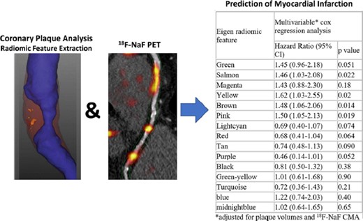
Figure 1

