


Abstract
The developmental impact of maternal diabetes on the fetal left atrium (LA) is unclear.
To determine if maternal diabetes mellitus (DM) impacts fetal LA size and function (fetal LA strain (LAS)).
We evaluated LA area (LAA) and LAS on fetuses of diabetic and control mothers who attended a mandated 24 week fetal morphology scan. Participants were excluded from the study if: there was a history of pre-eclampsia or if the fetus did not have adequate images for LAS analysis
We used fetal cardiac 4-chamber view for analysis. A region of interest was drawn along the LA endocardial border for tracking and was used for assessment of maximum LAA.
Baseline variables were compared using Student t test or Mann-Whitney U test and are presented as Mean ± Standard Deviation or Median (Interquartile range (IQR)). Body mass index (BMI), maternal age, gestational age, fetal heart rate (FHR), smoking status, estimated fetal weight (EFW) and Maternal DM were analysed in univariate and multivariate models with respect to LAA and LAS.
160 pregnant women (50 controls, 110 diabetics) were scanned. 9 were excluded due to poor image quality, resulting in 104 mothers with diabetes (T1DM 9, T2DM 8, and gestational DM 87) and 47 controls without diabetes.
The mothers were well matched for age, blood pressure, smoking prevalence and gestational age. The diabetic mothers had a significantly higher BMI: Median (IQR) ((30.4 kg/m2 (25.1–34.8) vs 20.8 kg/m2 (21.4–27.4), p<0.001) and had higher weight (77 kg (65–93) vs 64 kg (62–68), p<0.001).
FHR was higher in fetuses of diabetic mothers (147±10 vs 144±8, p 0.04). Maternal DM resulted in larger LAA 1.68 cm2±0.39 cm2 vs 1.56 cm2±0.36 cm2; p=0. 08, however the result was not significant. The LAS was significantly lower in fetuses with maternal DM compared to fetuses of controls: 28.8% ± 8.8% vs 32.3% ± 9.2%; p 0.033.
On multivariate analysis (Table 1), the predictors of LAS were Maternal DM and FHR and predictors of LAA were EFW and Maternal DM.
Maternal diabetes modulates both LA size and LA function. The association between LA function and FHR may provide an explanation for fetal tachycardia in Maternal DM.
Multivariate models for LAS and LAA
| Function, LAS (%) | Structure, LAA (cm2) | |||
|---|---|---|---|---|
| Beta | p | Beta | p | |
| Maternal DM | −0.72 | 0.03 | 0.15 | 0.03 |
| FHR | −0.21 | 0.01 | – | NS |
| EFW | – | NS | 0.001 | <0.001 |
| Function, LAS (%) | Structure, LAA (cm2) | |||
|---|---|---|---|---|
| Beta | p | Beta | p | |
| Maternal DM | −0.72 | 0.03 | 0.15 | 0.03 |
| FHR | −0.21 | 0.01 | – | NS |
| EFW | – | NS | 0.001 | <0.001 |
DM, diabetes mellitus; EFW, estimated fetal weight; FHR, fetal heart rate; LAA, left atrial area; LAS, left atrial strain.
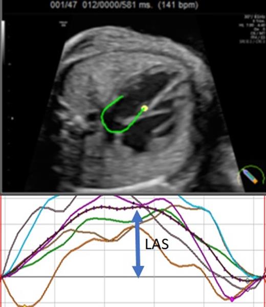
Fetal left atrial strain
Type of funding source: None

