


Abstract
To determine in a multicentre, multivendor trial the diagnostic performance for perfusion-cardiac magnetic resonance (perfusion-CMR) in comparison with coronary X-ray angiography (CXA) and single-photon emission computed tomography (SPECT).
Of 241 eligible patients from 18 centres, 234 were randomly dosed with 0.01, 0.025, 0.05, 0.075, or 0.1 mmol/kg Gd-DTPA-BMA (Omniscan™, GE-Healthcare) per stress (0.42 mg/kg adenosine) and rest perfusion study. Coronary artery disease (CAD) was defined as diameter stenosis ≥50% on quantitative CXA. Five CMR and eight SPECT studies (of 225 complete studies) were excluded from analyses due to inadequate quality (three blinded readers scored per modality). The comparison of CMR vs. SPECT was based on receiver operating characteristic (ROC) analysis. Perfusion-CMR at the optimal CM dose (0.1 mmol/kg) had similar performance as SPECT, if only the SPECT studies of the 42 patients with this dose were considered [area under ROC curve (AUC): 0.86 ± 0.06 vs. 0.75 ± 0.09 for SPECT, P = 0.12]; however, diagnostic performance of perfusion-CMR was better vs. the entire SPECT population (AUC: 0.67 ± 0.05, n = 212, P = 0.013).
In this multicentre, multivendor trial, ROC analyses suggest perfusion-CMR as a valuable alternative to SPECT for CAD detection showing equal performance in the head-to-head comparison. Comparing perfusion-CMR with the entire SPECT population suggests CMR superiority over SPECT, which warrants further evaluation in larger trials.
Introduction
The management of patients with known or suspected coronary artery disease (CAD) is ideally guided by documentation of myocardial ischaemia for optimal planning of medical therapy and/or revascularization.1–4 MR first-pass myocardial perfusion imaging (perfusion-CMR) has emerged as a sensitive and patient-friendly diagnostic modality to detect ischaemia, and several single centre5–9 and multicentre10,11 studies have yielded excellent results for CAD detection as defined by conventional coronary X-ray angiography (CXA). In particular, the high spatial resolution of perfusion-CMR allows detection of small, even subendocardial perfusion deficits.5,11,12 In a single centre study, perfusion-CMR was superior for detection of CAD in comparison with single-photon emission computed tomography (SPECT),13 and perfusion-CMR was similar in performance in comparison with positron emission tomography.5 In patients with acute chest pain, a normal perfusion-CMR study also had an excellent negative predictive value of 100% for subsequent diagnosis of CAD or an adverse outcome.14 However, the diagnostic performance of perfusion-CMR has not been compared with other established non-invasive diagnostic techniques in a large multicentre multivendor trial. Accordingly, in 18 centres in Europe and the US, perfusion-CMR was performed with five CM doses for the detection of CAD using CXA as the standard of reference and the diagnostic performance of perfusion-CMR at the optimum CM dose was compared with SPECT imaging.
Methods
Study design and patient population
This double-blind, randomized, phase II clinical trial was conducted at 18 centres in Europe and the USA. Eligible patients were those scheduled for a conventional CXA and/or a SPECT examination for clinical reasons. Before study entry, all patients had to agree to undergo all three tests (CXA, SPECT, and CMR). Patients were included if they had undergone CXA (being either positive or negative for the presence of CAD) or if they had undergone positive SPECT with scheduled CXA (a positive SPECT was required to guarantee adequate sensitivity calculations). The order of testing was determined by the availability of the infrastructure. SPECT was performed as first test in 32% of patients. Both, the CXA and the SPECT examinations must be performed within 4 weeks before or after the CMR examination. Exclusion criteria were acute myocardial infarction (MI) (≤1 week prior to study enrollment), a history of coronary bypass surgery, unstable angina pectoris, decompensated heart failure, any interventions on the coronary arteries in the time period between CXA, SPECT, and the perfusion-CMR examinations, and arrhythmias (considered to compromise quality of CMR imaging such as atrial fibrillation or frequent ectopic beats of >20 min−1). Patients in stable condition with a history of MI and/or percutaneous coronary interventions were not excluded from the study (see also Table 1). Additional exclusion criteria were any contraindications for adenosine (second or third AV-block, sick sinus syndrome, symptomatic bradycardia, severe asthma bronchiale or obstructive pulmonary disease), CM (known allergy), or CMR examination (implanted electronic devices, metallic foreign bodies in the eye, severe claustrophobia, and others according local regulations and manufacturer’s recommendations). For the CMR examination, all patients were randomly assigned (randomization over all subjects, not balanced by centre, using ClinPro/LBL system, Independent Review Center, GE Healthcare, USA) to one of five dose groups (0.01, 0.025, 0.05, 0.075, and 0.1 mmol/kg per stress and rest injection) of a conventional extracellular CM (Gd-DTPA-BMA, Omniscan™, GE Healthcare). (For more details on randomization, see Supplementary material online, Appendix in the online version of the article). For the patients having SPECT as their second or third examination (i.e. not first examination=unbiased SPECT population; n = 153), a sub-analysis was performed. The study was conducted according to the Declaration of Helsinki, the principles of Good Clinical Practice, and was approved by the Health Authorities and the local Ethics Committee of each participating institution. All patients gave written informed consent before study participation.
Demographics of population available for safety evaluation
| Dose 1 | Dose 2 | Dose 3 | Dose 4 | Dose 5 | |
|---|---|---|---|---|---|
| 0.01 (mmol/kg) | 0.025 (mmol/kg) | 0.05 (mmol/kg) | 0.075 (mmol/kg) | 0.10 (mmol/kg) | |
| Number | 45 | 50 | 48 | 45 | 45 |
| Male sex—no (%) | 32 (71.1) | 40 (80.0) | 33 (68.8) | 33 (73.3) | 32 (71.1) |
| Age—year | |||||
| Mean ± SD | 61.3 ± 9.4 | 60.6 ± 10.1 | 59.7 ± 10.3 | 61.3 ± 10.8 | 60.5 ± 10.6 |
| Range | 43.8–79.6 | 38.6–82.3 | 36.1–79.9 | 41.1–81.0 | 39.7–78.2 |
| BMI—kg/m2 | |||||
| Mean ± SD | 27.6 ± 3.6 | 26.7 ± 4.2 | 27.5 ± 4.4 | 27.6 ± 4.3 | 27.4 ± 3.9 |
| Range | 21.0–36.0 | 19.0–39.0 | 19.0–38.0 | 16.0–42.0 | 20.0–38.0 |
| Angina pectoris—n (%) | 38 (84) | 38 (76) | 38 (79) | 37 (82) | 36 (80) |
| Hypertens.—n (%) | 35 (78) | 35 (70) | 32 (67) | 31 (69) | 28 (62) |
| MI—n (%) | 19 (42) | 24 (48) | 14 (29) | 17 (38) | 16 (36) |
| PCI—n (%) | 12 (27) | 20 (40) | 14 (29) | 16 (36) | 10 (22) |
| CHF—n (%) | 6 (13) | 4 (8) | 6 (13) | 7 (16) | 8 (18) |
| QCA—n (%) | 45 (100) | 49 (98) | 47 (98) | 44 (98) | 45 (100) |
| CAD—n (%) | 35 (78) | 41 (84) | 36 (77) | 32 (76) | 33 (73) |
| MVD—n (%) | 20 (44) | 32 (65) | 22 (47) | 20 (48) | 20 (44) |
| LM—n (%) | 1 (2) | 2 (4) | 1 (2) | 2 (5) | 2 (4) |
| LAD—n (%) | 22 (49) | 35 (71) | 18 (38) | 25 (60) | 25 (56) |
| LCX—n (%) | 17 (38) | 24 (49) | 20 (43) | 18 (43) | 12 (27) |
| RCA—n (%) | 23 (51) | 31 (63) | 23 (49) | 16 (38) | 20 (44) |
| Any drugs—n (%) | 44 (98) | 49 (98) | 47 (98) | 42 (93) | 45 (100) |
| Beta-blockers—n (%) | 30 (67) | 37 (74) | 38 (79) | 36 (80) | 37 (82) |
| Statins—n (%) | 36 (80) | 34 (68) | 34 (71) | 27 (60) | 30 (67) |
| ACEI—n (%) | 21 (47) | 29 (58) | 21 (44) | 23 (51) | 20 (44) |
| Diuretics—n (%) | 9 (20) | 12 (24) | 13 (27) | 10 (22) | 5 (11) |
| Ca-CB—n (%) | 13 (29) | 11 (22) | 8 (17) | 10 (22) | 4 (9) |
| Antithromb.—n (%) | 34 (76) | 42 (84) | 40 (83) | 34 (76) | 38 (84) |
One patient received 0.037 mmol/kg Gd-DTPA-BMA and is therefore not represented in this table (but was included in the safety analysis and in Figure 2: dose group 4).
BMI, body mass index; MI, myocardial infarction; PCI, percutaneous coronary intervention; QCA, quantitative coronary angiography; CAD, coronary artery disease; MVD, multivessel disease; LM, left main; LAD, left anterior descending coronary artery; LCX, left circumflex coronary artery; RCA, right coronary artery; ACEI, angiotensin-converting enzyme inhibitors; Ca-CB, calcium channel blockers.
Efficacy measures
For CAD definition a ≥50% diameter stenosis in two orthogonal planes (≥75% area reduction) on quantitative coronary angiography was used as in previous studies5,11,15 present in ≥1 coronary artery. Quantitative assessment of stenosis severity was performed by a blinded reader in the core laboratory (Cleveland Clinic Foundation, Cleveland, USA). Only vessels of ≥2 mm diameter were considered for definition of CAD, since smaller vessels are rarely revascularized (e.g. no stents available for <2 mm vessels). Vessels with a history of percutaneous coronary interventions performed successfully prior to study entry (i.e. residual stenosis of <50% diameter reduction on quantitative CXA of the present study) were not defining the presence of CAD (even though such patients had a known positive history of CAD). The comparison of perfusion-CMR vs. SPECT for CAD detection was performed by means of receiver operating characteristic (ROC) analyses16–18 using CXA as the reference. Additional ROC analyses were performed in patients with multivessel disease (MVD) and in the gated-SPECT population.
Another efficacy measure was the accuracy (proportion of correct diagnoses per patient) for CAD detection by perfusion-CMR for the five CM doses (for details, see Supplementary material online, Appendix).
Cardiac magnetic resonance examination
In 1.5 T scanners (Siemens: 8, GE Healthcare: 7, Philips: 3), a breath-hold MR first-pass perfusion examination was performed. Three short-axis slices of 8–10 mm thickness were acquired every heart beat at 1/4, 1/2, and 3/4 of the left ventricular (LV) long axis to evenly space the slices along the long axis. The pulse sequences of all vendors used a non-slice selective 90°-preparation, a saturation—read-out delay time of ~120 ms, and read-out was achieved by a fast gradient-echo sequence (with an echo-planar component where available, whereas parallel imaging was not allowed) yielding a spatial resolution of 2–3 × 2–3 mm. Stress data were acquired following 3 min of adenosine (0.14 mg/min/kg intravenous). For the accuracy evaluation, a second CM injection was performed 20 min after the stress study allowing for a rest perfusion study (for details see Supplementary material online, Appendix). These additional acquisitions should enable differentiation between ischaemia and scar tissue, but these aspects are not specifically addressed in the current report.
CMR data were analysed visually by three blinded readers in an independent core laboratory (Independent Review Center, GE Healthcare, former Nycomed Amersham Imaging, Princeton, USA). All readers were experts in CMR and were blinded with respect to clinical information, the results of the other tests, as well as the order, in which these tests were performed. A 16-segment model (17-segment model19 minus the apical segment) was applied, since the vessels supplying the apex are <2 mm in diameter in most cases.20 Accordingly, no long axis acquisition for the apex was acquired.21,22 The stress perfusion data were analysed by ROC curves in order to assess test sensitivity and specificity simultaneously (without such a simultaneous specificity assessment, a high sensitivity and accuracy would be obtained simply by performing a highly sensitive reading in this population with relatively high disease prevalence).16,17 For the ROC analysis, the extent of CAD, i.e. the extent of perfusion abnormalities, was assessed on the stress perfusion data by grading the perfusion deficits in each of the 16 segments as abnormal with high confidence (3; defined as myocardium being black at the peak bolus), or abnormal with low confidence (2; myocardium being dark grey), or normal with low confidence (1; myocardium being light grey), or normal with high confidence (0; myocardium being bright; Figure 1A–C). Additional criteria indicative for true hypoperfusion vs. artifacts were subendocardial signal reduction persisting longer than the CM first-pass through the LV cavity, signal reduction in several slices and neighbouring regions, and absence of breathing motion and triggering artifacts during CM first-pass. For each patient, the gradings of all 16 segments were then summed up and the resulting scores of the three readers were averaged. These summed scores of the stress perfusion-CMR data (representing extent and severity of perfusion abnormalities) were analysed by ROC curves. If all three readers assessed ≥1 segment as non-diagnostic in a patient with all other segments normal, this patient was excluded from analyses resulting in a drop-out rate of 2.2% (five cases) of the entire study population (Figure 2). In order to keep as many examinations in the trial as possible (i.e. to avoid a selection bias towards high-quality data), only those examinations were excluded, in which all three readers found non-diagnostic segments (if only one or two readers found non-diagnostic segments, the readings of these readers were excluded for averaging the scores, but the examination was kept within the analyses). During the localization procedure of the CMR study, functional imaging of the left ventricle was performed, but these functional images were not presented to the blinded readers.
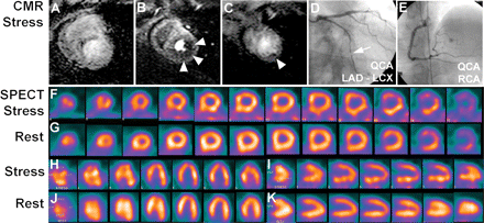
An example of a 47-year old patient is shown 2 months after successful stenting of the left anterior descending coronary artery and mild angina. The perfusion-CMR study during hyperemia (at 0.1 mmol/kg Gd-DTPA-BMA) demonstrates a perfusion deficit in the subendocardium of the lateral wall (B/C; arrow heads) appreciated by all three readers (mean score of 13; scores of readers 1–3: 15/9/15). Single-photon emission computed tomography in this patient was positive for the presence of CAD for one reader (mean score: 2; scores of readers 1–3: 0/6/0). Coronary X-ray angiography demonstrated a significant stenosis in the circumflex coronary artery (D, arrow). Perfusion in the anterior wall was assessed correctly by both techniques (normal perfusion) despite a stent in the left anterior descending coronary artery
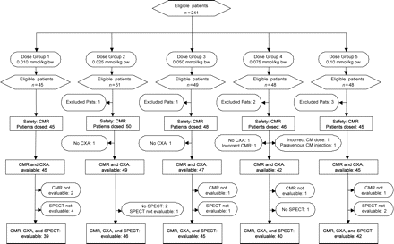
Flow chart demonstrating number of eligible patients and reasons for drop-out per dose group. CM: contrast medium (Gd-DTPA-BMA); CXA: coronary X-ray angiography; Pats: patients
Single-photon emission computed tomography examination
Stress and rest SPECT examinations were performed according to generally accepted guidelines19 on machines of different vendors (two or three head cameras) with 99mTc- or 201Tl-tracers, adenosine dose as for perfusion-CMR, or physical stress, and using 1 or 2 days protocols. Gated-SPECT using 99mTc-tracers was strongly recommended, but ungated acquisitions and/or 201Tl-tracers were accepted if part of the performing institution’s clinical routine. Three blinded readers, all experts on SPECT (and not identical with the CMR readers) analysed the SPECT data visually using a core laboratory (Beacon Bioscience, Inc., Doylestown, USA). All readers were blinded with respect to clinical information, the results of the other tests, as well as the order, in which these tests were performed. Each reader was presented with 10–12 short-axis as well as 6–9 vertical and horizontal long-axis images for both, stress and rest condition (Figure 1F–K). Gated-SPECT data were also presented to the readers, if they had been acquired. On the same 16-segment model used for CMR, perfusion deficits were graded in each segment as fully reversible (3), partially reversible (2), fixed defect (1), or normal (0), and scores were calculated as for the CMR analyses. Patients with ≥1 segment graded as non-diagnostic were treated as for the CMR examination, resulting in 3.6% (eight cases) excluded from the analyses (Figure 2). According to the CAD definition on CXA (not considering stenoses in <2 mm vessels), pure apical ischaemia was not considered a finding, which defines the presence of CAD.
Safety analysis
Measures of safety included physical findings obtained 1–36 h before and 24 h after the first CM injection. Vital signs (heart rate, blood pressure, oxygen saturation), respiratory rate, and body temperature were documented 0–2 h before the first CM injection and at predefined intervals during the CMR examination and up to 24 h later. 12-lead ECG’s were acquired 0–2 h before the first CM injection and at 1, 2, and 24 h later. Two-lead ECG’s were acquired at predefined intervals during the CMR examination. Samples for serum chemistry (including creatine kinase, aspartate and alanine aminotransferase, lactate dehydrogenase, creatinine, urea nitrogen, and others) and hematology (including hemoglobin, red blood cell count, white blood cell count and differential, platelet count, and others) were collected 0–36 h before and 24 h after the first CM injection. Safety data were assessed using a core laboratory for ECG and blood samples.
Statistical analysis
The main efficacy outcome was the comparison of perfusion-CMR at best CM dose vs. SPECT using ROC analysis. Additional study outcomes were performance of CMR in MVD patients and CMR comparison vs. gated-SPECT. For the comparison of perfusion-CMR vs. SPECT by ROC analysis, estimates suggested a required sample size of ~184 patients to yield an 90% power to detect a difference in the area under the ROC curve (AUC) of 0.15 (0.70 vs. 0.85 for SPECT and CMR, respectively) at a one-sided P-value of 0.05.18 ROC analyses were performed for both, CMR and SPECT on a patient basis in order to test the diagnostic performance of CMR and SPECT over the entire range of the summed scores calculated as the average scores of all three readers (Rockit 0.9.1 Beta). AUCs (as area ± SE) for CMR and SPECT were compared by a univariate z-score test (null hypothesis: data sets arose from binormal ROC curves with equal areas beneath them) taking correlation of the CMR and SPECT data into account (repeated measurements). Owing to the low performance of the doses ≤0.075 mmol/kg (see Figure 3A and which is also in line with published data),11 statistics were performed for CMR at the 0.1 mmol/kg dose only (head-to-head comparison and vs. entire SPECT population). Performance in MVD patients (2–3 vessel disease) was analysed after exclusion of patients with single vessel disease. Since the SPECT study must be positive if performed first (which could introduce a bias towards false positives), SPECT performance (AUC) was also calculated for SPECT being the second or third test (unbiased SPECT population).
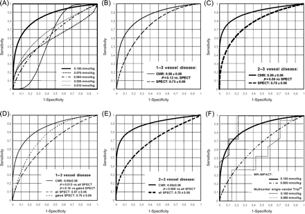
(A) Shows the diagnostic performance (receiver operating characteristics) for the different contrast medium doses ranging from 0.01 to 0.10 mmol/kg Gd-DTPA-BMA. At the highest dose of 0.10 mmol/kg (thick line, dose 5), best performance is achieved, which is compared vs. single-photon emission computed tomography in (B) (head-to-head comparison) showing no significant difference in the area under the receiver operating characteristic curve (0.86 ± 0.06 vs. single-photon emission computed tomography with 0.75 ± 0.09, P = 0.12). Similarly, for the 2–3 vessel disease population in (C), no significant difference between the two techniques is observed in the head-to-head comparison (P = 0.09). When comparing the perfusion-CMR performance vs. the entire single-photon emission computed tomography population in (D), the area under the receiver operating characteristic curve for CMR is larger than for single-photon emission computed tomography (0.86 ± 0.06 vs. 0.67 ± 0.5, P = 0.013). Difference between perfusion-CMR and gated-single-photon emission computed tomography did not reach statistical significance. For multivessel disease in (E), performance of perfusion-CMR is superior vs. the entire multivessel disease single-photon emission computed tomography population (area under the receiver operating characteristic curve: 0.89 ± 0.06 vs. 0.70 ± 0.5, P = 0.006). The performance of perfusion-CMR in this trial is in good agreement with an earlier smaller multicentre single-vendor trial as shown in (F) assessing the doses of 0.10 and 0.05 mmol/kg (thin dotted lines, Giang et al.11). Numbers indicate mean ± SE of the area under the receiver operating characteristic curve
The agreement rate between the independent CMR and SPECT readers was analysed by the κ coefficient. All tests were two-sided and a P-value <0.05 was considered statistically significant. For statistical comparisons of effect of CM dose on accuracy of perfusion-CMR vs. CXA see Supplementary material online, Appendix in the online version.
Results
Patient characteristics
From the 241 patients enrolled during 12 months, 234 entered the safety analysis (patients dosed). Of the seven patients not receiving CM, two refused informed consent (cancellation of given consent), one had a contraindication to receive adenosine, one experienced claustrophobia, in two patients invasive X-ray coronary angiography was cancelled, and in one patient the procedure during the MR study was incorrect. Evaluable CXA and correctly performed perfusion-CMR studies were available in 228 patients. Since three patients had no SPECT study, 225 patients were available for the perfusion-CMR vs. SPECT comparison. From these 225 patients, 5.8% (13 patients) had either non-evaluable perfusion-CMR (five patients; 2.2%) and/or SPECT examinations (eight patients; 3.6%, see also Figure 2). Of all study participants, 73% were male, 77% had CAD, and 31% had a history of percutaneous coronary interventions. Further demographics are given in Table 1.
Comparison of perfusion-cardiac magnetic resonance at the highest CM dose to single-photon emission computed tomography using receiver operating characteristic analysis
Head-to-head analysis
This comparison showed no superiority of perfusion-CMR over SPECT (Figure 3B, AUC 0.86 ± 0.06 for CMR vs. 0.75 ± 0.09 for SPECT, P = 0.12, n = 42 for both, CMR and SPECT) for the dose of 0.1 mmol/kg, which yielded the best performance for perfusion-CMR as shown in Figure 3A (largest AUC for 0.1 mmol/kg). From the perfusion-CMR ROC curve in Figure 3B, it can be seen that, for example, a sensitivity for CAD detection of 85% (95% CI: 69–93%), which is deemed clinically relevant, corresponds to a specificity of 67% (95% CI: 35–89%). In the MVD population (2–3 vessel disease), there was also no superiority for CMR (0.89 ± 0.06 vs. 0.78 ± 0.09 of SPECT, P = 0.09, n = 31, for dose 0.1 mmol/kg, Figure 3C).
Comparison vs. entire single-photon emission computed tomography population
When comparing perfusion-CMR at 0.1 mmol/kg vs. the entire SPECT population (Figure 3D), the ROC analysis demonstrates a better performance for perfusion-CMR (AUC: 0.86 ± 0.06, n = 42) vs. SPECT (n = 212, AUC: 0.67 ± 0.05, P = 0.013 vs. CMR). The CMR performance at 0.1 mmol/kg was also superior in the MVD population (n = 32 and 161 for CMR and SPECT, respectively, AUC: 0.89 ± 0.06 vs. 0.70 ± 0.05, P = 0.006, Figure 3E).
Gated-SPECT was performed in 42% (n = 95) of all SPECT studies yielding an AUC of 0.75 ± 0.08 (not different vs. ungated SPECT: AUC 0.65 ± 0.06, P = 0.12, and not different vs. perfusion-CMR: AUC 0.86 ± 0.06, P = 0.18, Figure 3D). For CMR at the CM doses of 0.01, 0.025, 0.05, 0.075 mmol/kg, the visual inspection of the ROC curves shows similar AUCs (0.69 ± 0.09, 0.60 ± 0.14, 0.64 ± 0.10, 0.60 ± 0.09, respectively) as for the SPECT population (0.67 ± 0.05, Figure 3A).
The study design requiring a positive SPECT result in patients undergoing SPECT as the first examination could introduce a bias towards false-positive SPECT studies. In the 153 patients with SPECT being the second or third examination, i.e. SPECT did not influence the referral for CXA (unbiased SPECT population), AUC for SPECT was 0.67 ± 0.05, which is identical to the entire SPECT population (also, the CMR result in these 153 patients did not influence the referral for CXA since the patients had to be scheduled for CXA before study enrollment). The portion of CAD in the population with SPECT first was 64% (46/72) and was 82% (125/153) for those with SPECT as second or third study.
A comparison of the five CM dose groups for accuracy of perfusion-CMR (proportion of correct diagnoses vs. CXA) yielded the lowest dose as inferior to the other doses (overall P < 0.001; for details see Supplementary material online, Appendix).
Reader agreement
The quality of the 225 CMR data sets was adequate with only 2.2% (five patients) having non-evaluable perfusion-CMR. For dose group 5 (0.10 mmol/kg), one patient (out of 45 patients) was non-evaluable by all three readers (2.2%). Reader 1 (R1) achieved 80% correct diagnoses (after elimination of another eight cases), whereas reader 2 (R2) and reader 3 (R3) achieved similar accuracies of 73 and 78%, respectively (with elimination of another three patients by R3). Agreement was best between R2 and R3 (κ = 0.39). Overall, the κ values for the CMR studies were fair with 0.32 (R1 vs. R2), 0.30 (R1 vs. R3), and 0.39 (R2 vs. R3), whereas for the SPECT studies, the κ values were 0.47 (R1 vs. R2), 0.58 (R1 vs. R3), and 0.48 (R2 vs. R3). The CMR data quality was similar for the three different perfusion territories as indicated by the similar AUCs for detection of stenoses in the left anterior descending (10 segments assigned), left circumflex (two segments assigned), and right coronary artery (four segments assigned) of 0.68 ± 0.08, 0.72 ± 0.09, and 0.70 ± 0.08. It is well known that vascular anatomy shows individual differences from patient to patient (e.g. left or right coronary artery dominance) which is an explanation for the lower AUCs for vessel-based analyses vs. patient-based analyses.
Safety
In the study, no deaths and no serious adverse events (AE) occurred. Twenty of 23 AE were mild and the only severe AE was angina pectoris, which was not related to CM and resolved within minutes. Angina was the most commonly reported AE occurring in three patients, followed by chest pain (2), flushing (2), and hyperpnea (1), which were primarily considered to be due to the adenosine administration. All 23 AE resolved and none was skin-associated (except flushing during adenosine infusion and one haematoma at the site of the intravenous line). No AE resulted in subject withdrawal. The safety profile of Gd-DTPA-BMA was comparable across all dose groups in this study and was consistent with the known excellent safety profile of most CMs used for CMR. No clinically significant trends or tendencies were noted in the biochemistry and hematology values over time. There were no 12 lead ECGs available directly after adenosine infusion. For the time points at 1, 2, and 24 h after first CM injection, there were no clinically relevant mean changes from baseline of any ECG-parameter (PR, QRS, QTc, and RR), which is well in line with other studies on adenosine administration.23
Discussion
Early detection of myocardial perfusion abnormalities is crucial for an optimal management of patients with suspected CAD and could potentially reduce the rate of fatal MIs.24 In this prospective, randomized, multicentre study, a high diagnostic performance of perfusion-CMR for the detection of CAD in ≥2 mm coronary vessels was found at a CM dose of 0.1 mmol/kg, which was equal to SPECT in the head-to-head comparison. At this CM dose, the diagnostic performance of perfusion-CMR was superior vs. SPECT imaging when comparing with all 212 SPECT studies, i.e. gated and ungated SPECT combined.
Perfusion-cardiac magnetic resonance and single-photon emission computed tomography: comparison with previous studies
MR-IMPACT is the largest multicentre perfusion-CMR study performed so far, and even more important, it also evaluates its test performance in a multivendor design, which is expected to reflect true diagnostic performance of widely applied perfusion-CMR more appropriately than single centre, single vendor studies. Similar considerations also apply for SPECT, where for example slightly less than half of all studies were performed with ECG-gating according their clinical routine. Diagnostic performance of perfusion-CMR was high with an AUC of 0.86 in this 18 centre, multivendor trial, while performance was as high as 0.91 in a previous smaller three centre, single-vendor trial.11 It is not surprising that high data quality is more difficult to preserve with a higher number of participating sites and involving various machine types, but more accurately reflects the routine clinical application of perfusion-CMR. Therefore, the goal was to homogenize the most important CMR imaging parameters for the different vendors (which is important since standardization for CMR is less advanced than for SPECT). The current CMR results are in line with earlier single-vendor CMR findings with best results at 0.1 mmol/kg,11 although the current study cannot exclude a potential further increase of performance at doses higher than 0.1 mmol/kg. The proposed perfusion-CMR approach meets several aspects of an optimal perfusion test. The perfusion-CMR examination is short (∼1 h) and safe, it lacks ionizing radiation, and the CM is well tolerated. Further, MR-IMPACT indicates that perfusion-CMR allows to study myocardial perfusion after interventions since signal response in the myocardium is not affected by stents located at the epicardium (see also Figure 1). Once hypoperfusion is detected by perfusion-CMR, additional information can be obtained by late enhancement CMR on whether hypoperfusion is caused by scar tissue.25–28 Therefore, a comprehensive work-up of patients with CAD in the future by CMR will most likely consist of a combined perfusion and late enhancement study.
A comparison of the current SPECT results determined in 212 patients (all with CXA as reference) from 18 centres with one of the largest multicentre 99mTc-based SPECT studies published so far15 including 112 patients (all with CXA) from seven centres can be made. Their sensitivity and specificity of 78 and 44%, respectively, is located on the ROC curve of the current SPECT study. Very close to the current ROC curve is the sensitivity and specificity of 87 and 36%, respectively, obtained in another multicentre SPECT study using 99mTc-sestamibi (and no attenuation and scatter correction).29 Similar sensitivities of 77–85% and specificities of 50–58% have also been reported for other multicentre SPECT trials.30,31 Thus, it can be concluded that the current SPECT results are in line with previous reports, and this holds despite a considerably larger number of participating sites in the present study. This high agreement between the performance of the entire SPECT population of the current study with that of previous multicentre SPECT studies supports the comparison of the perfusion-CMR results not only with the dose group 5 results (head-to-head comparison), but also with the results of the entire SPECT population. In support of the current study results, a recent smaller single centre study demonstrated superiority of perfusion-CMR over SPECT.13
Limitations of the study
It is important to note that gated-SPECT was not available in approximately half of patients. Perfusion-CMR did not outperform gated-SPECT with regard to CAD detection (Figure 3D; the reduced sample size for this subanalysis may partly explain the lack of statistical significance). Similarly, the higher AUC of gated-SPECT32,33 did not reach statistical significance over ungated-SPECT. Therefore, larger multicentre trials are warranted to confirm the trend of perfusion-CMR superiority over gated-SPECT.
The apical segment 17 was not included in the analysis, since the aim was to detect stenosed vessels suitable for revascularization, i.e. with a minimal diameter of ≥2 mm. Assuming the apex to represent a 17th of the LV myocardial mass translates into a vessel diameter of ∼0.8 mm,20 thus, detection of pure apical ischaemia would add potential false positives to both, perfusion-CMR and SPECT (since stenoses in vessels <2 mm on quantitative CXA were not considered to define CAD). On the other hand, the sensitivity for detection of any small ischaemic territory by both, SPECT and perfusion-CMR could be underestimated by applying this CAD definition. Finally, a pure apical ischaemia present in segment 17 only is assumed to be rare, although data in the literature on occurrence and diagnostic relevance of isolated apical ischaemia is lacking.
In the current study patients with decompensated heart failure, after bypass surgery, and with significant arrhythmias were excluded, and thus, statements cannot be made for these patient populations. Finally, in MR-IMPACT diagnostic performance of perfusion-CMR and SPECT was determined in patients undergoing CXA and therefore could differ in a population at a lower risk for CAD, e.g. when applied for exclusion of CAD in a screening setting. Also, the quantitative assessment of stenosis degree has its limitations to be used as a standard of reference, since myocardial perfusion is not only determined by epicardial coronary stenoses, but also by collateral flow and microcirculatory conditions.
Conclusions
In the MR-IMPACT, the ROC analyses suggest, that perfusion-CMR at 0.1 mmol/kg CM is a valuable technique for CAD detection in vessels of 2 mm or larger. Thus, in specialized centres perfusion-CMR may be considered as an alternative for SPECT imaging for the work-up of selected patients with known or suspected CAD. The comparison of perfusion-CMR with the entire SPECT population suggests CMR superiority over SPECT, which warrants further evaluation in larger trials.
Supplementary material
Supplementary material is available at European Heart Journal online.
Funding
The study was supported by GE Healthcare (former Amersham Buchler GmbH & Co. KG).
Acknowledgements
On behalf of the MR-IMPACT Study Group, the excellent efforts of K. Meurer, A. Mueller-York, C. Blankenstein, and G. Torheim, employees of GE Healthcare Buchler GmbH & Co. KG (former Amersham Buchler GmbH & Co. KG) is acknowledged for coordinating the trial, organizing site training for CMR readers, and for logistical support for data transfer from individual sites to the core laboratories.
Conflict of interest: J.S., and N.A.-S., report having served as consultants for GE Healthcare Buchler GmbH & Co. KG (former Amersham Buchler GmbH & Co. KG). M.M., is the chief statistician and an employee of GE Healthcare Buchler GmbH & Co. KG (former Amersham Buchler GmbH & Co. KG). The study was sponsored by GE Healthcare. Data analyses were performed in independent core laboratories. Statistical analyses were coordinated by the chief statistician, M.M., a GE Healthcare employee and co-author of this article.

