


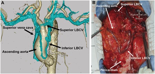
Figure 1:
(A) A preoperative 3-dimensional-reconstructed computed tomographic image showing an anomalous double LBCV. (B) The intraoperative image showing the inferior LBCV passing across the heart in front of the pericardium in a 79-year-old woman undergoing aortic valve replacement and coronary artery bypass grafting. LBCV: left brachiocephalic vein.
ACKNOWLEDGEMENTS
The authors thank Editage (www.editage.jp) for English language editing.
Reviewer information
European Journal of Cardio-Thoracic Surgery thanks Krishnasamy Arunkumar and Marko Ivan Turina for their contribution to the peer review process of this article.
Author notes
†
Tetsuro Uchida, Yoshinori Kuroda and Mitsuaki Sadahiro contributed equally to this work.
© The Author(s) 2020. Published by Oxford University Press on behalf of the European Association for Cardio-Thoracic Surgery. All rights reserved.
This article is published and distributed under the terms of the Oxford University Press, Standard Journals Publication Model (https://dbpia.nl.go.kr/journals/pages/open_access/funder_policies/chorus/standard_publication_model)
