


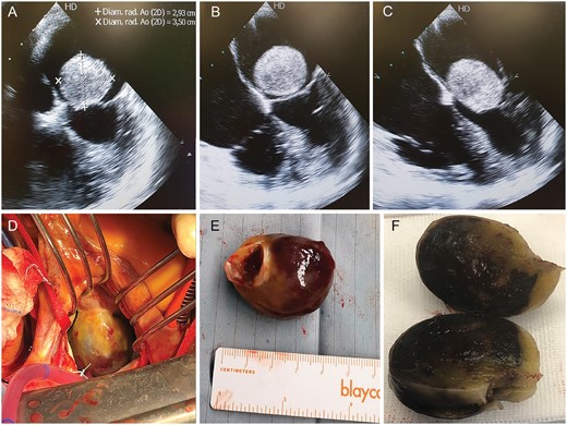
Figure 1:
Echocardiogram images showing a mobile giant left atrial mass determining mitral stenosis in a 50-year-old woman with a history of syncope and dyspnoea (A–C). Intraoperative view (D). The mass (E and F) was adjacent to the anterior mitral annulus and was eradicated through a standard direct left atrial surgical approach.
© The Author(s) 2019. Published by Oxford University Press on behalf of the European Association for Cardio-Thoracic Surgery. All rights reserved.
This article is published and distributed under the terms of the Oxford University Press, Standard Journals Publication Model (https://dbpia.nl.go.kr/journals/pages/open_access/funder_policies/chorus/standard_publication_model)
