


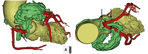
Figure 1
Computed tomography showing coronary artery fistulas to pulmonary artery originating from the right coronary, the left main trunk and the circumflex arteries (arrows).
Video 1
After injection of indocyanine green, intraoperative fluorescence imaging using photodynamic eye (Hamamatsu Photonics, Hamamatsu, Japan) clearly demonstrated fistulous vessels.
Video 2
No abnormal vessels were visualized after ligation and resection of fistulous vessels.
© The Author 2017. Published by Oxford University Press on behalf of the European Association for Cardio-Thoracic Surgery. All rights reserved.
