


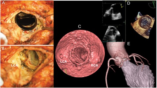
Figure 1
Pannus did not interrupt the valve (A). The pannus was revealed after taking the aortic valve with the narrowing of the outflow tract (B). Virtual angioscopy with multidetector computed tomography detected the pannus at the subvalvular position (C), which was difficult to detect by fluoroscopy, transoesophageal echocardiography (D) and 3D computed tomography (E). LCA: left coronary artery; RCA: right coronary artery.
© The Author 2017. Published by Oxford University Press on behalf of the European Association for Cardio-Thoracic Surgery. All rights reserved.
