


A 31-year old woman was referred to our institute at 37 weeks' gestation due to dyspnoea and a cough. The radiological finding revealed bilateral intralobar pulmonary sequestration (Fig. 1).
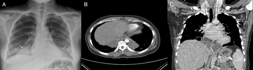
(A) Chest X-ray showed a round and well-defined mass consolidation of the right lower lung field and a similar one of the left lower lung field. (B and C) A computed tomographic scan of the chest revealed bilateral, large-sized pneumonic consolidation in the paravertebral region of the left and right lower lobe (B, arrow) with a possible bridging tunnel behind the heart and showed that an aberrant artery, which originated from the coeliac trunk, supplied the sequestration of the right lower lobe, and that a branch from the aberrant artery traversed to the sequestration in the left lower lobe through a bridging isthmus (C, arrows).
A staged thoracoscopic-assisted lobectomy was performed and the preoperative bronchial lavage detected Mycobacteria avium and Mycobacteria intracelluare (Fig. 2).
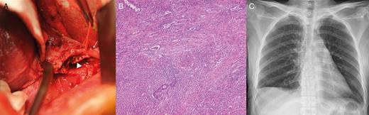
(A) In the operative field (1 week after caesarean section delivery), 7 days after the first operation (right lower lobectomy), the previously resected bridging isthmus (arrow head) during left lower lobectomy, and aberrant systemic artery (arrow), supplied the sequestration of the right lower lobe showed. (B) Histopathology of the lung showed multiple caseating granulomata with necrosis (HE stain, ×100). (C) The postoperative chest X-ray after 12 months.
Funding
This paper was supported by research funds from Chonbuk National University in 2013.
