


A 14-year old boy with neurofibromatosis type I underwent surgery for severe kyphoscoliosis (Fig. 1A). Impaired ventilation led to the termination of the procedure. Investigation revealed tracheal compression by the innominate artery and a tumour (Fig. 1B–D). The latter was excised via right thoracotomy (Fig. 2). Histology revealed neurofibroma.
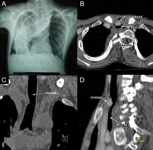
Figure 1:
(A) Chest X-ray: severe kyphoscoliosis; (B–D): computed tomography scan: trachea compressed between the innominate artery and a tumour, identified after surgery as neurofibroma.
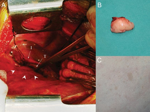
Figure 2:
(A) Operative view. Tumour (arrowheads); (B) excised tumour; (C) skin neurofibromatosis lesions. I: innominate artery; T: trachea.
© The Author 2014. Published by Oxford University Press on behalf of the European Association for Cardio-Thoracic Surgery. All rights reserved.
