-
PDF
- Split View
-
Views
-
Cite
Cite
Chung-Hang Wong, Ming-Shian Lu, Li-Wen Lee, Yao-Kuang Huang, Intraoperative airway occlusion during endovascular aortic repair for ruptured ductal arteriosus aneurysm: rescue by jet ventilation, European Journal of Cardio-Thoracic Surgery, Volume 44, Issue 5, November 2013, Pages e345–e346, https://doi.org/10.1093/ejcts/ezt414
Close - Share Icon Share
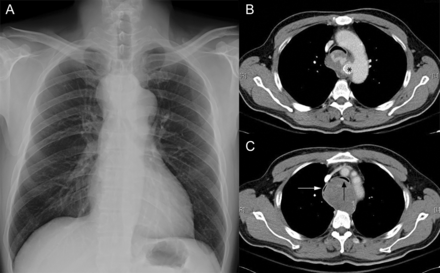
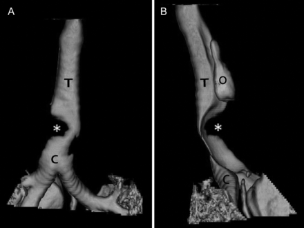
A 51-year old with an aneurysm of ductus arteriosus received thoracic endovascular aortic repair under general anaesthesia (Fig. 1). Intraoperative airway occlusion (suffocation) occurred owing to the distal tracheal compression by ruptured thoracic aortic aneurysms (Fig. 2). We applied jet ventilation to rescue the airway. Jet ventilation should be available even during endovascular aortic repair.

Preoperative plain film and chest computed tomographic (CT) scan. (A) Chest X-ray shows a widening mediastinum. Trachea is not deviated, and the distal tracheal is not well opacified. (B) Horizontal view demonstrates a contained rupture of the thoracic aorta, from a calcified pouch around the ligament arteriosus. Black asteroid indicates the calcified ring near the ligament arteriosus. (C) The contained rupture of thoracic aorta compressed the trachea and oesophagus (white arrow: oesophagus; black arrow: trachea).

Airway reconstructions from the chest CT. (A) Anterioposterior view of the trachea: a significant compression at the distal trachea (C: carina; T: trachea; white asteroid: external compression of the trachea). (B) Lateral view of the trachea: the compression from the posterior of the trachea (C: carina; O: oesophagus T: trachea; white asteroid: external compression of the trachea).


