


Abstract
This preliminary study evaluates, by qualitative score, the efficacy of the dynamic compression system (DCS) with a pressure-measuring device in the treatment of pectus carinatum (PC) as an alternative to surgery.
A total of 68 patients (infants, adolescents and young adults) presenting with typical PC (64 males and 4 females) were evaluated in our Chest Wall Deformities Unit, between October 2011 and February 2013. The criteria for including subjects were: patients with typical condrogladiolar PC and pressure for initial correction (PIC) ≤ 9 PSI (pound square inch). Seven patients were excluded in this study: three typical PC were treated by minimal invasive surgery (Abramson technique) due to highly elevated PIC and four atypical PC, hybrids forms (PE and PC) were treated by cup suction for pectus excavatum and by the DCS for the PC. The management protocol included: adjustment of the DCS, strengthening exercises and monthly clinical follow-up. The partial and final results were evaluated by the patients, by their parents or by both, using a qualitative scoring scale that was measured in a three-step grading system, where C is a low or very low result, B is acceptable and A is a very good or excellent result.
A total of 61 patients (59 males and 2 females) presenting with typical PC were treated by the DCS and included: symmetric PC in 43 cases and asymmetric PC in 18 cases. The mean age was 13.5 years (5–25). The mean PIC was 6.3 PSI (3–9 PSI). The mean utilization time was 19 h daily. The patients were divided into three groups. In Group I, consisting of 35 cases, all the patients have already completed the treatment with excellent aesthetic results (A). In 12 cases, Group II, the normal shape of the thorax has been obtained; all the patients in this group rated their results as excellent (A); however, these patients are still wearing the brace as a retainer for 3 additional months. Fourteen patients, Group III, are progressing and improving under active treatment, and surgeons and patients are very satisfied with the initial results. None of the 61 patients in this study abandoned the treatment and no complications were found.
This preliminary study demonstrated that the DCS with a pressure-measuring device is a minimal invasive system effective for treatment of PC in patients where the anterior chest wall is still compliant. The control of different pressure measurements could be used as the inclusion criterion as well as a predictive factor for aesthetic results and treatment duration.
INTRODUCTION
The mainstay of treatment of pectus carinatum (PC) over the past 50 years has been open surgery. The procedure involves the removal of abnormal costal cartilage and sternal osteotomy with reconfiguration of the chest wall [1–4]. Long-term results have been mixed, with reports of worsening cosmetic outcomes and decreased chest wall compliance over time [5–7]. Traditionally, surgical repair has been reserved for the most severe cases, and, as a result, many patients have been left with an untreated, mild-to-moderate deformity.
The concept of external compression for the treatment of PC was first described in the last century [8]. It was based on the fact that the chest wall is still compliant during puberty and allows the reshaping of the sternum and costal ribs by external compression applied over the deformity. Subsequently, this principle has been widely applied with reports showing favourable outcomes [9–16].
More recently, the evolution of the concept of external compression was described in 2008 by Martinez-Ferro et al. [17], who have advanced this technology with the implementation of the dynamic compressor system (DCS), using a custom-fitted, cushioned aluminium brace. The compression plate is attached to the anterior segment of the brace and placed at the level of the protrusion. By pushing the deformity backward, the compression gradually reshapes the chest into normal position. The DCS allows measurement of the pressure for initial correction (PIC) by an electronic pressure-measuring device, and this, in turn, helps establish the criteria for the use of the brace and avoids complications such as skin problems and patient non-compliance. PIC is used as a predictive factor for the evaluation of the results and of the treatment duration. This preliminary study evaluates, by qualitative score, the efficacy of the dynamic compression system (DCS) with a pressure-measuring device in the treatment of PC as an alternative to surgery for these patients.
MATERIALS AND METHODS
Between October 2011 and February 2013, 68 patients with typical condrogladiolar PC were treated in our Chest Wall Deformities Unit, after obtaining IRB approval for all patients. The criteria for including subjects were: patients with typical condrogladiolar PC and PIC ≤ 9 PSI (pound square inch). Seven patients were excluded in this study: minimal invasive surgery (Abramson technique) was performed in 3 cases due to very highly elevated PIC superior to 14 PSI and 4 patients with atypical PC, hybrid forms (PE and PC) were treated simultaneously by cup suction for pectus excavatum and by the DCS for PC, with very good results so far. The remaining 61 cases were included in this study and were treated by the DCS, 4 of whom had a relevant medical history: cardiac surgery by sternotomy, Ravitch procedure with recurrence of PC, Marfan syndrome and previous bracing failure. In 9 patients (14%), a scoliosis was found during the PC deformity physical evaluation. At the first consultation, PIC is obtained by applying the electronic device over the deformity until normal shape. As stated in the ‘Introduction’ section, PIC helps establish the criteria for the inclusion of patients likely to benefit from the use of the brace. It is worth noting that patients with a PIC of <9 PSI were enrolled in this study. Further measurements were then taken to customize the brace. On placing the brace, it was duly adjusted. For the regulation of the pressure of the treatment, the pressure device was attached to the anterior segment of the brace, thus permitting the adjustment of the pressure to the desired level to minimize the risk of skin lesions. The pressure of the treatment used in our study was ≤3 PSI. Patients were instructed to wear the brace during sleep and as much as possible during the day and were allowed to remove it only during sports or in the shower. At the same time, the patients were instructed by physiotherapists to do appropriate exercises to optimize results and they were advised to do certain physical activities, for example, swimming and weights. Patients are checked every month and when complete correction is achieved, the brace is used progressively for fewer hours as a retainer mode for 4–6 months. Partial and final results are evaluated by the patients, their parents or by both using a qualitative scoring scale that was measured in a three-step grading system, where C is a low or very low result, B is acceptable and A is a very good or excellent result. Given the age of the patients, CT scan before and at the end of the treatment was not suitable in such a short period of treatment. It was performed only in a few patients.
RESULTS
Sixty-one patients (59 males and 2 females) were treated by the DCS. The mean age of the patients was 13.5 years (5–25 years). The mean time of use per day in all patients was 19 h (17–22 h). Forty-three of the patients were symmetric deformities, and 18 asymmetric deformities. The mean PIC in pounds per square inch (PSI) was 6.3 PSI (3–9 PSI). Patient characteristics are presented in Table 1. The 61 patients were divided into 3 groups. In Group I, consisting of 35 patients (57.3%), all patients have already completed the treatment and were: 23 symmetric cases and 12 asymmetric cases. The mean age in this group was 13.3 years (5–25 years), and the mean PIC was 6 PSI (3–7.5 PSI). The mean time of treatment was 10 months (2.6–11.4 months). In symmetric cases, the mean time of treatment was 8.9 vs 11.3 months in asymmetric cases. The follow-up was 5 months (3–15 months). Only 1 of them experienced a minimal recurrence 2 months after finishing the treatment and needed to wear the brace during 3 additional months, after which there was no recurrence. Both patients and parents in this group rated the results as excellent (A). In Group II, consisting of 12 patients (19.6%), there were: 7 symmetric deformities and 5 asymmetric deformities. All the patients exhibited flattening of the sternum and are currently wearing the brace as a retainer ∼12 h every 2 days during 3 additional months. The mean age in this group was 14 years (5–19 years old) and the mean PIC was 6.2 PSI (3.7–9 PSI). The mean time of treatment until normal shape was achieved was 10.5 months (3–18 months). Both patients and parents in this group rated the results as excellent (A). To date, there has been no recurrence. In Group III, consisting of 14 patients (22.9%), there were 13 symmetric and 1 asymmetric deformity. All patients are progressing and improving under active treatment. The mean age in this group was 13.6 years (5–18 years old) and the mean PIC was 6.8 PSI (3–14 PSI). In 2 of them, the PIC was 14 PSI and both had an improved result with brace treatment after 6 months. Both parents and patients are very satisfied with the initial results. No patients abandoned the treatment and no complications were found. On comparing symmetric PC vs the asymmetric PC patients in Group I, that is, in patients who have completed the treatment, the mean time of treatment was 8.94 months in symmetric cases (Fig. 1) vs 11.3 months in asymmetric cases (Fig. 2). It would seem, therefore, that the patients with unilateral protrusion are more difficult and their mean time of treatment to reshape the chest with the brace takes is longer than that of those that have the symmetric defect.
Patient characteristics
| Group I | Group II | Group III | |
|---|---|---|---|
| Treatment | Completed | Flattening of the sternum ‘retainer mode’ | Active progressing and improving in all |
| Patients | 35 | 12 | 14 |
| Mean age (years) | 13.3 (5–25) | 14 (5–19) | 13.6 (5–18) |
| Pressure for initial correction | 6 (3.5–7.5) | 6.2 (3.7–9) | 6.8 (3–14) |
| Symmetric | 23 | 7 | 13 |
| Asymmetric | 12 | 5 | 1 |
| Mean time of treatment (months) | 10 (2.6–11.4) | 10.5 (5–18) + retainer mode | In treatment |
| Results | A | A | Patients and parents are very satisfied |
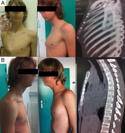
Symmetric case. Same patient with symmetric PC. 16 years old, PIC of 7.5 PSI. History of cardiac surgery by sternotomy. Group I: (A) before treatment and (B) after 11 months of treatment.
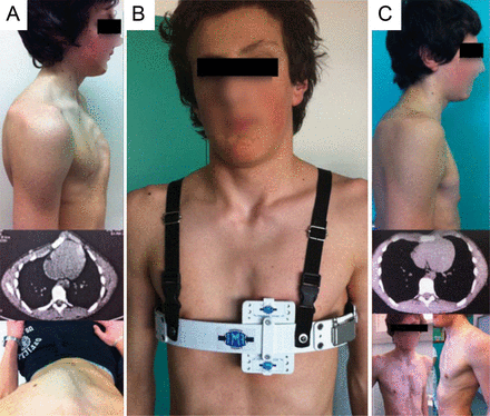
Asymmetric case. Same patient, with asymmetric PC 14 years old. PIC of 8.8 PSI. Group II: (A) before: lateral view, CT scan and upper view, (B) during the treatment and (C) after 7 months of treatment, CT scan, lateral and front view.
DISCUSSION
The first report in the English literature for correction of PC by compressive orthotic bracing was made by Haje and Bowen [16] in 1992. Since then, subsequent reports have been described and the majority of the trials have shown good results [15–17]. Several authors have suggested a variety of non-operative approaches based on the concept that the anterior chest wall is still compliant during puberty and permits remodelling by applying external compression [18, 19]. Compared with open surgery, brace treatment eliminates risks such as anaesthesia and major surgery, decreases the complication rate, leaves no visible scar, avoids hospital admission and reduces the cost of treatment. The final cosmetic results are similar to those of open surgery [13, 14, 17]. Compressive orthotic bracing is becoming a well-recognized therapy and an effective non-operative treatment for PC, improving both the objective protrusion and appearance, as judged by the patient. The advantages of the DCS over other orthotics brace are: it involves the use of pressure device measurements, it helps to establish the criteria for inclusion, and acts as a predictive factor to evaluate the results and treatment duration. Regulation of the pressure during the treatment helps to avoid complications such as skin problems and patient non-compliance [17]. Brace treatment is becoming the treatment of choice for almost all patients, particularly in mild-to-moderate defects where surgical treatment could be excessive. In 2010, Martinez-Ferro [20] proposed a decisional algorithm for PC. In patients with PIC ≤ 7.5 PSI, the DCS should be the treatment of choice. If the patients tolerate the DCS, no surgery will be needed. Our initial results in patients with PC treated by the DCS were excellent, within this limit of pressure. Fifty-one of 65 patients with PC had PIC ≤ 7.5 PSI and were: 35 in Group I, 6 in Group II, and 10 in Group III. As can be appreciated in the ‘Results’ section of Table 1, all patients and parents rated the results of the treatment as excellent (A). Our decision was to increase the limit of pressure to 9 PSI. This allowed us to include a number of patients in the prescription of the DCS, in an attempt to give them the benefits of minimal invasion. This alternative to surgical treatment was well received by our patients with greater PIC. In our study, 14 patients had PIC ≥ 7.5 PSI; 2 of them had PIC of 14. All of these patients are still undergoing the treatment. Eight of them, from Group II, exhibited flattening of the sternum (Fig. 2), and 6 from Group III are progressing and improving under active treatment. Our feeling is that the use of the DCS could be extended to patients with PIC ≤ 9, although a greater sample of this kind of patient and longer follow-up would be necessary. The time of the treatment is related to the age of the patient, the type of PC, PIC and the hours of use per day of the DCS. It is interesting to observe the response of the patients; once they begin to see results, which typically occur in 1–2 months, they enthusiastically increase the force used and the corresponding chest compliance. As Martinez Ferro et al. [17] claim, in younger patients, a more malleable chest wall responds more rapidly to the treatment. However, to validate this, objective evaluation methods are required. In conclusion, this preliminary study supports the principle that the DCS with pressure measuring of the deformity is a minimal invasive system, with minimal invasion and an effective non-operative alternative for treatment of PC to improve self-image in patients where the anterior chest wall is still compliant. The control of different pressure measurements could be used as the inclusion criteria as well as a predictive factor for aesthetic results and the duration of the treatment. In asymmetric deformities, the treatment duration is longer than symmetric cases.
Conflict of interest: none declared.
