


Abstract
Thoracoscopic sympathectomy at levels T2 or T2–T3 is a treatment for focal hyperhidrosis and facial blushing. These levels of the sympathetic trunk innervate the heart, and consequently, the procedure is reported to change the heart rate variability due to changes in efferent cardiac autonomic activity. Our objective was to investigate the effects of thoracoscopic sympathectomy on global autonomic control, including baroreceptor sensitivity.
Eight patients (6 F, median age 28 years [range 20–58 years]) were exposed to the tilt-table test and cardiopulmonary exercise test before, and 3 months after, thoracoscopic sympathectomy. Eight healthy age-, gender- and BMI-matched controls were used as controls and underwent the same tests once. During tilt-table testing electrocardiogram, blood pressure, impedance cardiography and respiration were measured continuously, and efferent cardiac autonomic balance was estimated.
The heart rate measured during orthostatic stress test was lowered after thoracoscopic sympathectomy (between-group; P = 0.01) due to a change in autonomic tone, with increased vagal (high-frequency power n.u.; P = 0.001), and reduced sympathetic efferent cardiac activity (low-frequency power n.u.; P < 0.001). Baroreceptor sensitivity measured during rest was increased (26 ± 13 vs 44 ± 19 ms/mmHg; P = 0.01), and diastolic blood pressure reduced after surgery (P = 0.01). The increases in systolic blood pressure and the sympathetic marker CCV-LF in response to orthostatic stress were higher before sympathectomy, with almost no increases post-surgically (condition × group interaction; P = 0.01 and P = 0.001, respectively). We found no change in post-procedure exercise capacity, although patients had a lower peak VO2 and maximal cardiac index than controls.
Thoracoscopic sympathectomy changes the autonomic tone towards increased vagal activity; this is potentially cardioprotective. To our knowledge, this is the first study to show increased baroreceptor sensitivity after thoracoscopic sympathectomy.
INTRODUCTION
Thoracoscopic sympathectomy (TS) is used to treat severe focal hyperhidrosis and facial blushing and may be used for peripheral vascular disorders and angina pectoris [1]. TS at level T2 relieves blushing and facial hyperhidrosis, while combined T2–T3 TS diminishes palmar hyperhidrosis [2, 3]. The success rate is reported to be about 90% for facial blushing and 99% for focal hyperhidrosis [2, 3]. The aetiology of focal hyperhidrosis and blushing is unknown. However, both entities are associated with abnormal regulation of the autonomic nervous system [4, 5]. The heart is innervated by vagal and sympathetic fibres. The right vagus nerve primarily innervates the sinoatrial node, whereas the left vagus innervates the atrioventricular node; however, there can be significant overlap in the anatomical distribution. Levels T2–T3 of the sympathetic trunk innervate the heart, and cardiac complications after TS have been reported in three cases (non-lethal asystolic cardiac arrest and symptomatic bradycardia, requiring pacemaker), possibly induced by a postoperative increase in vagal tone [6]. Therefore, the effect of T2–T3 TS on the cardiac function is an issue of investigation and discussion. There are few side effects; the most common is compensatory sweating from other body parts, seen in up to 89% of the patients [7].
Heart rate variability is a measure of the activity in the cardiac autonomic system. It describes the variations in the interval between consecutive heart beats and is caused by the continuous interplay between the efferent cardiac parasympathetic and sympathetic activities. The interplay between these two systems can be evaluated non-invasively in the time and frequency domains. Baroreceptor sensitivity (BRS) is involved in short-term blood pressure regulation. Heart rate variability and BRS are used to study the general balance of autonomic activation. Reduced heart rate variability is related to increased cardiac and overall mortalities, while decreased BRS is related to cardiac death in patients after acute myocardial infarction and cardiac and all-cause death in patients with hypertension and renal failure [8–10].
T2–T3 TS reduces the heart rate both at rest and during exercise, but work capacity is unchanged [11]. There are no available data on maximal cardiac output levels after TS.
The primary aim of this study was to determine whether TS changes the global autonomic regulation. The secondary aim was to investigate whether maximal cardiac output is lowered after TS. Global autonomic regulation can be evaluated through measurements of heart rate variability and BRS.
MATERIAL AND METHODS
Participants
Eight patients referred to the Department of Cardiothoracic Surgery, Aarhus University Hospital, Denmark, were included. Two patients had palmar hyperhidrosis, 2 facial hyperhidrosis, 3 facial blushing and 1 combined facial hyperhidrosis and blushing. Eight healthy volunteers were recruited by advertising at Aarhus University Hospital. They were matched to the patients concerning age, gender and four body mass index (BMI) intervals (<21, 21–26, 26–30, >30 kg/m2).
General exclusion criteria
General exclusion criteria were previous sympathectomy, severe pulmonary or cardiovascular disease, diabetes mellitus, abnormal electrocardiogram, cerebrovascular stenosis, pharmacological treatment affecting cardiovascular or autonomic function, pregnancy, lactation and age <18 years.
Surgical techniques
Patients were placed in the supine position with 90° abduction of both arms. Single-lumen intubation anaesthesia was used. Two ports were placed in the axillary hairline, one anteriorly and one posteriorly. A 30° videothoracoscope (Olympus Winter & Ibe, Hamburg, Germany) was used through the anterior port and a harmonic scalpel (Ultracision, Ethicon Endo Surgery, Cincinnati, OH) through the posterior port. The sympathetic chain was transected over the middle portion of the second rib, and the incision was extended laterally to include accessory nerve fibres. After the procedure, the lung was reinflated and the air exsufflated through a small chest tube placed through the 5-mm port.
Experimental set-up
The study was carried out in accordance with the Declaration of Helsinki, and approved by the Local Ethics Committee (No 20090004). The same researcher (E.B.) and an experienced laboratory technician or a research nurse performed the tilt-table experiments. All cardiopulmonary exercise tests were performed by E.B. To minimize external autonomic influences, tilt tests were performed in a quiet room with dim lights and mean room temperature of 23.2°C (SD 0.8°C). The patients were exposed to tilt-table testing and cardiopulmonary exercise test 2 weeks preoperatively, and 3 months postoperatively.
Orthostatic stress (tilt-table test)
The patient and the matched control were investigated on separate days but at the same hours of the day, either between 8 and 10 a.m. or between 10 and 12 a.m. Subjects rested in the supine position for 30 min before recording. The Task Force Monitor (CNSystems Medizintechnik AG, Graz, Austria) non-invasively recorded electrocardiogram, beat-to-beat blood pressure, impedance cardiography and respiration during: (i) 10-min supine rest (baseline), (ii) two times 10 min during tilt-table testing and (iii) 10-min supine recovery [12, 13].
QRS detection and heart rate variability expressed in the time and frequency domain
Raw data from electrocardiogram, lead II (sample rate: 1000 Hz) were exported to software (Aalborg University, Denmark) to manually visualize QRS complexes, verify their correctness and correct for noise or arrhythmic behaviour [12]. QRS detection was done with a Pan-Tompkins-like algorithm [13]. In the present material, 16 ectopic QRS complexes were adjusted by interpolation.
Heart rate variability was expressed in the time domain as the distance in ms between consecutive normal R waves in the QRS complexes (mean RR interval), the SD of all normal RR intervals (SDNN), and the square root of the mean squared differences of successive RR intervals (RMSSD). RMSSD is not influenced by mean resting heart rate and estimates high-frequency (HF) variation in heart rate [12].
Power spectral analysis was performed with a 20th-order auto-regression model [14] and expressed in the frequency domain as HF power (0.15–0.4 Hz), an index of cardiac vagal activity and low-frequency (LF) power (0.04–0.15 Hz) expressing both sympathetic and parasympathetic activity [12]. Each power spectral component was expressed in (i) absolute units, (ii) normalized units (n.u., power/(total power – very low (<0.03 Hz)-frequency oscillations)) and (iii) the coefficient of component variance in the LF and HF bands (CCV-LF/CCV-HF) (square root of LF or HF power/mean RR interval) [15]. CCV-LF and CCV-HF adjusts for influences of different RR intervals on the power amplitude because saturation of the sinus node by a very high sympathetic or parasympathetic drive is proposed to make the sinus node less capable of maintaining a rhythmic modulation [16].
Baroreceptor sensitivity
The spontaneous baroreflex activity was analysed off-line by the Task Force Monitor by means of the Sequence-Method [17] analysing and displaying rising/falling sequences (progressive increase/decrease in systolic blood pressure and lengthening/shortening of mean RR intervals separately over more than three consecutive beats). The minimum changes accepted as a spontaneous increase or decrease in systolic blood pressure and RR interval were 1 mmHg and 4 ms, respectively. The mean slope of all regression lines between mean RR intervals and systolic blood pressure sequences represented the BRS. In the present material, the BRS could not be estimated in 1 patient in the test after TS during the supine period due to absent events.
Cardiopulmonary exercise test
A photo-acoustic gas-rebreathing technique was combined with a bicycle fatigue test.
The rebreathing system consists of a three-way respiratory valve connected to a mouthpiece, and a rubber bag connected to an infrared photo-acoustic gas analyser (Innocor®, Innovision A/S, Odense, Denmark). To ensure a closed system, the participant was equipped with a nose-clamp. Pulse oxymetry was used to determine % SpO2 (saturation of haemoglobin with oxygen in arterial blood). A gas-mixture containing known concentrations of O2, N2O (soluble) and SF6 (insoluble) was used during rebreathing. By infrared photo-acoustic gas analysis during sessions of five rebreathings, gas concentrations were decided, and pulmonary blood flow (PBF) and VO2 (ventilation of oxygen, i.e. oxygen uptake) were estimated. Cardiac output was estimated through calculations based upon PBF, VO2 and SpO2. The method is described in detail elsewhere [18].
Rebreathing was performed at rest and during exercise of increasing intensity until exhaustion (30W, 60W, 90W etc.). Three-minute intervals between rebreathing sessions were applied to ensure sufficient washout of N2O.
Statistical analysis
Measurements were summarized as arithmetic mean and SD, except some from the supine position, and raw data for parameters analysed after log transformation, using median and range. To accommodate the assumptions of normal distributions, SDNN, RMSSD and the frequency domain parameters were log-transformed before analysis. Unpaired Student's t-tests were used to test differences in age, weight, height and BMI between patients and controls. Patients’ pre- and post-TS were compared with respect to haemodynamic and autonomic parameters in the supine period of tilt-table test using paired Student's t-tests and Wilcoxon signed-rank test. Supine measurements in patients and control subjects were compared by the unpaired t-tests and Mann–Whitney test. In 6 patients exposed to the tilt-table test both before and after TS, haemodynamic and autonomic parameters were compared using two-way repeated measures analysis of variance (ANOVA). The condition × group interaction and the main effect of the group were assessed. If the condition × group interaction was statistically significant (non-parallel profiles), this analysis was, for the secondary effect parameters, supplemented by a comparison of the absolute differences and the relative changes between groups. In the same manner, the 6 patients before the TS were compared with six matched control subjects. CPX measurements before and after TS were compared by the paired Student's t-test; patients and controls were compared by unpaired Student's t-test.
All statistical tests were two sided, and the level of significance was 5%. Stata 12.0 (StataCorp. 2011, Stata Statistical Software: College Station, TX, USA: Stata Corporation) and Graphpad Prism version 5, Graphpad Software, Inc., were used in the basic statistical analyses.
RESULTS
Participant characteristics and effect of surgery
Eight patients (6 F, 2 M, mean age 32 years, range 20–58 years, mean BMI 23.6 kg/m2 (SD 2)) and eight healthy control subjects (6 F, 2 M, mean age 32 years, range 18–59 years, mean BMI 22.8 kg/m2 (SD 3)) participated. Three patients and no controls were active smokers. The patients with focal hyperhidrosis all had warm, dry hands post-surgically. The patients with facial blushing reported the effects of the surgical procedure.
Orthostatic stress test
Two of the patients had tilt-induced near-syncopal symptoms before TS, and were only evaluated and retested in the supine position at follow-up. None of the controls fainted during tilt test. The 2 patients who did not tolerate the tilt-test were young (24 and 26 years), non-smoking females with a blood pressure within normal range (100/65 and 117/80 mmHg) and with normal electrocardiograms.
Heart rate variability during baseline conditions and orthostatic stress test
Comparing patients before and after TS showed significant between-group effects with increased post-surgical mean RR interval and HF power n.u. and reduced LF power n.u. during baseline, upright position and recovery (Fig. 1). There was a tendency towards increased RMSSD after TS (Table 1). Preoperatively, the patients had increases in CCV-LF during orthostatic stress, which were significantly diminished after surgery both for the first and second upright period (Table 1). There were no statistically significant changes during the tilt-table test in CCV-HF or SDNN (Table 1). After TS, there were no differences between patients and controls, except a lower CCV-LF (P = 0.046) and a higher HF power in n.u. in patients (Fig. 1).
Heart rate variability during tilt-table testing and recovery in patients (n = 6) and controls (n = 6) before and after sympathectomy
| Baseline | Upright 1 | Upright 2 | Recovery | ANOVA (P-value) | ||
|---|---|---|---|---|---|---|
| Condition × group | Between-group | |||||
| LF power (ms2/Hz) | ||||||
| Patients pre | 556 (206–1487) | 535 (259–1358) | 600 (259–1486) | 674 (290–1266) | ||
| Patients post | 586 (226–1622) | 276 (65–826) | 324 (59–593) | 642 (384–1972) | 0.0001* | |
| Controls | 994 (104–3841) | 753 (336–1044) | 480 (319–1117) | 984 (153–5109) | 0.73** | 0.46** |
| CCV-LF (%) | ||||||
| Patients pre | 2.6 (0.8) | 3.3 (1.2) | 3.7 (1.4) | 2.6 (0.4) | ||
| Patients post | 2.2 (1.0) | 1.9 (0.9) | 2.0 (0.9) | 2.6 (1.2) | 0.001* | |
| Controls | 3.3 (1.7) | 3.3 (0.7) | 3.2 (0.8) | 3.2 (2.0) | 0.28** | 0.63** |
| HF power (ms2/Hz) | ||||||
| Patients pre | 809 (53–2095) | 183 (33–208) | 148 (39–155) | 745 (61–2302) | ||
| Patients post | 1317 (227–5246) | 227 (61–341) | 93 (55–253) | 1288 (207–4107) | 0.34* | 0.03* |
| Controls | 1123 (329–5442) | 122 (49–716) | 117 (25–248) | 1216 (493–4379) | 0.55** | 0.30** |
| CCV-HF (%) | ||||||
| Patients pre | 2.9 (1.3) | 1.5 (0.6) | 1.5 (0.5) | 2.8 (1.2) | ||
| Patients post | 3.5 (2.0) | 1.6 (0.6) | 1.3 (0.4) | 3.3 (1.7) | 0.45* | 0.43* |
| Controls | 3.9 (1.9) | 1.7 (1.0) | 1.5 (0.6) | 3.6 (1.7) | 0.48** | 0.32** |
| Total power (ms2/Hz) | ||||||
| Patients pre | 1434 (277–3735) | 745 (335–1637) | 757 (390–1714) | 1694 (370–3461) | ||
| Patients post | 1803 (954–7056) | 602 (137–1230) | 554 (128–744) | 1850 (918–6422) | 0.0000* | |
| Controls | 2212 (626–9665) | 995 (425–1947) | 724 (425–1384) | 2618 (795–9857) | 0.44** | 0.32** |
| RMSSD (ms) | ||||||
| Patients pre | 49 (14–83) | 17 (12–25) | 16 (11–25) | 53 (15–93) | ||
| Patients post | 68 (29–113) | 24 (13–31) | 17 (13–26) | 74 (28–120) | 0.17* | 0.05* |
| Controls | 57 (36–170) | 19 (12–43) | 19 (11–27) | 73 (41–117) | 0.49** | 0.18** |
Heart rate variability measured during: 10-min supine rest (baseline), two times 10 min in 60° upright position (upright 1, upright 2) and during 10-min supine recovery (recovery). Values presented as mean (SD) and median [range]. Responses to tilt test were compared by using two-way repeated measures ANOVA. Bold values are statistically significant P-values (<0.05).
*Patients pre-TS vs patients post-TS.
**Patients pre-TS vs controls.
HF power and LF power: high- and low-frequency power expressed in absolute values [ms2/Hz] and coefficient of component variance (CCV) of LF or HF; RMSSD: the square root of the mean squared differences of successive RR intervals. RR intervals and normalized LF and HF power are presented in Fig. 1.
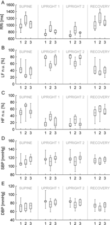
Results from (1) pre-TS, (2) post-TS and (3) controls during four consecutive events (supine rest, two times upright, supine recovery). (A) RR intervals. (B) LF power n.u. (C) HF power n.u. (D) continuous SBP. (E) continuous DBP. For the mean RRI (P-value 0.01), LF n.u. (0.0009), HF n.u. (0.001) and DBP (0.01) the main effect of the group was significantly different in patients pre-TS compared with patients post TS. For SBP, there was a significant condition by group interaction when patients pre-TS were compared with post-TS (0.01) and when patients pre-TS were compared with controls (0.03). The relative increase in SBP during upright position was diminished after TS.
Baroreceptor sensitivity and blood pressure during baseline conditions and orthostatic stress test
Comparing patients before and after TS showed a significant between-group effect with lower continuous DBP (contDBP) after TS for all three conditions: baseline, upright and recovery (Fig. 1). For contSBP, the relative increase in SBP during the upright position was higher before than after TS. We found no significant changes in total peripheral resistance, cardiac index (CI, cardiac output/body surface area) or BRS after TS during tilt-table testing, and no differences between patients and control subjects.
After TS, BRS was higher in the supine position compared with both before TS and with controls (Table 2). This was apparent in 7 of 8 patients. In the last patient, BRS could not be calculated due to missing events. BRS did not change in the upright position after TS.
Tilt-table test baseline values in patients (n = 8) and controls (n = 8)
| Pre-TS | Post-TS | P-value | Controls | P-value | ||
|---|---|---|---|---|---|---|
| Mean RRI (ms) | 1007 (150) | 1189 (165) | 0.01* | 1060 (113) | 0.43** | 0.09*** |
| BRS (ms/mmHg) | 26.0 (13) | 44.2 (19) | 0.01* | 25.1 (9) | 0.88** | 0.03*** |
| DBP (mmHg) | 73 (11) | 68 (7) | 0.02* | 72 (10) | 0.88** | 0.28*** |
| LF (n.u.) | 43 (11–74) | 23 (14–72) | 0.02* | 44 (17–51) | 0.77** | 0.11*** |
| HF (n.u.) | 49 (19–87) | 70 (24–84) | 0.02* | 51 (34–77) | 0.89** | 0.05*** |
Heart rate variability, baroreceptor sensitivity (BRS) and diastolic blood pressure (DBP) measured during 10-min supine rest in all included subjects before and after TS. Values presented as mean (SD). t-test is used to calculate P-value. For LF power n.u. and HF power n.u., median [range] is presented and Wilcoxon or Mann–Whitney used to compare groups. Bold values are statistically significant P-values (<0.05).
*Patients post-TS compared with before TS.
**Controls compared with patients before TS.
***Controls compared with patients after TS.
TS: Thoracoscopic sympathectomy; RRI: mean time between consecutive normal R waves in the QRS complexes; HF and LF power n.u.: high- and low-frequency power expressed in normalized units.
Cardiopulmonary exercise test
In the CPX, there were no significant differences between pre- and post-TS tests. Compared with controls, patients had lower maximal CI and VO2-uptake before and after TS, and after TS, maximal arterio-venous oxygen difference (AVO2) was also lower in patients than controls (Table 3).
Cardiopulmonary exercise test
| Patients pre-TS | Patients post-TS | P-value | Controls | P-value | ||
|---|---|---|---|---|---|---|
| Max CO (l/min) | 12.8 (2.6) | 12.1 (3.7) | 0.39* | 14.9 (2.4) | 0.11** | 0.10*** |
| Max CI (l/min/m2) | 7.2 (1.0) | 6.6 (1.4) | 0.19* | 8.2 (0.7) | 0.05** | 0.01*** |
| Max AVO2 difference (%) | 70.6 (8.6) | 66.6 (6.8) | 0.10* | 78.0 (7.6) | 0.09** | 0.007*** |
| Peak VO2 uptake/kg (ml/min/kg) | 25.1 (6.9) | 22.2 (5.2) | 0.05* | 32.5 (5.5) | 0.03** | 0.002*** |
Maximal exercise values in patients (n = 8) and controls (n = 8), presented as mean (SD). Paired t-tests used to compare pre and post-TS, unpaired t-test to compare with controls. TS: thoracoscopic sympathectomy. Bold values are statistically significant P-values (<0.05).
*Patients post-TS compared with before TS.
**Controls compared with patients before TS.
***Controls compared with patients after TS.
TS: thoracoscopic sympathectomy; CO: cardiac output; CI: cardiac index = cardiac output/body surface area; VO2: oxygen uptake; AVO2 difference: arterio-venous oxygen difference.
DISCUSSION
Our main findings are that TS caused a general reduction in heart rate during rest and orthostatic stress, probably due to a change in the autonomic balance towards increased vagal and reduced sympathetic activity (higher HF power n.u. and reduced LF power n.u.). Moreover, the BRS at rest was increased after TS. The diastolic blood pressure was in general lower after TS and the increases in systolic blood pressure and CCV-LF in response to orthostatic stress was higher before TS, with almost no increase post-surgically.
In the present study, two females were close to syncope during tilt-table testing, in accordance with the fact that about 10% of healthy adults will experience syncope or near-syncope in a tilt-table test [19].
Heart rate variability
The general reduction in heart rate during rest and orthostatic stress was probably due to a change in the autonomic balance towards increased vagal and reduced sympathetic activity (higher HF power n.u. and reduced LF power n.u.) as previously reported [20, 21]. The trend towards a predominant parasympathetic tone was also indicated by the higher BRS and by the fact that the increase in CCV-LF in response to orthostatic stress was higher before TS, with almost no increase post-surgically. In summary, the changes in heart rate variability after TS suggest that sympathetic activity was lowered, particularly during orthostatic stress, and the changes in BRS and HF power n.u. reflect a shift towards parasympathetic control. The changes in heart rate variability do not reflect a general decrease in heart rate variability, rather a smaller shift in autonomic regulation, without negative consequences for the overall haemodynamic control.
Baroreceptor sensitivity
This is the first study to show higher BRS in the supine position after TS—compared with both before TS and with controls. BRS is a marker of parasympathetic baromodulation, and may be referred to as cardiovagal BRS. Low BRS is a predictor of cardiac death in patients after myocardial infarction as well as a predictor of all-cause death in hypertensive patients with chronic renal failure [9, 10]. Considering this, an increase in BRS might have positive effects; at least increased BRS is unlikely to have a negative impact on the TS patients’ health. Our findings are in direct contrast to a previous report of reduced BRS after TS [22]. However, in the study by Kawamata reporting reduced BRS after TS, the patients were under general anaesthesia during the investigation both before and after TS. Anaesthesia and artificial ventilation have a profound impact on cardiac autonomic activity and the results are therefore not comparable to ours.
High vagal activity and reactivity (BRS) are consistent markers of good health and well-integrated cardiovascular reactivity. High vagal reactivity is, in principle, beneficial for the protection of the myocyte against the potential deleterious effects of high sympathetic activity in, i.e. ischaemic heart disease conditions [23]. The change in the autonomic balance towards increased vagal and reduced sympathetic activity very likely explains the increase in BRS after sympathectomy.
We did not find any difference in BRS in the 6 patients exposed to tilt-table testing. It could be explained by the change in the population because 2 patients had tilt-induced near-syncopal symptoms before TS, and were only evaluated and retested in the supine position at follow-up. Alternatively, cardiovagal BRS may have a relatively larger impact during rest than during orthostatic stress.
Blood pressure
The increase in SBP during tilt-table test was higher in the patients before TS compared with controls. It is known that SBP increases only minimally during tilt-table test in healthy subjects [24]. This could mean that an exaggerated blood pressure response to orthostatic stress in our patients with blushing or focal hyperhidrosis was normalized by TS. DBP was lowered after TS, both at baseline and during tilt-table test.
Cardiopulmonary exercise test
There were no significant changes in the CPX results after TS. This is consistent with earlier research on exercise capacity after TS [11]. The controls had a moderately better exercise capacity compared with patients. A possible explanation could be that the patients with hyperhidrosis would experience fatigue earlier because of dehydration, or more discomfort during testing because of excessive sweating. Another reason might be that our healthy volunteers were more willing to pressurize themselves in the CPX than the patients. The maximal VO2 levels indicate that our participants are either unfit or only reached submaximal exercise level.
Limitations
Our cohort was small, which might explain why we were unable to detect a change in exercise capacity. Two patients did not complete the tilt-table test; thus, we do not know if TS affected their ability to tolerate orthostatic stress. There was a difference in smoking habits between patients and controls, but it could not be accounted for because of the small sample size. Smoking can decrease heart rate variability and global autonomic control. This problem is reduced as patients acted as their own controls at follow-up. There are few data regarding long-term outcome following endoscopic procedures. However, sympathetic regeneration has been demonstrated in a case study [25]. To investigate whether these changes are permanent, it would be interesting to re-investigate the participants again years after the procedure.
In conclusion, TS induced a general reduction in the heart rate during rest and orthostatic stress and increased the BRS, probably due to a change in the autonomic balance towards increased vagal and reduced sympathetic activity. This change in autonomic activity is potentially beneficial in the protection against the potentially deleterious high levels of sympathetic activity.
Funding
This work was supported by the Danish Agency of Science, Technology and Innovation.
Conflict of interest: none declared.
ACKNOWLEDGEMENTS
Bo Martin Bibby (Associate Professor, Department of Biostatistics, Aarhus University, Denmark) is thanked for statistical advice. Medical laboratory technologists Charlotte Bæk and Karin Kirketerp, Department of Cardiology and research nurse Vibeke Laursen, Department of Cardiothoracic Surgery, Aarhus University Hospital, are thanked for assistance at tests.
