


A 62-year-old man was referred to our institute for an ovoid mass located in the right atrium and detected by transthoracic echocardiography. Transoesophageal echocardiography and computed tomography scan confirmed the diagnosis. The patient underwent total excision of the yellow and well-encapsulated mass, suggestive of a lipoma. Histology confirmed the diagnosis (Fig. 1).
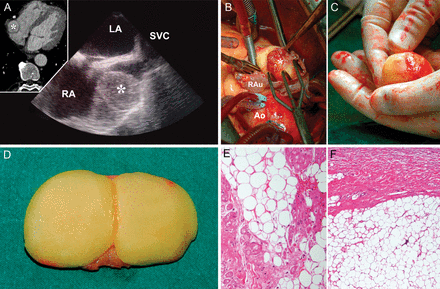
(A) Transoesophageal echocardiography and 64-slice multidetector computed tomography angiogram (small window) showing the lipoma as a 30 × 35-mm capsulated mass attached to the lateral wall of the right atrium. (B) Surgical view: a lipoma bulging from the right atrium as a spherical mass. (C) A lipoma in the surgeon's hand. (D) Lipoma divided after removal. (E and F) Histological section at ×150 and ×200 magnification, respectively, showing adipose tissue intermixed with myocardium (haematoxylin and eosin stain). Asterisks denote the lipoma; RA: right atrium; LA: left atrium; SVC: superior vena cava; Ao: aorta; RAu: right auricle.
