


A 49-year old woman with a history of hysterectomy 4 years previously was found to have intravenous leiomyomatosis. Enhanced computed tomography (CT) revealed that the tumour had reached the right atrium from the right iliac vein through the inferior vena cava. (Fig. 1) The tumour was successfully removed by a single-stage operation (Fig. 2).
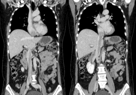
Figure 1:
Multi-detector CT showed the tumour arising from the right internal iliac vein and extending into the right atrium through the inferior vena cava.
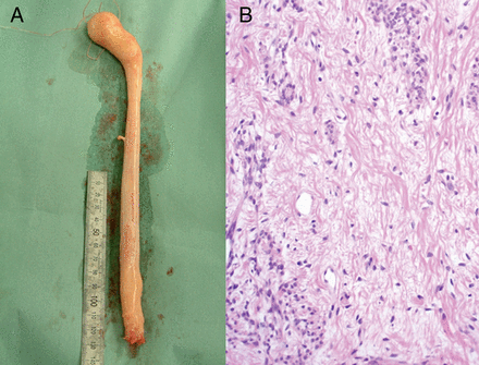
Figure 2:
(A) The 43 cm-long white tumour removed from the right atrium. (B) Histology image of the excised specimen showed benign smooth muscle cells and stroma cells with myometrial vessels (haematoxylin and eosin stains, original magnification ×100).
© The Author 2013. Published by Oxford University Press on behalf of the European Association for Cardio-Thoracic Surgery. All rights reserved.
