


Abstract
Surgical adhesives are frequently used after pulmonary resection to prevent or reduce pulmonary air leakages, since leakages may cause complications delaying the removal of chest drainage tubes and prolonging in-hospital stay. In this paper, we present 2 patients who underwent curative-intent pulmonary resection for non-small-cell lung carcinoma, in which the biological adhesive BioGlue® was used. Follow-up fluoro-2-deoxy-d-glucose positron emission tomography/computed tomographic (FDG-PET/CT) imaging revealed hypermetabolic pulmonary nodular lesions. Subsequent surgical exploration showed that the lesions were foreign body reactions to the bioadhesive. To our knowledge, this is the first study to examine false-positive follow-up FDG-PET/CT scans caused by the use of BioGlue® in pulmonary resection procedures.
Surgical adhesives such as BioGlue®, which is a bioadhesive composed of purified bovine serum albumin and glutaraldehyde, are frequently used after pulmonary resection to control air leakages. Fluoro-2-deoxy-d-glucose positron emission tomography/computed tomographic (FDG-PET/CT) imaging is a precise tool for detecting non-small-cell lung carcinoma (NSCLC) recurrence in asymptomatic patients after a curative-intent surgical procedure. The presence of bioadhesives should be taken into account when performing differential diagnosis of false-positive follow-up FDG-PET/CT imaging [1, 2], since a foreign body reaction to the bioadhesive may lead to unnecessary biopsy or surgery.
CASE REPORTS
Patient 1
A 66-year old man with a history of smoking was found to have a pulmonary nodule in June 2008. FDG-PET/CT imaging revealed a hypermetabolic nodule (maximum standardized uptake value, SUVmax 7.32) in the left upper lobe (Fig. 1A). Left thoracotomy and atypical pulmonary resection were performed. Stapler suture was performed, followed by the application of BioGlue® to the suture line. The final diagnosis was squamous cell carcinoma of 1.5 cm diameter, pT1aN0M0 (Stage IA) according to the seventh edition of the IASLC TNM Classification of malignant tumours. Adjuvant treatment was not administered. FDG-PET/CT imaging performed 6 months after surgery revealed the presence of hypermetabolic foci in the resected area of the left upper lobe, with a SUVmax of 4.66 suggestive of tumour recurrence (Fig. 1B). Re-thoracotomy was required, revealing the presence of fibrotic tissue in the surgical site; we found an encapsulation surrounding the area where the bioadhesive was applied (Fig. 1C). Histological study showed a lesion compatible with foreign body granuloma (Fig. 1D). In May 2012, the patient remains disease free.
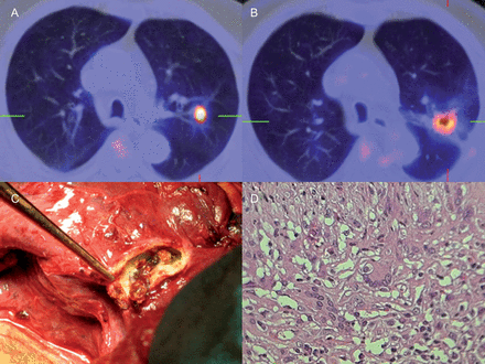
(A) FDG-PET/CT before tumour resection. (B) Hypermetabolic foci in the upper lobe of the left lung in the resected area 6 months after surgery. (C) Fibrotic tissue surrounding the BioGlue® area. (D) Histological section of the fibrous scar tissue showing a combined infiltration composed of polymorphonuclear neutrophils, eosinophils, histiocytes, plasma cells and foreign body multinucleated giant cells.
Patient 2
A 49-year old woman with a history of smoking was found to have a nodule in the right lung. FDG-PET/CT revealed a hypermetabolic focus in the medium lobe with SUV of 2.88. In July 2009, the patient underwent right thoracotomy and atypical pulmonary resection for the removal of the nodule. The diagnosis of the intraoperative frozen section was squamous cell carcinoma. Lobectomy of the middle lobe was completed followed by the application of BioGlue® to the parenchymal laceration site adjacent to the pulmonary hilum. The tumour was 1.8 cm in diameter and involved the visceral pleura (pT2aN0M0, Stage IB). Adjuvant treatment was not provided. Follow-up FDG-PET/CT was performed 6 months after surgery, revealing an uptake focus in a nodule in the surgical site with a SUV of 3.27. Malignancy was not suspected because of the wide surgical margins achieved and the recent resection, which were not suggestive of tumour recurrence. FDG-PET/CT was performed at 3 months, revealing an increase in the metabolic rate (SUV of 3.91) suggestive of tumour recurrence. Re-intervention was required. We found BioGlue® rests surrounding the fibrotic tissue adjacent to the pulmonary hilum. Biopsy revealed giant multinucleated cells caused by a foreign body reaction. In May 2012, the patient was disease-free.
COMMENT
The detection of new hypermetabolic pulmonary foci on FDG-PET/CT in asymptomatic patients with resected NSCLC is a diagnostic challenge. The reason is that physicians may take these hypermetabolic foci for a new tumour, which may lead to unnecessary biopsies or surgery.
Persistent air leakages after pulmonary resection are a common complication. They are generally parenchymal leakages (alveolar leakages) induced by surgery, or they may be caused by the use of metal staples in emphysematous areas. Air leakagess are generally minor complications that are spontaneously resolved in a few days. However, they may prolong in-hospital stay and require re-intervention. Although surgical adhesives or sealants such as BioGlue® Surgical Adhesive (CryoLife, Inc., Kennesaw, CA, USA) will never replace appropriate surgery, they are extensively used in surgical procedures. BioGlue® is a surgical adhesive composed of purified bovine serum albumin and glutaraldehyde [3]. In the first case, the adhesive was used as an adjunct to suture with staples, while in the second, it was applied to the lacerated parenchyma.
Following the application of BioGlue® to a surgical area, there is an inflammatory response consisting of polymorphonuclear neutrophils, macrophages and granulation tissue. Granulation tissue progressively becomes fibrous scar tissue [3]. In exceptional cases, healing is complicated by a chronic foreign body inflammatory reaction to the bioadhesive.
PET provides metabolic information by the injection of radiotracers, the most common being an analogue of glucose (18F-FDG). PET-CT is a multimodality technique where a PET and a CT are superposed to provide both anatomical and metabolic information. FDG-PET/CT is highly sensitive in the detection of NSCLC recurrence. However, its specificity is limited because of the elevated FDG concentrations caused by postoperative inflammation. To prevent misinterpretations, it is recommended that FDG-PET/CT is performed 2–3 months after radiotherapy or chemotherapy, or up to 1–2 months after surgery. In these cases, careful examination of the CT is essential, as it can provide useful information on postoperative inflammation [1]. FDG-PET/CT can also be used in postoperative follow-up evaluation for early recurrence in asymptomatic patients with resected NSCLC. To minimize false-positive results, some authors recommend performing another scan at 90 min after radiotracer injection (‘dual-time point imaging’) because inflammatory cells progressively suffer an FDG washout, which results in an SUV decrease with respect to the 60-min scan [4].
Also, false-positive results have been reported to be caused by a foreign body inflammatory reaction to surgical materials—some left inside inappropriately, such as textile or metal elements—and other materials or agents appropriately used such as non-absorbable sutures, metal staples, Teflon, talc, etc. [5]. In the cases presented, false-positive results were obtained at least 6 months after surgery. In any case, false-positive FDG-PET/CT obtained as a result of a foreign body reaction can occur at any time during the follow-up period. The 2 patients exhibited granulomatous foreign body reactions to BioGlue®, which was applied to the sutured or lacerated pulmonary parenchyma.
In conclusion, operative reports should detail the surgical materials employed and the area where they were used. Pathological findings on follow-up FDG-PET/CT for early recurrence in asymptomatic patients with resected NSCLC should be interpreted with caution, especially if these findings are located in areas adjacent to the resected zone, in order to avoid unnecessary biopsies or surgical procedures. However, on suspicion of malignancy, surgery is indicated.
Conflict of interest: none declared.
