


Abstract
Optimized temporary bi-ventricular (BiV) pacing may benefit heart failure patients after on-pump cardiac surgery compared with conventional dual-chamber right ventricular (RV) pacing. An improvement in haemodynamic function with BiV pacing may reduce the duration of ‘Level 3’ intensive care.
Thirty-eight patients in sinus rhythm, ejection fraction ≤35%, undergoing on-pump surgical revascularization, valve surgery or both were enrolled in this study. Before closing the sternum, temporary epicardial pacing wires were attached to the right atrium, RV outflow tract and basal posterolateral wall of the left ventricle. Patients were randomly assigned to postoperative BiV pacing with the optimization of the atrio- (AV) and inter-ventricular (VV) pacing intervals (Group 1) or conventional dual-chamber right AV pacing (Group 2). The primary end-point was the duration of ‘Level 3’ intensive care. Secondary end-points included cardiac output which was measured by thermodiluation at admission to the intensive care unit and at 6 and 18 h later, in five different pacing modes.
The duration of ‘Level 3’ care was similar between groups (40 ± 35 vs 54 ± 63 h; Group 1 vs 2; P = 0.43). Cardiac output was similar in all pacing modes at baseline. At 18 h, cardiac output with BiV pacing (5.8 l/min) was 7% higher than atrial inhibited (5.4 l/min) and 9% higher than dual-chamber RV pacing (5.3 l/min; P = 0.02 and 0.001, respectively). Optimization of the VV interval produced a further 4% increase in cardiac output compared with baseline settings (P = 0.005).
Postoperative haemodynamic function may be enhanced by temporary BiV pacing of high-risk patients after on-pump cardiac surgery.
INTRODUCTION
Left ventricular (LV) function may be optimized after cardiac surgery with acute bi-ventricular (BiV) pacing. This is achieved by attaching temporary pacing wires to the right atrium (RA), right ventricle (RV) and LV before closing the sternum. Previous studies of permanent BiV pacing have reported acute improvements in haemodynamic function [1–4] but attempts to reproduce these findings acutely after cardiac surgery have produced mixed results [5–10]. In addition, one postoperative study has reported no improvement in coronary conduit flow with BiV pacing compared with atrial synchronous RV pacing [11].
There are several potential explanations for these findings. Most studies have used a fixed atrio-ventricular (AV) delay. Only one relatively small study (n = 11) investigated the effect of inter-ventricular (VV) optimization, which was beneficial [12], and earlier studies in stable (non-surgical) heart failure patients have suggested benefits from AV and VV optimization [13–17]. The site of LV wire may also influence the haemodynamic effect of BiV pacing. Flynn et al. [18] reported the superior cardiac index in patients paced from the posterior wall of the LV compared with the anterior wall.
Therefore, it is possible that temporary BiV pacing after cardiac surgery may improve postoperative haemodynamics and clinical outcomes when pacing algorithms are optimized. It remains unclear whether the benefits of temporary BiV pacing after cardiac surgery are restricted to patients with a preoperative bundle branch block.
The aim of this study was to investigate if temporary BiV pacing with the optimization of AV and VV intervals improves the outcomes after on-pump cardiac surgery in subjects with severe LV systolic dysfunction [ejection fraction (EF) ≤ 35%], irrespective of preoperative QRS width. We investigated the impact of temporary BiV pacing on the duration of ‘Level 3’ intensive care, acute haemodynamic function and major adverse events after cardiac surgery.
MATERIALS AND METHODS
We performed a two-centre, prospective, randomized, single-blinded, intervention controlled trial in patients with severely impaired LV systolic function (EF ≤ 35%) undergoing on-pump cardiac surgery for revascularization, valve surgery or both. The principal exclusion criteria were permanent atrial fibrillation, permanent pacemaker/defibrillator, infective endocarditis, hypertrophic cardiomyopathy and dialysis-dependent renal failure. The trial was registered at clinicaltrials.gov (NCT01027299) and approved by the local Research Ethics Committee. All patients provided written informed consent. A detailed description of the rationale and design of this study has been published [19].
After enrolment, a 12-lead electrocardiogram was performed to confirm sinus rhythm and to measure QRS duration. A transthoracic echocardiogram was performed (GE, Vivid 7, Horten, Norway) to measure LVEF (biplane Simpson), VV mechanical delay and intraventricular dys-synchrony (time difference to peak velocity in the basal septum and the basal lateral wall using myocardial velocity imaging). Blood samples were sent for the calculation of an estimated glomerular filtration rate and assays of troponin T and N-terminal brain natriuretic peptide (NT-pro-BNP).
Before surgery, all patients received a pulmonary arterial catheter (either 7.5F CCO: 139HF75P or 7F TD: 131 F7, Edwards Lifesciences, Irvine, USA). Cardiac output was also monitored from a radial arterial line (Flotrac MHD8R, Edwards Lifesciences, Dominican Rep.). Patients were randomized, using open access software that matched patients for key variables, into two groups: Group 1, BiV pacing and Group 2, standard pacing [atrial inhibited (AAI) pacing or dual-chamber RV pacing] [19].
Before weaning from the cardiopulmonary bypass circuit, temporary unipolar epicardial pacing wires were sutured to the roof of the RA, the RV outflow tract and the posterolateral-free wall of the LV (first obtuse marginal territory) avoiding scar tissue. The wires were attached via extension cables to a temporary external triple-chamber pacemaker with independently programmable AV and VV intervals, (Osypka PACE 300, Germany; Figs 1 and 2).
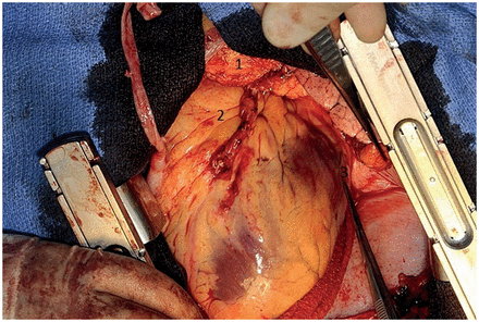
Position of temporary pacing wires (1, right atrium; 2, right ventricular outflow tract; 3, left ventricle, basal region of OM1). Two unipolar wires were attached at each site.
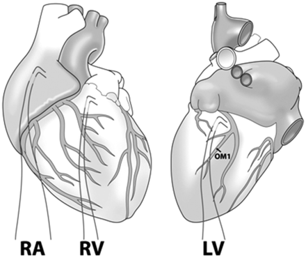
Position of temporary pacing wires (RA: right atrium; RV: right ventricular outflow tract; LV: basal region of OM1).
Pacing was initiated before separation from the cardiopulmonary bypass circuit. Group 1 received rate responsive BiV pacing, with the AV interval set at 120 ms and the VV interval set at the LV preactivation of −5 ms, a baseline rate of 90/min and a maximum tracking rate of 110/min. Group 2 received AAI pacing at a baseline rate of 90/min unless ventricular pacing was indicated for a postoperative AV block in which case rate responsive dual chamber (RV) pacing was programmed with the AV interval set at 150 ms. In both groups, pacing was continued for 48 h or until the transition from Level 3 to Level 2 intensive care (the primary end-point), whichever occurred first.
Acute haemodynamic studies were performed using the PA catheter on admission to cardiac intensive care, and then at 6 and 18 h later. At 18 h, AV and VV interval optimization was performed. A range of AV delays from 80 to 180 ms and VV delays from −40 to 40 ms were interrogated at 20 ms increments; the optimal settings were selected using the Flotrac device. The glomerular filtration rate, troponin T and NT-pro-BNP were measured immediately after the operation and at 18, 48 and 72 h later.
The prespecified primary end-point was the transition from Level 3 to Level 2 intensive care. This was defined as the time point after extubation, when weaning haemodynamic support to either a single inotrope/vasoconstrictor or an intra-aortic balloon pump. Prespecified secondary end-points were the comparison of haemodynamic data, duration of ventilation/inotropic support, arrhythmias, change in renal function/troponin T/NT-pro-BNP and in-hospital mortality.
The estimated total sample size was 34 [19]. The data were analysed for normality and unpaired t-tests were used to compare the two groups for the primary end-point. Within-group comparisons of haemodynamic data were performed using paired t-tests.
The temporary wires were removed on the cardiac surgical ward. In patients receiving anticoagulation, the INR was ≤2.5 and each patient was monitored for at least 1 h after the procedure.
RESULTS
Between January 2010 and July 2011, 55 patients were enrolled in the study and 38 patients completed the protocol. Four patients were excluded at the request of the cardiac surgeons, three patients could not be included when operations were conducted simultaneously at different hospitals, two patients had ‘off-pump’ surgery, two had angioplasty, in two patients EF improved >40% and four patients were excluded because of emergency surgery, palliative care, failure to float a pulmonary arterial catheter or latex allergy (total, n = 17).
Preoperative data
The baseline demographics for the 38 patients who completed the trial are illustrated in Table 1. The two groups were well matched. For all participants, the mean age was 67.4 ± 11.4 years and 79% were male. Prior to surgery, 28 patients (74%) had a narrow QRS duration ≤120 ms and 24% required a preoperative intra-aortic balloon pump as directed by local protocols or the surgeon's preference.
Clinical characteristics of the patients who completed the trial protocol
| Demographic | Group 1: BiV pacing (n = 19) | Group 2: Standard pacing (n = 19) | P-value |
|---|---|---|---|
| Age (years) | 69.2 ± 9.3 | 65.5 ± 12.7 | 0.32 |
| Male (%) | 79 | 79 | 1.0 |
| New York Heart Association functional classification system score | 2.5 ± 0.7 | 2.4 ± 0.9 | 0.18 |
| Canadian Cardiovascular Society score | 1.9 ± 0.9 | 1.8 ± 0.8 | 0.71 |
| Angiotensin converting enzyme inhibitor (%) | 58 | 47 | 0.52 |
| Beta blocker (%) | 63 | 84 | 0.14 |
| LVEF (%) | 26.1 ± 5.4 | 28.0 ± 7.3 | 0.38 |
| End-diastolic volume (ml) | 173.93 ± 66.5 | 161.0 ± 39.2 | 0.47 |
| End-systolic volume (ml) | 132.6 ± 65.0 | 115.5 ± 29.0 | 0.30 |
| Prior myocardial infarction (%) | 80 | 89 | 0.37 |
| Chronic kidney disease score | 2.2 ± 0.9 | 1.9 ± 0.6 | 0.28 |
| PR interval (ms) | 176 ± 30 | 174 ± 28 | 0.87 |
| QRS duration (ms) | 113.3 ± 29.3 | 115.2 ± 24.4 | 0.84 |
| Left bundle branch block (%) | 24 | 29 | 0.40 |
26% of the patients had an intraventricular mechanical delay (≥65 ms difference between the time-to-peak septal and lateral wall velocities) and 26% had a VV mechanical delay (≥40 ms difference from the onset of pulmonary to the aortic ejection period). Overall, 61% of the patients had evidence of either electrical or mechanical dys-synchrony before surgery. Only 11% of the patients had an EF of <20%.
Surgical data
Twenty-three (61%) patients received CABG only, 10 (26%) had CABG and valve surgery and 5 (13%) had valve only surgery. Thirty-three (87%) patients received surgical revascularization with 3.2 ± 1.2 grafts including an arterial conduit in 70%. Valve replacements included 13 aortic valves, 3 mitral valves and 1 double-valve operation (Table 2). The duration of cardiopulmonary bypass and the aortic cross clamp time were similar between groups.
Surgical details
| Demographic | Group 1 (n = 19) | Group 2 (n = 19) | P-value |
|---|---|---|---|
| Coronary revascularization (n) | 15 | 17 | 0.37 |
| Number of grafts (n) | 3.2 ± 1.2 | 3.3 ± 1.3 | 0.89 |
| Arterial conduit (n) | 13 | 15 | 0.46 |
| Valve surgery (n) | 7 | 8 | 0.73 |
| Cardiopulmonary bypass time (min) | 127 ± 41 | 144 ± 46 | 0.25 |
| Aortic cross clamp time (min) | 86 ± 34 | 90 ± 28 | 0.63 |
| Intra-aortic balloon pump (n) | 5 | 4 | 0.70 |
The LV wires were sutured to the basal posterolateral wall adjacent to the first obtuse marginal artery in 37 cases. One lead was placed in a more anterior position due to LV scar tissue at the intended site of implantation. Phrenic nerve stimulation was observed in three cases but this was successfully terminated by programming a lower LV output while maintaining BiV capture. The duration of BiV pacing was 22.7 ± 8.8 h in Group 1.
Duration of Level 3 care
The primary end-point was the duration of Level 3 care. For the majority of patients, this was achieved after extubation, and when inotropic support was weaned to a single infusion. The duration of Level 3 care was 40.4 ± 35.0 h in the BiV pacing group and 54.1 ± 63.0 h in the standard pacing group (P = 0.42; Fig. 3). Two patients in Group 2 had a prolonged duration of Level 3 care: one patient had postoperative tamponade requiring resternotomy and the removal of clots with subsequent haemofiltration for acute renal insufficiency, and the other required prolonged inotropic support with two infusions for 238 h despite being extubated at 10 h. One patient in Group 1 developed atrial fibrillation and required 129.5 h of Level 3 care. Excluding these outliers would reduce the duration of Level 3 care to 35.5 ± 28.3 h in the BiV and 35.3 ± 28.9 h in the standard pacing group (P = 0.99).
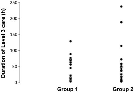
Duration of Level 3 care for BiV (Group 1) and standard pacing (Group 2).
Haemodynamic data
The haemodynamic measurements taken at baseline, 6 and 18 h, in five different pacing modes are shown in Fig. 4. The baseline measurements of cardiac output were similar, with the exception of VVI pacing which reduced cardiac output by 15% compared with the AAI mode (P < 0.001). At 18 h, BiV pacing increased cardiac output by 16% compared with baseline measurements (P = 0.007). Comparison of the various pacing modes at 18 h demonstrated that BiV pacing produced the greatest augmentation of cardiac output (7%) compared with standard AAI pacing (P = 0.02). No other pacing mode was better than AAI pacing.
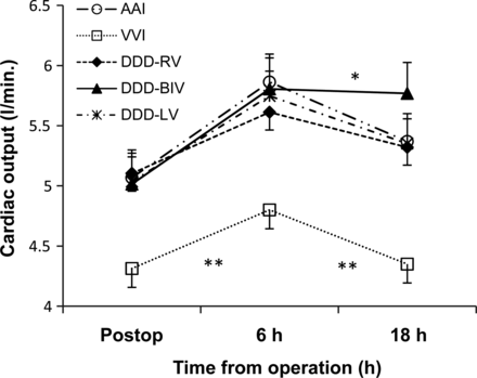
Cardiac output in different pacing modes after cardiac surgery (mean value ± SEM). *P < 0.05 BiV pacing compared with all pacing modes. **P < 0.001 VVI pacing compared with all pacing modes.
Optimization of AV/VV intervals
AV optimization did not increase cardiac output compared with the standard default (AV interval of 120 ms); the mean change was 0.13 ± 0.22 l/min (P = 0.19). Optimization of the VV delay augmented cardiac output by 0.22 ± 0.22 l/min (P = 0.005). The modal optimal setting was LV pre-activation by 20 ms.
Inotropes
The duration of inotropic/vasoconstrictor support was similar between groups: phosphodiesterase inhibitor 37.5 ± 31.2 vs 49.1 ± 59.9 h (P = 0.47) and noradrenaline 50.4 ± 37.8 vs 54.3 ± 60.3 h (P = 0.81) in Group 1 compared with Group 2, respectively. Likewise, the doses of enoximone, milrinone and noradrenaline adjusted for weight during the study did not differ between the two groups: 3.5 ± 1.7 vs 5.0 ± 3.2 mg/kg (P = 0.25), 0.6 ± 0.6 vs 0.6 ± 0.6 mg/kg (P = 0.99) and 0.3 ± 0.4 vs 0.3 ± 0.5 mg/kg (P = 0.97), respectively, for Groups 1 and 2.
Biochemical markers
There were no differences between the groups with respect to changes in postoperative renal function or biochemical markers of myocardial injury assessed using troponin T (Table 3). The postoperative change in NT-pro-BNP showed no difference between the pacing groups (Fig. 5). The baseline measurements were similar in Groups 1 and 2 (5434 ± 6874 vs 4577 ± 5447 pg/ml; P = 0.71, respectively).
Renal function and cardiac biomarkers after cardiac surgery
| Time | Renal function: estimated glomerular filtration rate (ml/min/1.73 m2) | Troponin T(ng/ml) | ||||
|---|---|---|---|---|---|---|
| Group 1 | Group 2 | P-value | Group 1 | Group 2 | P-value | |
| Preoperative | 70.1 ± 22.7 | 77.9 ± 24.6 | 0.31 | 0.05 ± 0.01 | 0.21 ± 0.66 | 0.26 |
| Postoperative | 80.0 ± 23.8 | 78.8 ± 22.0 | 0.22 | 0.70 ± 0.48 | 0.82 ± 0.79 | 0.66 |
| 24 h | 73.7 ± 26.0 | 71.6 ± 30.0 | 0.82 | 0.60 ± 0.28 | 0.84 ± 0.70 | 0.28 |
| 48 h | 75.9 ± 36.5 | 82.2 ± 37.7 | 0.61 | 0.37 ± 0.21 | 0.46 ± 0.51 | 0.59 |
| 72 h | 83.2 ± 40.0 | 91.6 ± 44.7 | 0.53 | 0.29 ± 0.20 | 0.63 ± 0.70 | 0.13 |
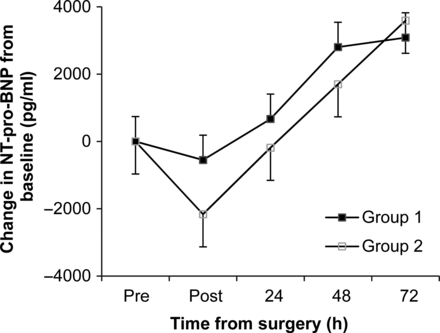
NT-pro-BNP: change in the measurement compared with the baseline. Values are given as the mean ± SEM (pg/ml).
Markers of dys-synchrony
There was no change in the VV mechanical delay after surgery in the entire cohort (preoperative: 22.7 ± 31.1 ms vs postoperative: 27.0 ± 27.1 ms; P = 0.94) or within each group. Likewise, there was no increase in the intraventricular delay (preoperative: 100 ± 19 ms vs postoperative: 70 ± 40 ms; P = 0.80). The paradoxical motion of the upper ventricular septum was observed in all postoperative transthoracic echocardiograms, and the pericardium was left open in all patients.
Complications
There were no deaths in the first 30 days after surgery. One patient in each group had a cerebrovascular accident and one subject in Group 1 and two subjects in Group 2 required haemofiltration. There were eight episodes of atrial fibrillation in both groups.
The additional pacing wire did not cause any adverse events or haemorrhagic complications. During the study, one patient developed a transient second-degree heart block after aortic valve surgery, and no permanent pacing systems were required before hospital discharge.
DISCUSSION
Optimized temporary BiV pacing improved cardiac output at 18 h compared with all other pacing modes but did not influence the duration of Level 3 care. BiV pacing did not influence the frequency of postoperative arrhythmias, renal dysfunction or biomarkers of myocardial function.
Length of stay in cardiac intensive care (Level 3)
Despite the improvements in haemodynamic function with optimized temporary BiV pacing compared with AAI pacing, there was no overall reduction in the duration of Level 3 care (40.4 ± 35.0 vs 54.1 ± 63.0 h; P = 0.43, respectively). This observation is consistent with previous studies of temporary BiV pacing after cardiac surgery [6, 8]. We elected to use the duration of Level 3 care rather than the duration of intensive care to reduce the potential for confounding variables in the primary end-point. Currently, only one trial (n = 178) has reported a significant reduction in intensive care stay after cardiac surgery with permanent BiV pacing compared with no BiV pacing [5]. However, the duration of Level 3 care in the BiV pacing group in our study was significantly shorter than in the previous study (40.4 ± 35.0 vs 60.0 ± 12 h; P = 0.02) [5].
Haemodynamic changes
In this study, optimized temporary BiV pacing significantly improved cardiac output compared with AAI pacing, at 18 h. Although a difference in these two pacing modes was not apparent immediately after cardiac surgery, acute changes in fluid status, invasive ventilation, heart rate, inotropes and vasopressor infusions may in part explain these findings. At 18 h, 29 of 38 (76%) patients were extubated compared with all subjects requiring ventilation at baseline assessment.
The 7% improvement in cardiac output at 18 h was observed in a heterogeneous population of surgical candidates requiring revascularization and/or valve surgery irrespective of baseline QRS duration. Despite only 10 of 38 (26%) patients having a QRS duration of over 120 ms, 8 of 38 (21%) had a VV mechanical delay and 10 of 38 (26%) had an intraventricular mechanical delay. Preoperative assessment identified that only 23 of 38 (61%) patients had evidence of electromechanical dys-synchrony. In addition, all subjects developed a paradoxical septal motion after surgery which is related to regional myocardial oedema after bypass [20] and invasive ventilation after chest wall closure [21].
AV/VV delay optimization
Optimization of the VV delay produced a significant improvement in cardiac output compared with simultaneous pacing (0.22 ± 0.22 l/min; P = 0.005). This may explain the beneficial effect of BiV pacing compared with AAI pacing in our study compared with previous trials [6, 8, 9]. There are only limited data on postoperative VV optimization after cardiac surgery. Hamad et al. [12] reported an additional response to postoperative BiV pacing after VV optimization (n = 11), using invasive LV dP/dT measurements. Optimization of the AV delay was limited in the postoperative period. The baseline PR interval measured 177.7 ± 30.4 ms and only 6 of 38 subjects were able to maintain BiV pacing with an AV delay ≥180 ms.
Limitations
The primary end-point of Level 3 care was selected, because it has a precise definition and it is used in the critical care environment. We consider this primary end-point to be more robust than measuring the length of intensive care stay. The limitations of using ‘Level 3 care’ are that a few patients will reach the end-point after extubation, but the majority of patients wait until they are supported by only one inotrope. In addition, the use of inotropes is a binary field and does not take into account the concentration of inotrope that is prescribed during ‘Level 3 care’.
Optimization of the VV interval produced a small but statistically significant improvement in cardiac output. Optimizing the VV interval at an earlier stage may have influenced the primary end-point as the duration of optimized BiV pacing was limited to 5 h.
We excluded patients with permanent atrial fibrillation from our study. However, it is possible that such patients may benefit from postoperative BiV pacing.
Clinical implications
Temporary BiV pacing should be considered for all patients with severe LV systolic impairment after cardiac surgery. Patients who require postoperative bradycardia pacing or who are likely to require a prolonged ITU admission have the most to gain from this treatment. Future studies should consider postoperative BiV pacing in patients with permanent AF and investigate the patterns of regional dyskinesis and septal dys-synchrony in more detail, in relation to the optimal site and mode of pacing.
Funding
This work was supported by an educational grant to Stuart J. Russell from St Jude Medical UK. Zaheer R. Yousef is an NISCHR research fellow.
Conflict of interest: none declared.
ACKNOWLEDGEMENTS
We would like to acknowledge the Cardiothoracic Directorates at the University Hospital of Wales, Cardiff, and Morriston Hospital, Swansea, for their support of this research trial. In addition to our research group, we would like to thank Mr Youhana, Mr Kumar, Miss Deglurkar, Mr Azzu, Mr Kulatilake and Mr Mehta for enrolling patients into our trial. David Gaze, St George's Hospital, London, assisted with the biochemical analysis. Matthew Briggs created the pacing diagrams (Royal Preston Hospital, Lancashire, UK) and Mr Mehta supplied the photograph. The Heart Research Fund for Wales kindly provided funds to purchase the external pacemakers.
