-
PDF
- Split View
-
Views
-
Cite
Cite
Frank Edwin, Robin H. Kinsley, Hendrick M. Mamorare, Kenny Govendrageloo, The spectrum of double-outlet right atrium including hearts with three atrioventricular valves, European Journal of Cardio-Thoracic Surgery, Volume 41, Issue 4, April 2012, Pages 947–949, https://doi.org/10.1093/ejcts/ezr073
Close - Share Icon Share
Abstract
Double-outlet right atrium (DORA) is characterized by simultaneous right atrial emptying into both ventricles. Ventriculoatrial septal malalignment is the cardinal morphological feature. Three cases are presented to depict two major types of DORA—DORA with a malaligned atrial septum and DORA with a malaligned ventricular septum. We describe two subtypes of each form of DORA: DORA with a malaligned atrial septum presents with either a common atrioventricular (AV) junction (guarded by a common AV valve) or with a single AV junction (due to the absence of the left AV junction). DORA with a malaligned ventricular septum may be associated with a right ventricle (RV) that is adequate for biventricular repair or a severely hypoplastic RV not compatible with biventricular repair. DORA with a malaligned ventricular septum is closely related to typical straddling of the tricuspid valve. Peculiarly, DORA with a malaligned ventricular septum presents three AV valves at the AV junction and is associated with an abnormal disposition of the AV conduction axis. Clear understanding of the morphology of these lesions is important in preventing a surgical misadventure at the crux of the heart.
INTRODUCTION
Double outlet atrium has been described as a condition in which the right or the left atrium empties into both ventricles [1]. Double-outlet right atrium (DORA) is a consequence of malalignment between the atrial and ventricular septa. In normal subjects, the atrial and ventricular septal planes intersect with the plane of the atrioventricular (AV) junction at the echocardiographic crux cordis. In DORA, either the atrial or the ventricular septum fails to intersect at this echocardiographic crux resulting in two distinct types of DORA. The first type is DORA with a malaligned atrial septum due to leftward rotation of the atrial septum; this is often associated with a common AV junction guarded by a common AV valve [2]. In a severer substrate of this type, the rotated atrial septum fuses with the left parietal AV junction resulting in an absent left AV junction and a single right AV valve; an inter-atrial communication decompresses the left atrium [3]. These two subtypes of DORA are distinct both morphologically and therapeutically—the former has a common AV valve and is amenable to biventricular repair; the latter has a single AV valve and a single ventricle repair is the treatment of choice. Kiraly et al. [3] suggested that the marked difference in surgical options means that it is better to separate the two entities in terms of anatomy and classification.
Rightward deviation of the ventricular septum without an associated ventricular septal defect (VSD) results in the second type of DORA (DORA with the malaligned ventricular septum) in which the right AV valve is divided into two components, one on either side of the deviated ventricular septum. The right-sided component of the valve opens from the right atrium into a small right ventricle (RV), while the left-sided component opens into the larger left ventricle (LV). The mitral valve opens separately into the LV, resulting in three AV valves in this type of DORA [4]. First reported by Büchler et al. [5], this type of DORA is closely related to a typical straddling tricuspid valve with the important difference that the latter features a VSD which is absent in the former. We report our experience and propose a management-based classification of these extremely rare anomalies.
CASE 1
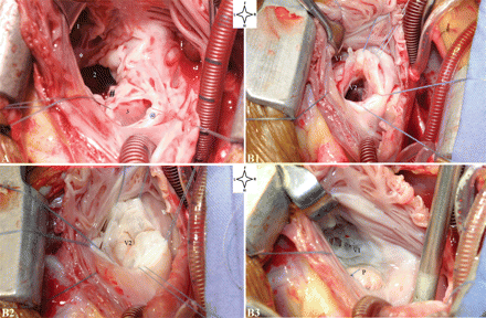
Echocardiography demonstrated a common AV junction with a leftward rotation of the atrial septum and a restrictive primum atrial septal defect (ASD) creating DORA in a 13-year-old girl. Angiocardiography showed reversible hypertensive pulmonary vascular disease. Under cardiopulmonary bypass (CPB), the malpositioned atrial septum (Fig. 1A) was excised; a two-patch reconstruction of the common AV junction was performed restoring a normal ventriculoatrial septal alignment. The post-operative course was uneventful.

(A) The operative view from the right atrium (case 1) showing the right AV valve (1), the left AV valve (2) and the ventricular septal crest (*) between the two. The lower rim (@) of the posteriorly deviated atrial septum has been retracted to show the restrictive primum ASD (3) which was in proximity to the posterior rim (#) of the left AV valve. (B1) Operative view from right atrium (case 3) showing the two components (V1 and V2) of the right AV valve and an intact ventricular septum; V1 opens into the mildly hypoplastic RV; V2 (∼40% of the right AV valve) opens into the LV establishing a right atrium-to-left ventricle communication; (B2) demonstrates a competent V2 after saline injection of the LV; (B3) shows appearance of V1 (∼60% of the right AV valve) after patch closure (P) of V2. The mitral valve (not shown) opened separately into the left ventricle so that three distinct AV valves were present at the AV junction. Picture orientation: F: foot; H: head; L: left; R: right.
CASE 2
In a 6-year-old girl, echocardiography showed a primum ASD with the leftward rotation of the atrial septum creating DORA. There was no evidence of pulmonary hypertension. The atrial septum was refashioned under CPB to restore a normal ventriculoatrial septal alignment and close the ASD. The left AV valve zone of apposition was closed. Post-operative recovery was uneventful.
CASE 3
A 2-year-old boy was evaluated on account of easy fatigue and a heart murmur. Echocardiography showed a secundum ASD, rightward deviation of the ventricular septum with a suspected straddling tricuspid valve but no VSD. Intraoperative findings are shown in Fig. 1 (B1–B3). Using CPB, bovine pericardial patches were used to close both the right atrium-to-left ventricular valved communication (V2) and the ASD. Recovery was uneventful.
DISCUSSION
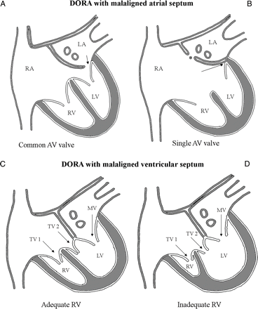
Earlier workers classified DORA morphologically on the basis of atrial or ventricular septal deviation and the number of AV valves present [6, 7]. We adopted a management-based classification incorporating those of the earlier workers and recognize two major types of DORA (one with the malaligned atrial septum and the other with the malaligned ventricular septum); each has two subtypes (Fig. 2).

(Upper panel) Leftward rotation of the atrial septum results in DORA with the malaligned atrial septum. Two subtypes are described: one with a common AV junction (guarded by a common AV valve) shown in (A), and the other with a single AV junction (due to the absence of the left AV junction) shown in (B). A restrictive primum ASD (arrow, A) is usually present in the first subtype. An interatrial communication (asterisk, B) decompresses the LA in the second subtype. (Lower panel) Rightward deviation of the intact ventricular septum results in DORA with the malaligned ventricular septum. Two subtypes result depending on the adequacy of the RV for biventricular repair. In the first subtype (C), the RV is adequate (though mildly hypoplastic) for biventricular repair. In the second subtype (D), the RV is inadequate (from severe hypoplasia) and only a single ventricle or one-and-half ventricle repair is possible. LA: left atrium; LV: left ventricle; MV: mitral valve; RA: right atrium; RV: right ventricle; TV: tricuspid valve.
In DORA with the malaligned atrial septum (Fig. 2, upper panel), the atrial septum is rotated leftward (as in cases 1 and 2). Importantly, the AV conduction axis arises from a regularly positioned AV node as is usual for AV septal defect. In the first subtype of this variant, two (common) AV valves guard a common AV junction, making biventricular repair possible. In the second subtype, there is only a single AV valve (due to absent left AV connection) making a single ventricle repair the treatment of choice.
In DORA with the malaligned ventricular septum (Fig. 2, lower panel), there is a rightward deviation of the ventricular septum associated with varying degrees of RV hypoplasia. Depending on the severity of RV hypoplasia, two subtypes are identified—the first is characterized by mild RV hypoplasia so that biventricular repair is possible [8] as in case 3, whereas in the second marked RV hypoplasia makes only single ventricle (or one-and-half ventricle) repair feasible [6].
In contrast to DORA with the malaligned atrial septum, an abnormal disposition of the conduction tissues occurs in DORA with the malaligned ventricular septum. Because of the ventricular septal malalignment, the AV conduction axis is unable to take origin from the regular AV node in the triangle of Koch. The conduction axis is rather located in the posterior margin of the muscular ventricular septum after taking origin from an anomalous AV node situated in the atrial myocardium where the muscular septum meets the right AV junction [9].
Peculiarly, DORA with the malaligned ventricular septum features three AV valves at the AV junction. The report of Gnanapragasam et al. [10] would suggest that in such hearts, the right AV junction straddled the malaligned ventricular septum during development. Then as the valvar leaflets developed, a dual orifice was formed within the straddling valve such that one orifice was committed to the left ventricle and the other orifice to the RV. The ventricular septal malalignment and associated abnormal disposition of the conduction tissues create a potential for surgical misadventure if not recognized.
The two forms of DORA are thus distinguished by the malalignment of either the atrial or the ventricular septum. The subtypes have prognostic implications depending on whether a biventricular or univentricular repair is the feasible option.
Conflict of interest: none declared.
REFERENCES
Author notes
Presented at the third Walter Sisulu Pediatric Cardiac Center for Africa (WSPCCA) International Cardiac Symposium at the Sunninghill Hospital, Johannesburg, 24–25 March 2010.


