


A 43-year-old man was hit in the left anterior thoracic wall while using a bush cutter. No hemodynamic changes were detected but computed tomography (CT) image revealed peri-aortic hematoma and a metallic foreign body (Fig. 1 and Video 1). Patient underwent surgery and was discharged on the 7th day post-operation (Fig. 2 ).
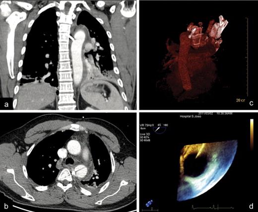
CT image revealed a peri-aortic hematoma, minimal left pleural effusion, and a 3 cm-long linear metallic foreign body in tight contact with the descending aorta (a)–(c). However, because of image artifacts, aortic wall integrity could not be ensured. Transesophageal echography unequivocally showed that one of the tips of the foreign body was inside the lumen of the vessel (d).
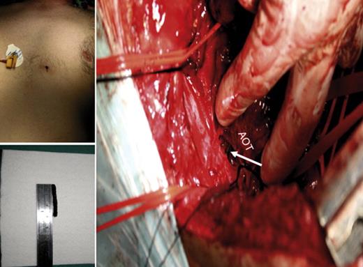
A postero-lateral thoracotomy was performed with removal of the object (arrow) and repair of the aortic wall perforation. The patient did not identify the piece as belonging to the bush cutter. AOT – thoracic descending aorta.
Appendix A Supplementary data
Supplementary data associated with this article (Video 1) can be found, in the online version, at doi:10.1016/j.ejcts.2011.05.033.
