


A 64-year-old man presented with effort angina, inferior T-wave depression, and normal troponin-I and echocardiograph.
Angiography showed left-main aneurysm, stenoses on left anterior descending and marginal, occluded left circumflex and right coronary, collateral circulation, and fistula connecting the left main and the diagonal branch, draining into pulmonary artery with two separate orthogonal dye-jets (Fig. 1 , Videos 1 and 2).
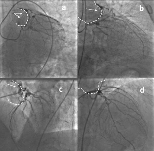
Left coronary angiography showing left coronary to pulmonary artery fistula, connecting left main to diagonal branch, draining with two dye-jets (white arrows) into pulmonary artery (dashed line), distal aneurysm of left main, severe stenosis on left anterior descending and marginal branch, occlusion of left circumflex and collateral circulation for right coronary. (a) Caudal view, (b) right caudal view, (c) left view and (d) right cranial view.
Appendix A Supplementary data
Supplementary data associated with this article can be found, in the online version, at doi:10.1016/j.ejcts.2011.05.040.
