-
PDF
- Split View
-
Views
-
Cite
Cite
Taner Sen, Yesim Guray, Edjon Hajro, Burcu Mecit Demirkan, Giant unruptured noncoronary sinus of Valsalva aneurysm with ascending aorta dissection, European Journal of Cardio-Thoracic Surgery, Volume 36, Issue 1, July 2009, Page 187, https://doi.org/10.1016/j.ejcts.2009.03.006
Close - Share Icon Share
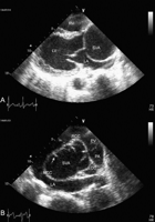
A 48-year-old woman was admitted with dyspnea. Echocardiography showed noncoronary sinus of Valsalva aneurysm (4.5 cm × 5.1 cm) compressing the left atrium. A flap-like appearance was visualized in this unruptured aneurysm (Fig. 1 , Videos 1 and 2). Computerized tomography revealed the aneurysm and the Debakey type 2 dissection flap (Fig. 2 ).

(A) In transthoracic echocardiography, the parasternal long axis view shows a cystic mass, a noncoronary sinus of Valsalva aneurysm, compressing the left atrium. (B) In the parasternal short axis view, the noncoronary sinus of Valsalva is aneurysmatic and a flap-like appearance (white arrow) can easily be seen. LV, left ventricle; LA, left atrium; RV, right ventricle; SVA, sinus of Valsalva aneurysm; NCC, noncoronary cusp; RCC, right coronary cusp; LCC, left coronary cusp; PV, pulmonary valve; PA, pulmonary artery.

Parts A, B and C are the computerized tomographic angiography images of different segments of the ascending aorta. The ascending aorta is dilated. The black arrow shows Debakey type 2 dissection flap at different segments of the ascending aorta.
Appendix A Supplementary data
Supplementary data associated with this article can be found, in the online version, at doi:10.1016/j.ejcts.2009.03.006.


