


Abstract
Objective: Prognosis of primary thymic carcinomas is poor due to advanced stage progression at diagnosis and highly malignant behavior. We retrospectively evaluated patients with thymic carcinoma to determine the prognostic factors. Methods: Sixty patients diagnosed and treated for thymic carcinoma from 1986 to 2005 were reviewed retrospectively. Influences of demographic characteristics, Masaoka stage, histologic grade, completeness of resection and adjuvant treatment on survival were evaluated. We defined complete resection as macroscopically and microscopically total resection of a tumor (R0 resection) and incomplete resection was subdivided into microscopic incomplete resection (R1 resection) or macroscopically incomplete resection (R2 resection). Results: There were 42 male and 18 female patients and mean age was 53.9 (±14.4) years old. The 5-year overall survival rate was 38.8% and median survival time was 35.6 months. The most common histologic type was squamous cell carcinoma (n = 29). In our study, 5 patients (8.3%) were in Masaoka stage I, 5 (8.3%) were in stage II, 19 (31.7%) were in stage III, 15 (25.0%) were stage in IVa, and 16 (26.7%) were in stage IVb. Among 40 patients who underwent surgical resection, complete resection was achieved in 14 patients. The 5-year survival rate after complete resection was 85.1% and was considered significantly better than those after incomplete resection (29.0%, p = 0.001) and non-surgical treatment (16.7%, p ≪ 0.001). But, no survival difference could be found between the incomplete resection group and non-surgical treatment group (p = 0.15). The 5-year survival rates of early Masaoka stage patients were significantly higher than advanced Masaoka stage (90.0% vs 28.3%, p = 0.001). The recurrence rates within 3 years after R1 resection (75.0%) were significantly higher than that after R0 resection (14.9%, p = 0.008). Conclusions: In thymic carcinoma, complete resection of early Masaoka stage lesions is the most important factor for disease control and long-term survival of patients.
1 Introduction
Primary thymic carcinomas are rare [1], and already have invaded neighboring organs, pleural or pericardial dissemination, and lymph node metastasis or distant metastasis at diagnosis. Compared to other thymic tumors, thymic carcinomas have malignant cytologic features and frequent metastases with subsequent poor prognosis [1–4]. However, there are few studies to determine prognostic factors or to evaluate the efficacy of treatment modalities for patients with thymic carcinoma due to the rarity of this tumor. Some studies suggest that tumor respectability, tumor stage, tumor histology grade, great vessel invasions, or postoperative radiotherapy have an impact on prognosis of thymic carcinoma [1,5–9]. Nevertheless, the clinicopathologic characteristics are still unclear. In this study, we retrospectively evaluated patients with thymic carcinoma at our institute to determine prognostic factors.
Materials and methods
Data of 60 patients, who were diagnosed and treated for thymic carcinoma from February of 1986 to January of 2005, were reviewed retrospectively and influences on survival of demographic characteristics, Masaoka stage, histologic grade, completeness of resection and adjuvant treatment were evaluated.
All pathological specimens and surgical reports were reviewed by a pathologist. Thymic carcinomas according to WHO classification, excluding cases with WHO classification type B3 thymoma (well-differentiated thymic carcinoma, WDTC), were included in this study, but cases with neuroendocrine tumor (n = 5) were also included in this series. Tumors except for neuroendocrine tumors are classified broadly as low or high-grade histology [2]. Low-grade histology includes well-differentiated squamous cell carcinoma, mucoepidermoid carcinoma, and basaloid carcinoma. All other types of tumor are classified as high-grade histology. Clinical or pathological staging in this study was performed based on Masaoka’s staging system [10].
Pretreatment evaluation of tumors was done by physical examination, chest radiography, chest computed tomography (CT) scan, abdominal CT scan, and bone scan. The criteria for considering a tumor to be unresectable were (1) apparent pleural or pericardial dissemination, (2) massive invasion of the great vessels or apparent invasion of the heart, or (3) hematogenous metastasis of another organ.
Preoperative chemoradiation therapy using cisplatin-based regimens (a combination of cisplatin, doxorubicin, vincristine, and cyclophosphamide [ADOC]) along with radiation therapy was performed to downstage unresectable tumors. If the tumor showed progressive downstage, operative procedures were subsequently performed.
For surgical resection, sternotomy was performed in most of the patients, who were treated for curative purpose, and thoracotomy or clamshell incision for the rest of the patients. Also, the thymus was completely resected, but extended thymectomy which was our routine practice for surgically treated myasthenia gravis patients was not performed routinely. We dissected loco-lesional lymph node that was located only in the anterior mediastinum and that was resected en-bloc with the thymus and the tumor.
Adjuvant treatment was considered an option for cases that underwent complete resection, and was performed routinely in cases with incomplete resection.
To determine the relationship between resectability and prognosis, clear definitions of complete resection and incomplete resection were necessary. We defined complete resection as macroscopically and microscopically total resection of a tumor (R0 resection) and incomplete resection was subdivided into microscopic incomplete resection (R1 resection) or macroscopically incomplete resection (R2 resection). Pleural seeding which was all removed macroscopically was also defined as R1 resection.
Statistical analysis of survival was performed using the Kaplan–Meier and univariate log-rank test. Statistical significance was defined as p ≪ 0.05. All statistical analyses were performed with the Statistical Package for Social Science (SPSS 12.0, Chicago, IL, USA).
2 Results
The patient population consisted of 42 males and 18 females with mean age of 53.9 ± 14.4 years. Forty-three out of 60 patients had clinical symptoms such as chest pain or discomfort (n = 17), dyspnea (n = 9), cough (n = 6), or SVC syndrome (n = 4). Except for one patient with Cushing’s syndrome, none of the subjects in this study had concomitant paraneoplastic syndromes such as myasthenia gravis or pure red cell aplasia.
Twenty-six patients were diagnosed with low-grade histology and 27 patients were in the high-grade histology. Five patients were diagnosed as having neuroendocrine tumors of the thymus. In two patients who underwent needle aspiration biopsy, histologic classification was not determined.
Twenty patients underwent definite chemoradiotherapy after tissue diagnosis. Forty patients out of 60 patients underwent surgical resection. Twenty-nine patients were treated with surgical resection followed by radiotherapy with or without chemotherapy. Nine patients were treated with chemotherapy following surgical resection. No adjuvant treatment was performed in two patients with R0 resection.
According to the Masaoka staging system, there were 5 patients with stage I disease (8.3%), 5 stage II (8.3%), 20 stage III (33.3%), 15 stage IVa (25.0%), 15 stage IVb (25.0%). Complete resection (R0) was achieved in 14 (35.0%) out of 40 patients who underwent surgical resection. There were 12 patients (30.0%) in R1 resection and 14 patients (35.0%) in R2 resection (Table 1 ).
Characteristics of patients.
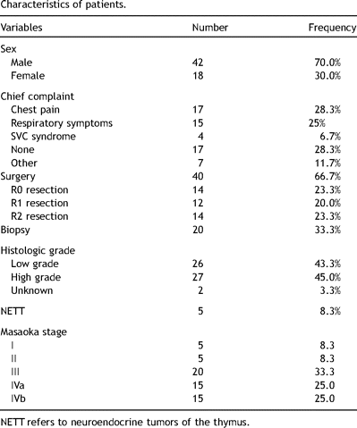
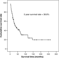
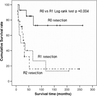
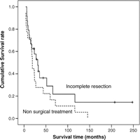
Fifty-nine patients were followed during a median of 35.6 months (range, 3–261 months). The overall 5-year survival rate was 38.8% (Fig. 1 ). Complete resection group showed significantly better prognosis than incomplete resection group (p = 0.001) (Fig. 2 ). However, 5-year survival rate of incomplete resection group was not different than non-surgical treatment group (p = 0.15) (Fig. 3 ). There were significant differences in prognosis among early Masaoka stage (I or II) and advanced Masaoka stage (III, IVa or IVb) (p = 0.001). But, no significant differences in prognosis could be found between stage III and stage IV (p = 0.18) (Fig. 4 ).
Overall survival rate curve of thymic carcinoma.
Completeness of resection and overall survival.
Overall survival of incomplete resection and non-surgical treatment group.
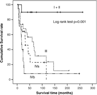
Survival rate curve according to Masaoka stage.
Mortality cases were mostly cancer related. But one patient with stage III died during the adjuvant chemotherapy after R1 resection due to pneumonia. Two deaths were related to cardiovascular complications, and both of them were stage III patients who have undergone R1 resection.
In R0 resection group, there was no significant difference in survival between surgery alone and surgery with adjuvant treatment (5-year survival, 50.0% vs 61.1%, p = 0.34).
3 Discussion
Complete resection and Masaoka stage are the determinant factors of the effectiveness in treating thymic carcinoma in this study. However, incomplete resection including debulking surgery was not significantly effective in treating thymic carcinoma compared with non-surgical treatment modalities.
Thymic carcinoma is a relatively rare malignant tumor among anterior mediastinal tumors. These tumors usually have poor prognosis due to their invasiveness, and high rate of recurrence. There are few studies that have analyzed the prognosis of thymic carcinoma in small numbers. Moreover, differences in prognostic factors of thymic carcinoma have been found in previous articles reported.
Complete resection in thymic carcinoma has been advocated as a prognostic factor in the majority of articles although some studies failed to show improved survival rates in complete resection group compared to incomplete resection group. Blumberg et al. reported that survival was dependent not on complete resection but on innominate vessel invasion [3]. This result, however, comes from the fact that 16 patients (37.2%) with well-differentiated thymic carcinoma (WDTC, WHO type B3 thymoma), which has shown better prognosis than thymic carcinoma, were included in that study. Although the concept of WDTC as a true primary thymic carcinoma remains controversial, WDTC was excluded in recent studies that contained analysis of prognostic factors in thymic carcinoma [11–14]. We also reported that survival rates of type B3 thymoma were statistically better than those of thymic carcinoma [15]. A review article by Chung suggested that WDTC may represent an intermediate stage of dedifferentiation of thymomas and has characteristics more in keeping with thymomas than with other thymic carcinomas. Chung also mentioned that the best survival trends were seen through analysis of 19 clinicopathological studies in completely resected lesions [16].
The role of incomplete resection including debulking surgery in thymic carcinoma has been questionable. Yano et al. reported that cases with subtotal resection demonstrated better prognosis than inoperable cases [12]. However, Kondo and Monden reported that debulking surgery is not valuable in thymic carcinoma [11]. Our series also showed that incomplete resection including R1 resection was not better in prognosis than inoperable cases. In this study, R1 resection unwillingly occurred in many instances. The presence of tumor in the resection margin of invading organ occurred in eight patients despite macroscopically complete resection and two patients who did not undergo extended thymectomy had presence of tumor in the cut surface of perithymic fat tissue. Two patients had pleural seeding which was all removed macroscopically. This result suggests that the microscopic confirmation of the resection margin during operation with subsequently more extended resection and radical resection of perithymic fat is necessary to prevent microscopic incomplete resection.
The Masaoka staging system [10], originally described for thymomas, has been adopted for thymic carcinomas but did not gain universal support. Tseng et al. reported that prognosis of thymic carcinoma seemed mainly dependent on tumor invasion of the great vessels, not on Masaoka staging [14]. Also, Ogawa et al. reported that the Masaoka staging system was not the prognostic factor of thymic carcinoma [13]. However, in those reports, any case in Masaoka stage I was not included. So, there were relatively fewer cases of early stage lesions than of advanced stage lesions in those studies. A meta-analysis in a review article including 10 patients with Masaoka stage I tumors revealed that stage I and stage II tumors have the best median survivals [16]. Our series including five patients (8.3%) in stage I also showed that the survival rate in early Masaoka stage is statistically higher than that in advanced Masaoka stage.
The role of adjuvant treatment in completely resected thymic carcinoma has not been established yet. Kondo et al. reported that there was no significant difference in survival between surgery alone and surgery with radiotherapy in completely resected thymic carcinoma. Our series also revealed that adjuvant treatment could not be found to be of significant effect in completely resected thymic carcinoma. However, considering selection bias and small number, no conclusion can be set on the efficacy of adjuvant treatment.
There are some limitations in this study. First, this study analyzed retrospectively only in a single center. Secondly, we could not identify any significant prognostic factors in resectable stage III thymic carcinoma patients as others did. This may be attributable to the fact that there were many unintended R1 resections. Conversely, this reminds us of the importance of microscopic complete resection once more.
4 Conclusions
In thymic carcinoma, completely resected lesions in the early Masaoka stages are the most important factors for disease control and long-term survival. However, incomplete resection including debulking surgery could not be proven to be of significant effect on thymic carcinoma compared with other treatment modalities. To prevent microscopic incomplete resection, histologic confirmation of the resection margin during operation and radical resection must be kept in mind.
Presented at the 22nd Annual Meeting of the European Association for Cardio-thoracic Surgery, Lisbon, Portugal, September 14–17, 2008.
Appendix A
Conference discussion
Dr D. Van Raemdonck (Leuven, Belgium): In your institution, in order to plan your treatment strategy in an individual case, is it important to have histology in advance? In other words, do you decide to give neoadjuvant therapy based on the Masaoka clinical staging or on the WHO classification according to the histology?
Dr Bae: There was not a treatment strategy, and to my knowledge, a lot of adjuvant treatment in thymic carcinoma has not been established yet.
Dr Van Raemdonck: You had a few patients with stage I?
Dr Bae: Yes, 5.
Dr Van Raemdonck: Grade C thymic carcinoma?
Dr Bae: Yes.
Dr Van Raemdonck: Did you know the histology in advance in these patients?
Dr Bae: No.
Dr Van Raemdonck: So you did primary surgery followed by adjuvant therapy in these patients?
Dr Bae: What is this?
Dr Van Raemdonck: You started with surgery?
Dr Bae: Oh, yes.
Dr Van Raemdonck: And then you gave adjuvant therapy, is that correct, in stage I?
Dr Bae: They were diagnosed from routine checkup. We don’t know preoperative histology. They are diagnosed with postoperative histologic confirmation.
Dr W. Klepetko (Vienna, Austria): I would like to ask you whether you have seen recurrence in these patients who were treated surgically and whether your decision to operate was dependent on the underlying histology.
Dr Bae: We did not analyze it according to histology. The recurrence rate within 3 years, there are definite differences in the incomplete resection group and the complete resection group. They have statistical significance. The incomplete resection group is higher than the complete resection group. The incomplete resection group showed 75% and the complete resection group 15% in 3 years.
Dr Klepetko: But did you see a place for surgery in primary complete resection patients when they had the localized recurrence?
Dr Bae: In the R0 resection group, local recurrence occurred in 2 cases, and overall, local recurrence occurred in a total of 9 cases.
Dr Klepetko: Did you operate on those patients?
Dr Bae: No.
